Point-of-Care Ultrasound Techniques for the Small Animal Practitioner
Здесь есть возможность читать онлайн «Point-of-Care Ultrasound Techniques for the Small Animal Practitioner» — ознакомительный отрывок электронной книги совершенно бесплатно, а после прочтения отрывка купить полную версию. В некоторых случаях можно слушать аудио, скачать через торрент в формате fb2 и присутствует краткое содержание. Жанр: unrecognised, на английском языке. Описание произведения, (предисловие) а так же отзывы посетителей доступны на портале библиотеки ЛибКат.

- Название:Point-of-Care Ultrasound Techniques for the Small Animal Practitioner
- Автор:
- Жанр:
- Год:неизвестен
- ISBN:нет данных
- Рейтинг книги:5 / 5. Голосов: 1
-
Избранное:Добавить в избранное
- Отзывы:
-
Ваша оценка:
Point-of-Care Ultrasound Techniques for the Small Animal Practitioner: краткое содержание, описание и аннотация
Предлагаем к чтению аннотацию, описание, краткое содержание или предисловие (зависит от того, что написал сам автор книги «Point-of-Care Ultrasound Techniques for the Small Animal Practitioner»). Если вы не нашли необходимую информацию о книге — напишите в комментариях, мы постараемся отыскать её.
New chapters in
cover ultrasound-guided nerve blocks, musculoskeletal, brain imaging, and applications of focused ultrasound techniques in cats, exotics and marine mammals—making it an essential purchase for veterinarians wanting to incorporate point-of-care ultrasound techniques into their veterinary practices.
Presents a standardized approach to point-of-care ultrasound as an extension of the physical exam, including trauma, non-trauma, and monitoring applications Includes coverage of new techniques for focused ultrasound exams, including lung, anesthesia and ultrasound guided nerve blocks, transcranial brain imaging, musculoskeletal, volume status evaluation, and rapid assessment for treatable forms of shock Adds cats, exotic and wildlife mammals, and marine mammals to the existing canine coverage Emphasizes the integration of point-of-care ultrasound techniques for optimizing patient care and accurate patient assessment Offers access to a companion website with supplementary video clips showing many clinically relevant didactic examples The second edition of
is an excellent resource for veterinary practitioners, ranging from the general practitioner to nearly all clinical specialists, including internal medicine, oncology, cardiology, emergency and critical care, anesthesiology, ophthalmology, exotics, and zoo medicine specialists, and veterinary students.
Point-of-Care Ultrasound Techniques for the Small Animal Practitioner — читать онлайн ознакомительный отрывок
Ниже представлен текст книги, разбитый по страницам. Система сохранения места последней прочитанной страницы, позволяет с удобством читать онлайн бесплатно книгу «Point-of-Care Ultrasound Techniques for the Small Animal Practitioner», без необходимости каждый раз заново искать на чём Вы остановились. Поставьте закладку, и сможете в любой момент перейти на страницу, на которой закончили чтение.
Интервал:
Закладка:
Potentially misses peritonitis and small‐volume bleeds in dehydrated or hypotensive patients. AFAST examinations should be applied minimally once after resuscitation and rehydration has taken place and as many times as necessary until you are assured that the patient is not surgical
Cannot replace a detailed complete abdominal ultrasound.
Cannot replace proper training.
Indications
In reality, AFAST and the Global FAST approach are everyday imaging modalities for nearly every patient as an extension of the physical examination. However, to better grasp this concept with baby steps to get to “an extension of the physical examination” mentality, we have bulleted the following.
All blunt and penetrating trauma cases as standard of care for screening for indirect evidence of intraabdominal injury.
All collapsed (both recovered and unrecovered) cases with unexplained hypotension, tachycardia, or mentation changes.
All anemic cases.
All “ain’t doing right” (ADR) cases.
All postinterventional, postsurgical cases, at risk for bleeding, infections, vascular complications.
All peritonitis suspects, including acute abdomen, for expedient diagnosis through the detection of free fluid (and sampling, fluid analysis testing as deemed appropriate).
Add‐on for all POCUS exams (abdomen, thorax, eye, brain) to make sure that forms of peritonitis and pleuritis, presence of bleeding, cardiac and pulmonary complications are not being missed that could have easily been detected with the Global FAST* approach.
*The Global FAST approach includes the combination of AFAST and its fluid scoring system and its target organ approach, the TFAST and Vet BLUE combined as part of the physical examination (see Chapters 36and 37).
Objectives
Perform the five acoustic windows of AFAST and accurately assign and abdominal fluid score.
Apply the “small‐volume bleeder versus large‐volume bleeder” principle to hemorrhaging subsets of small animal patients to better direct definitive therapy and decision making, including the need for blood transfusion and exploratory surgery and medical versus surgical management.
Recognize sonographic striation of the gallbladder wall referred to as the “halo effect,” “double rim effect” or “halo sign.”
Know additional rule‐outs (cardiac causes) for the collapsed or acutely weak dog with sonographic striation of the gallbladder wall.
Know additional rule‐outs for sonographic striation of the gallbladder wall in dogs and cats without acute collapse and weakness.
Recognize retroperitoneal free fluid and distinguish it from intraabdominal fluid.
Recognize pleural and pericardial effusion via the DH view by looking cranial to the diaphragm.
Know how to assess volume status through characterization of the caudal vena cava and its associated hepatic veins.
Know common artifacts and pitfalls at each respective AFAST view (see Chapter 6).
Final Note
The POCUS abdominal, thoracic, ocular, neurological, and musculoskeletal examinations described throughout this textbook, should as a general rule always include an AFAST and an assigned abdominal fluid score, and even better the Global FAST approach. By only performing a POCUS examination targeted at a specific organ or system, you risk missing obvious significant conditions to the patient's detriment. Integration of POCUS information is now similarly being advocated in human medicine like the author's Global FAST approach (Lichtenstein 2010; Narasimhan et al. 2016; Ha and Yo 2016).
Calculating the Abdominal Fluid Score
The AFAST‐applied fluid scoring system is a simple and easy to remember 0–4 scoring system developed over a decade ago (Lisciandro et al. 2009). Originally, each AFAST view was scored as 0 being negative or 1 being positive; however, the abdominal fluid scoring system has been further modified, explained subsequently based on more recent studies (Romero et al. 2015; Lisciandro et al. 2019). With this system, an abdominal fluid score (AFS) of 1 was defined as positive at any single AFAST view, an AFS of 2 was positive at any two views, an AFS of 3 was positive at any three views, and the maximum AFS of 4 was positive at all four views ( Figure 7.1). The impact of assigning an AFS is that in bleeding patients, an AFS of 1 and 2 is characterized as major injury/pathology, “small‐volume bleeder.” In contrast, an AFS 3 and 4 are characterized as major injury/pathology, “large‐volume bleeder” (Lisciandro et al. 2009; Lisciandro 2011, 2012). “Small‐volume” versus “large‐volume” effusions may also be semiquantitated regarding fluid loss (third spacing) into the abdominal cavity.
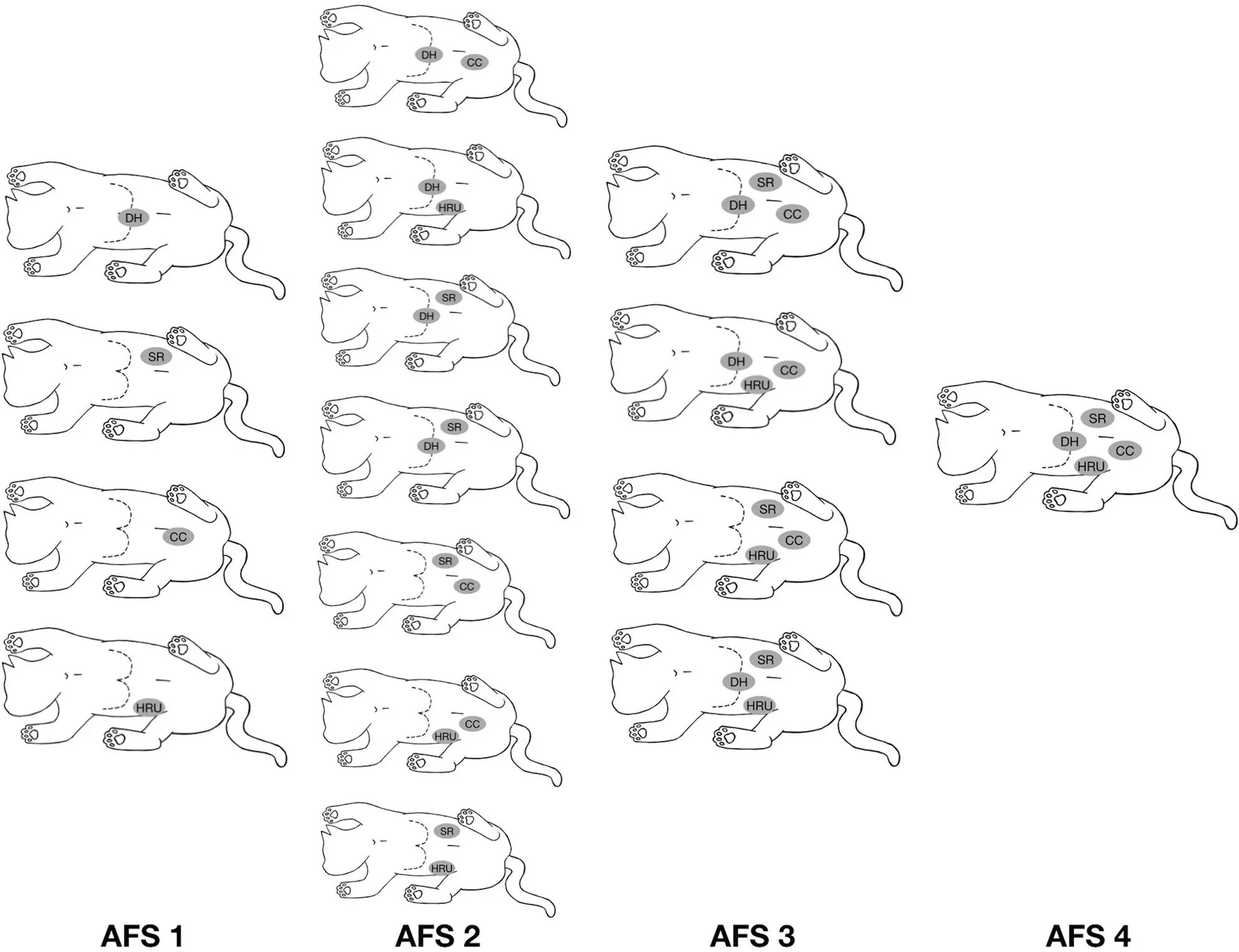
Figure 7.1. AFAST‐applied abdominal fluid scoring system. Calculating the abdominal fluid score (AFS) using the AFAST‐applied fluid scoring system shown on a cat placed in right lateral recumbency as a 0–4 system (the same system applies analogously to left lateral recumbency and to dogs). An AFS of 1 is a positive at any single view, an AFS 2 as any combination of two views, an AFS 3 as any combination of three views, and the maximum AFS of 4 when positive at all four views. Note the author has recently modified the AFS by assigning a score of ½ at views that have smaller pockets with maximum dimensions of ≤1 cm or >1 cm in dogs and ≤0.5 cm or >0.5 cm in cats based on more recent research (see Figure 7.2) (Romero et al. 2015; Lisciandro et al. 2019).
Source: Reproduced with permission of Dr Gregory Lisciandro, Hill Country Veterinary Specialists and FASTVet.com, Spicewood, TX. Illustration by Hannah M. Cole, Adkins, TX.
Modification of the Abdominal Fluid Scoring System – Using Maximum Dimensions
For several years the author has used a modified AFS system to better differentiate between small‐volume and large‐volume bleeder and effusions. The modified system now includes scores of 0, ½, and 1, which we refer to as the measurement approach (Lisciandro et al. 2015, 2019; Romero et al. 2015). The measurement approach categorizes positives as ½ or 1 dependent on the maximum dimension at each AFAST view ( Figure 7.2). Based on recent research since the first edition, we suggest an “under–over” of 1 cm (10 mm) in dogs and “under–over” of 0.5 cm (5 mm) in cats (Lisciandro et al. 2015, 2019; Romero et al. 2015). Thus, in a dog, when a maximum dimension at an AFAST view is ≤1 cm, it is scored as a ½, and if >1 cm, it is scored as 1; and in a cat ≤0.5 cm is scored as a ½ and >0.5 cm is scored as a 1 ( Figure 7.3; see also 7.8).
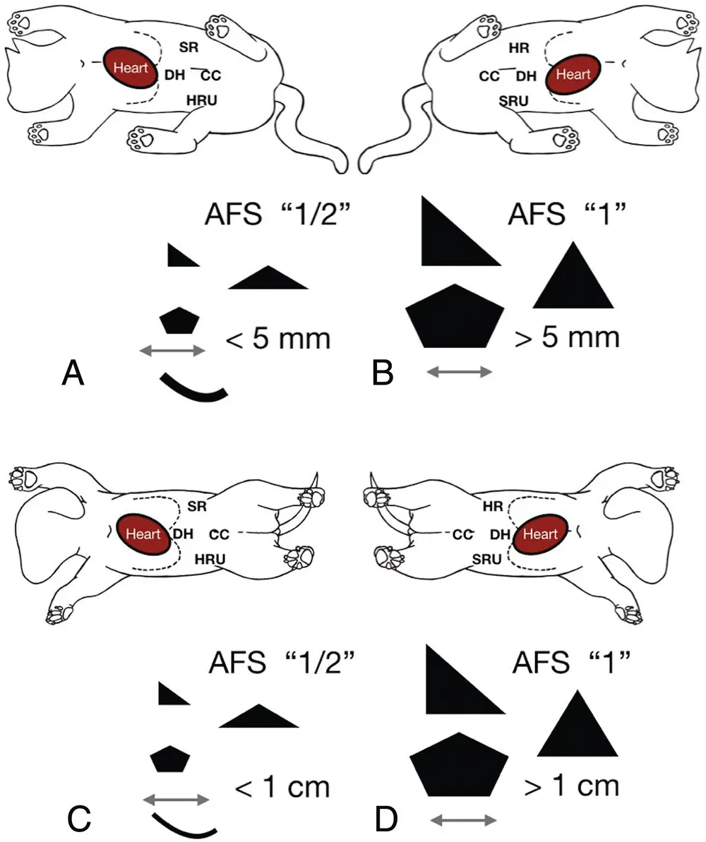
Figure 7.2. The “measurement” modification of the AFAST‐applied abdominal fluid scoring system. The figure shows the measurement abdominal fluid scoring (AFS) system. (A,B) Cartoons of a cat in right and left lateral recumbency and (C,D) cartoons of a dog in right and left lateral recumbency. The modification of the AFS better distinguishes between “small‐volume” and “large‐volume” bleeding (and other forms of nonhemorrhagic effusions) by assigning a score of ½ or 1 dependent on the maximum measured dimension at each AFAST view based on recently published studies (Lisciandro et al. 2015, 2019; Romero et al. 2015). The AFS is validated in either right or left lateral recumbency (Lisciandro et al. 2009). See also Figure 7.3.
Читать дальшеИнтервал:
Закладка:
Похожие книги на «Point-of-Care Ultrasound Techniques for the Small Animal Practitioner»
Представляем Вашему вниманию похожие книги на «Point-of-Care Ultrasound Techniques for the Small Animal Practitioner» списком для выбора. Мы отобрали схожую по названию и смыслу литературу в надежде предоставить читателям больше вариантов отыскать новые, интересные, ещё непрочитанные произведения.


Обсуждение, отзывы о книге «Point-of-Care Ultrasound Techniques for the Small Animal Practitioner» и просто собственные мнения читателей. Оставьте ваши комментарии, напишите, что Вы думаете о произведении, его смысле или главных героях. Укажите что конкретно понравилось, а что нет, и почему Вы так считаете.