Veterinary Endoscopy for the Small Animal Practitioner
Здесь есть возможность читать онлайн «Veterinary Endoscopy for the Small Animal Practitioner» — ознакомительный отрывок электронной книги совершенно бесплатно, а после прочтения отрывка купить полную версию. В некоторых случаях можно слушать аудио, скачать через торрент в формате fb2 и присутствует краткое содержание. Жанр: unrecognised, на английском языке. Описание произведения, (предисловие) а так же отзывы посетителей доступны на портале библиотеки ЛибКат.
- Название:Veterinary Endoscopy for the Small Animal Practitioner
- Автор:
- Жанр:
- Год:неизвестен
- ISBN:нет данных
- Рейтинг книги:4 / 5. Голосов: 1
-
Избранное:Добавить в избранное
- Отзывы:
-
Ваша оценка:
Veterinary Endoscopy for the Small Animal Practitioner: краткое содержание, описание и аннотация
Предлагаем к чтению аннотацию, описание, краткое содержание или предисловие (зависит от того, что написал сам автор книги «Veterinary Endoscopy for the Small Animal Practitioner»). Если вы не нашли необходимую информацию о книге — напишите в комментариях, мы постараемся отыскать её.
Covers diagnostic endoscopy, interventional endoscopy, and minimally invasive soft tissue surgery Includes thousands of images to illustrate endoscopy concepts for veterinarians Provides a clinically oriented reference book for using rigid and flexible endoscopy in a small animal practice Supports veterinarians who are seeking to increase their services and enhance their revenue streams Any practitioner who is using or preparing to use endoscopic techniques will find
an essential practice resource.
Veterinary Endoscopy for the Small Animal Practitioner — читать онлайн ознакомительный отрывок
Ниже представлен текст книги, разбитый по страницам. Система сохранения места последней прочитанной страницы, позволяет с удобством читать онлайн бесплатно книгу «Veterinary Endoscopy for the Small Animal Practitioner», без необходимости каждый раз заново искать на чём Вы остановились. Поставьте закладку, и сможете в любой момент перейти на страницу, на которой закончили чтение.
Интервал:
Закладка:
The most common telescope used for small animal endoscopy is a 2.7 mm diameter, 18 cm long, 30° angle of view arthroscope ( Figure 2.26and Table 2.2). This is called the 2.7 mm multipurpose rigid telescope (MPRT) because of its wide range of applications including cystoscopy, rhinoscopy, otoscopy, laparoscopy, thoracoscopy, bronchoscopy, laceroscopy, fistuloscopy, vaginoscopy, oculoscopy, neoscopy, transabdominal nephroscopy, transabdominal cholecystodocoscopy, and also arthroscopy. This telescope is supplied with multiple sheaths ( Figure 2.30and Table 2.3) allowing expansion of its use to include all the above listed procedures. Additional commonly used telescopes include those listed previously plus a 9.5 Fr one‐piece cystoscope ( Figure 2.31) and operating laparoscopes ( Figure 2.32). The length and angle of view of these telescopes vary based on their intended use with each having advantages, disadvantages, and specific best applications. The original intended use may or may not apply to its application in small animal practice and actual use is based on the diameter, length, angle of view, and sheath selection that fit our patient's needs. Current uses of individual telescopes will be presented in the individual chapters.
Table 2.2 Rigid telescopes used for small animal endoscopy.
| 10 mm diameter, 31 cm long, 0° laparoscope(Karl Storz #26003AA)5 mm diameter, 29 cm long, 0° laparoscope(Karl Storz #62046AA)3 mm diameter, 14 cm long, 0° laparoscope(Karl Storz #7220AA)2.7 mm diameter, 18 cm long, 30° multipurposeRigid telescope (MPRT) (Karl Storz #64029BA)2.4 mm diameter, 10 cm long, 30° arthroscope(Karl Storz #64300BA)1.9 mm diameter, 6.5 cm long, 30° arthroscope(Karl Storz #28305BA)1.0 mm diameter, 20 cm long, 0° semirigidNeedle scope (Karl Storz #62512) |
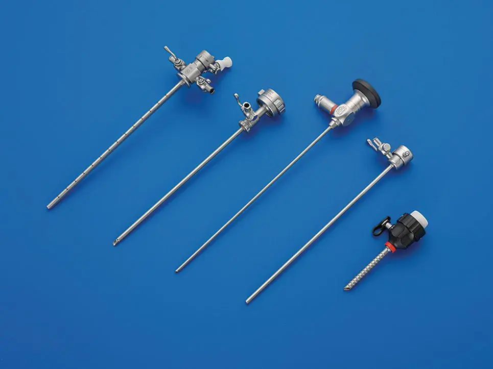
Figure 2.30 The 2.7 mm diameter, 18 cm long, 30° multipurpose rigid telescope (MPRT) that is the most widely used telescope in small animal practice with a selection of sheaths available for use with this telescope. From upper left to lower right: the 14.5 Fr, 15 cm working length operating/cystoscopy sheath with a 5 Fr working channel, and 2 Luer connectors with stopcocks; the arthroscopy sheath with 4.0 mm outside diameter, 13.5 cm working length, and 1 Luer connector with a stopcock; the 2.7 mm diameter telescope; the examination and protection sheath with 3.5 mm outside diameter, 17 cm working length, and 1 Luer connector with a stopcock; and a 3.9 mm laparoscopy trocar‐cannula with a working length of 5 cm and a silicone leaflet valve.
( Source: Photo courtesy of Karl Storz: ©Karl Storz SE & Co KG, Germany.)
Table 2.3 The 2.7 mm MPRT with available sheath selection.
| 2.7 mm diameter, 18 cm long, 30° telescope (Karl Storz #64029BA) Cystoscopy/operating sheath (Karl Storz#67065CK)14.5 Fr, 15 cm working length, 5 Fr operating channel, with 2 fluid portsArthroscopy sheath (Karl Storz #64132S)4 mm diameter, 13.5 cm working length, with 1 fluid port Guard or examination sheath (Karl Storz #64018US)3.5 mm diameter, 17 cm working length, with 1 fluid portLaparoscopy trocar cannula (Karl Storz #62117GK)3.9 mm diameter, 5 cm long, with a silicone leaflet valveLaparoscopy endotip cannula (Karl Storz #30117MT)3.9 mm diameter, 5 cm long, with an automatic valve |
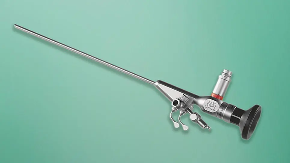
Figure 2.31 A one‐piece cystoscope incorporating the telescope and sheath into one unit with a 9.5 Fr diameter, a working length of 14 cm, a 3 Fr instrument channel, and 2 fluid connection ports with 2 stopcocks. This telescope has a better image than the 1.9 mm diameter cystoscope and sheath that it replaces plus it is much more robust.
( Source: Photo courtesy of Karl Storz: ©Karl Storz SE & Co KG, Germany.)
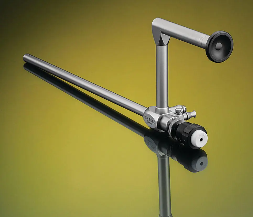
Figure 2.32 A 10 mm diameter, 0° operating laparoscope with a working length of 23 cm and with a 6 mm diameter working channel for use with 5 mm diameter minimally invasive surgery instruments.
( Source: Photo courtesy of Karl Storz: ©Karl Storz SE & Co KG, Germany.)
Most of the telescopes that we use in small animal practice are adapted from already available human instruments. One of the exceptions to this is the Veterinary Otoscope that was specifically developed and produced for small animal application ( Figure 2.33). This telescope is a one‐piece instrument for ear examination in the awake patient.
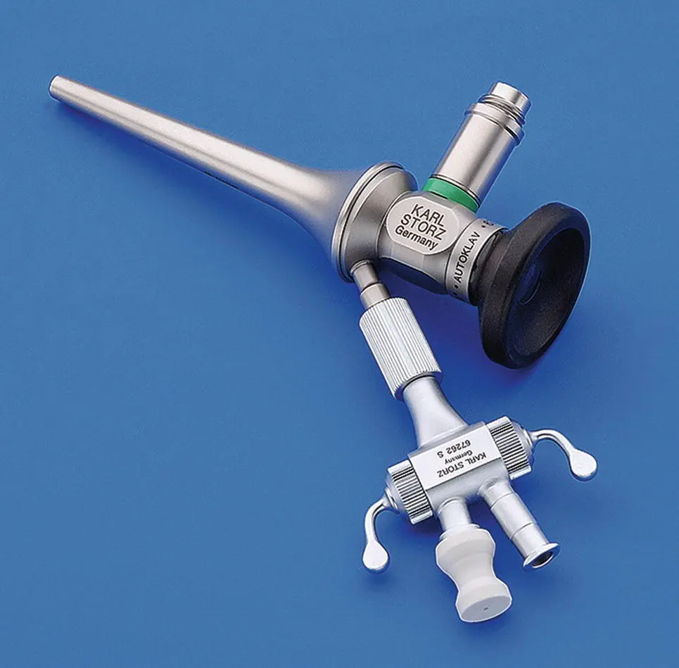
Figure 2.33 The Veterinary Otoscope for ear examination in awake patients with a tip diameter of 5 mm, an 8.5 cm length with a 5 Fr working channel, and a stopcock attachment allowing simultaneous irrigation and passing 5 Fr instruments for manipulations.
( Source: Photo courtesy of Karl Storz: ©Karl Storz SE & Co KG, Germany.)
A recent significant advance in telescope technology is that most telescopes are now autoclavable. The autoclavable telescopes are labeled as autoclavable. This feature greatly facilitates instrument turnaround and practice efficiency.
2.2.10 Flexible Endoscopes
Flexible endoscopes are described by insertion tube diameter, working length, working channel diameter, tip deflection control, and video or fiberoptic image ( Figure 2.34). For small animal practice, flexible endoscopes range in diameter from 2.8 to 9.7 mm, working lengths from 70 to 140 cm, two‐way and four‐way tip deflection control, 1–2.8 mm working channel diameter, and are available in fiberoptic and video options. Light transmission into the patient is through an incoherent fiberoptic bundle incorporated into the flexible endoscope insertion tube and flexible umbilical cord that connects to an external remote cold light source. Some flexible endoscopes use a flexible fiberoptic light guide cable as used with rigid telescopes to connect to the external light source. The advent of LED light sources has also made it possible to incorporate a light source into the endoscope ( Figure 2.10).
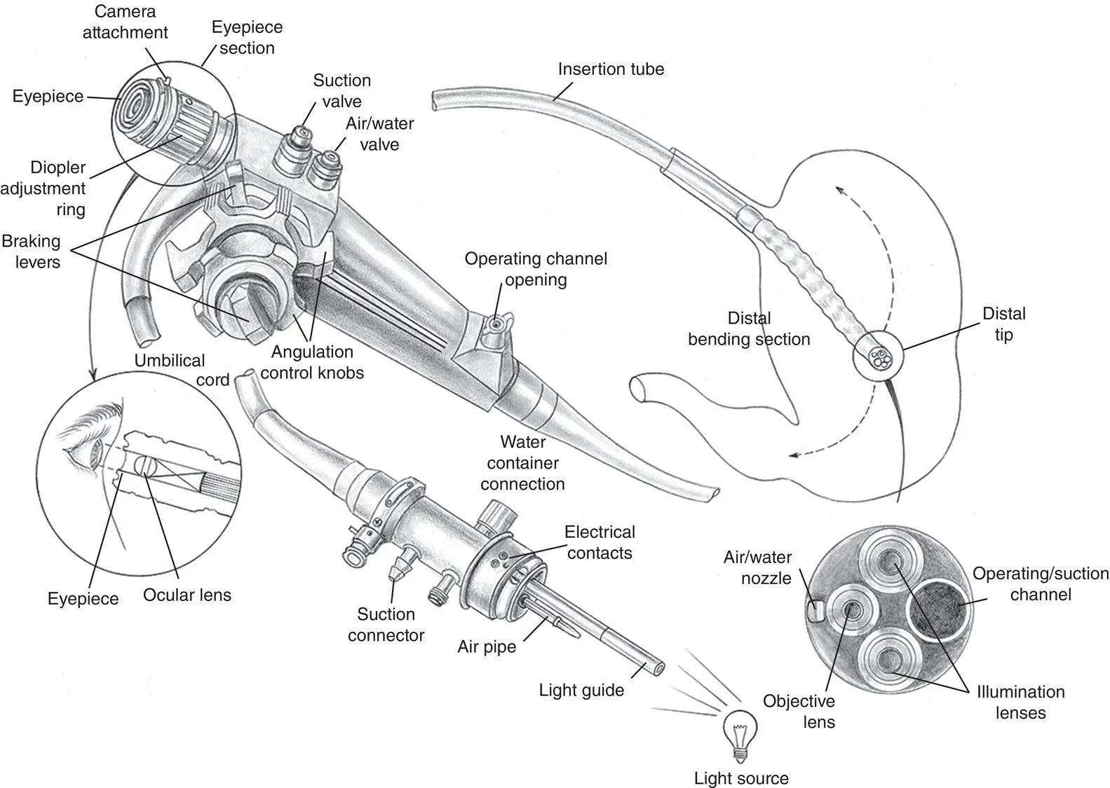
Figure 2.34 The anatomy of a flexible fiberoptic gastrointestinal endoscope. A connector that attaches to a light source with an air pump for insufflation is at the proximal end of the umbilical cord. This connector has a light guidepost, an air insufflation tube, suction connector, lens washing water container connection, a pressure compensation valve, and electrical contacts. The umbilical cord runs from this connector to the handpiece of the endoscope carrying the light guide cable, suction channel, and air insufflation/water channel. The handpiece has an eyepiece section with fiberoptic endoscopes, angulation control knobs with breaking levers, a suction valve, an air insufflation/water valve, and an operating channel port. The eyepiece has a focus ring and camera attachment mechanism. Video endoscopes do not have the eyepiece section. The insertion tube is the portion of the endoscope extending from the handpiece to the distal tip that is passed into the patient. The tip portion of the endoscope is called the bending section and is the portion of the endoscope that is bent using the angulation control knobs on the handpiece. Additional information of flexible gastrointestinal endoscopes is presented in Chapter 3.
Читать дальшеИнтервал:
Закладка:
Похожие книги на «Veterinary Endoscopy for the Small Animal Practitioner»
Представляем Вашему вниманию похожие книги на «Veterinary Endoscopy for the Small Animal Practitioner» списком для выбора. Мы отобрали схожую по названию и смыслу литературу в надежде предоставить читателям больше вариантов отыскать новые, интересные, ещё непрочитанные произведения.


Обсуждение, отзывы о книге «Veterinary Endoscopy for the Small Animal Practitioner» и просто собственные мнения читателей. Оставьте ваши комментарии, напишите, что Вы думаете о произведении, его смысле или главных героях. Укажите что конкретно понравилось, а что нет, и почему Вы так считаете.