Veterinary Endoscopy for the Small Animal Practitioner
Здесь есть возможность читать онлайн «Veterinary Endoscopy for the Small Animal Practitioner» — ознакомительный отрывок электронной книги совершенно бесплатно, а после прочтения отрывка купить полную версию. В некоторых случаях можно слушать аудио, скачать через торрент в формате fb2 и присутствует краткое содержание. Жанр: unrecognised, на английском языке. Описание произведения, (предисловие) а так же отзывы посетителей доступны на портале библиотеки ЛибКат.
- Название:Veterinary Endoscopy for the Small Animal Practitioner
- Автор:
- Жанр:
- Год:неизвестен
- ISBN:нет данных
- Рейтинг книги:4 / 5. Голосов: 1
-
Избранное:Добавить в избранное
- Отзывы:
-
Ваша оценка:
Veterinary Endoscopy for the Small Animal Practitioner: краткое содержание, описание и аннотация
Предлагаем к чтению аннотацию, описание, краткое содержание или предисловие (зависит от того, что написал сам автор книги «Veterinary Endoscopy for the Small Animal Practitioner»). Если вы не нашли необходимую информацию о книге — напишите в комментариях, мы постараемся отыскать её.
Covers diagnostic endoscopy, interventional endoscopy, and minimally invasive soft tissue surgery Includes thousands of images to illustrate endoscopy concepts for veterinarians Provides a clinically oriented reference book for using rigid and flexible endoscopy in a small animal practice Supports veterinarians who are seeking to increase their services and enhance their revenue streams Any practitioner who is using or preparing to use endoscopic techniques will find
an essential practice resource.
Veterinary Endoscopy for the Small Animal Practitioner — читать онлайн ознакомительный отрывок
Ниже представлен текст книги, разбитый по страницам. Система сохранения места последней прочитанной страницы, позволяет с удобством читать онлайн бесплатно книгу «Veterinary Endoscopy for the Small Animal Practitioner», без необходимости каждый раз заново искать на чём Вы остановились. Поставьте закладку, и сможете в любой момент перейти на страницу, на которой закончили чтение.
Интервал:
Закладка:
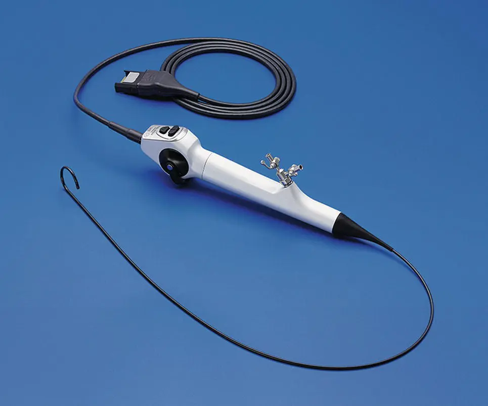
Figure 2.10 The FlexXC video cystourethroscope with an internal LED light source eliminating the need for a separate flexible light guide cable.
( Source: Photo courtesy of Karl Storz: ©Karl Storz SE & Co KG, Germany.)
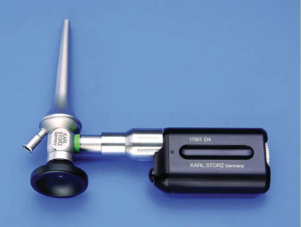
Figure 2.11 A battery‐powered LED light source attached to an otoscope.
( Source: Photo courtesy of Karl Storz: ©Karl Storz SE & Co KG, Germany.)
2.2.5 Documentation Equipment
Documentation is not required when performing diagnostic and operative endoscopy. Having the ability to capture still images is beneficial as a practice‐building tool for demonstrating to clients, pathology that is seen and procedures that are performed. Documentation is also needed for presentations and publications on endoscopic techniques and procedures.
Capture devices are available as separate modules ( Figures 1.10and 2.2) and as integral systems built into camera control boxes ( Figures 2.1, 2.3, and 2.6). They are incorporated into the video system tower with all their needed wire connections, so they are ready whenever the system is used. Video still image printers and video cassette recorders were used in the early days of video endoscopy but have been replaced with digital capture devices. Video and still images are captured directly off the camera during procedures. The captured images are transferred to a computer for editing, stored on disks or thumb drives, used for computer presentations, or printed for distribution to clients and referring veterinarians. The highest quality still images are obtained with digital capture systems. Capture of video during procedures is an optional method of documentation but short video clips are recommended as opposed to recording whole procedures. Recording entire procedures uses massive amounts of storage space and requires extensive editing to provide an acceptable end product. Most capture devices can be set to a predetermined video recording time.
2.2.6 Power Equipment
2.2.6.1 Radio‐Frequency Instrumentation
Monopolar or bipolar radio‐frequency instrumentation is used to cut tissue and cauterize bleeding vessels. Standard radio‐frequency systems for traditional open surgery are usable for minimally invasive surgery with appropriate monopolar or bipolar cords connected to minimally invasive surgery instruments. Minimally invasive surgery hand instruments have a connector pin for monopolar electrosurgery ( Figure 2.12). Adaptation of monopolar electrosurgery units for bipolar function is also possible with bipolar MIS instruments connected to the radio‐frequency generator with a special power cord that allows placement of one connector to the active socket and the other connector to the ground socket ( Figure 2.13). This drives the current between the two sides of the instrument creating a bipolar instrument. Bipolar generators are also used with a power cord for this application. Monopolar and bipolar radiofrequency is capable of sealing vessels up to 3 mm in diameter and is sufficient for most minimally invasive surgery in small animal practice.
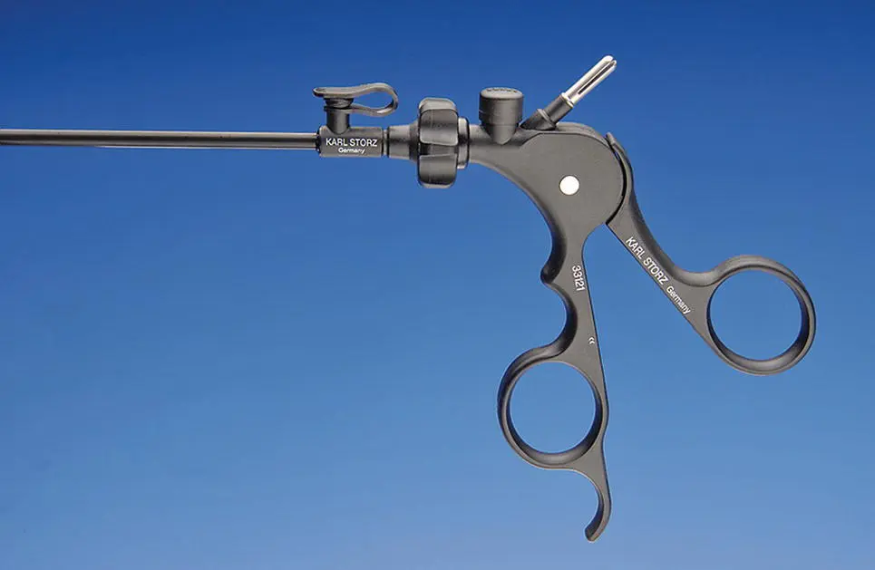
Figure 2.12 A minimally invasive surgery instrument with a connection post for monopolar electrosurgery. The connection post is the silver split projection at the top right of the pistol grip handle. This post is attached to the active side of the electrosurgery generator using a standard open‐surgery grounding plate for the inactive or ground connection.
( Source: Photo courtesy of Karl Storz: ©Karl Storz SE & Co KG, Germany.)
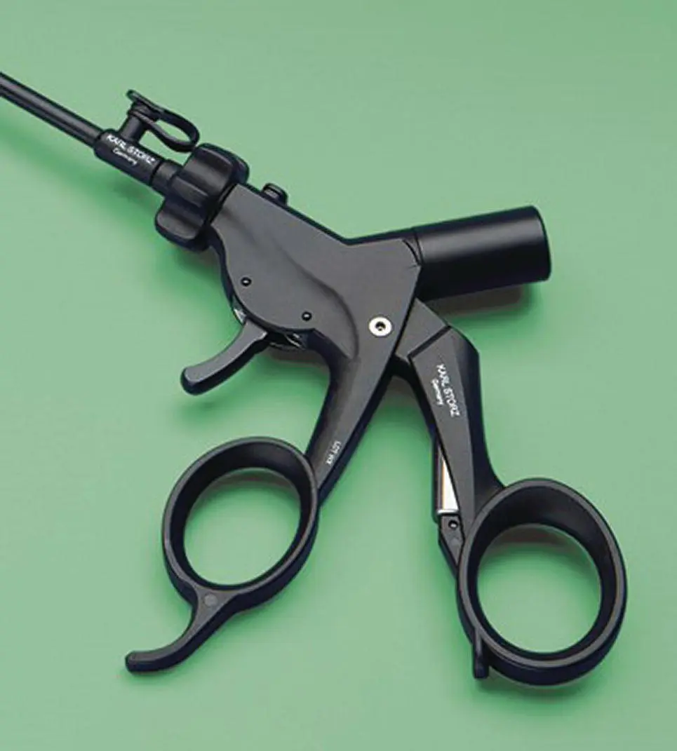
Figure 2.13 A minimally invasive surgery bipolar vessel sealing and cutting instrument, Karl Storz 5 mm diameter RobiPlus, with a connection post for bipolar cautery seen as a black cylinder projecting from the upper right of the instrument handle. This instrument is used with a monopolar generator and a power cord with two plugs, one that goes into the active socket, and the other goes into the ground socket. This instrument also works with a bipolar generator using a proper cord for this application.
( Source: Photo courtesy of Karl Storz: ©Karl Storz SE & Co KG, Germany.)
2.2.6.2 Vessel Sealing Devices
Systems capable of sealing larger blood vessels, up to 7 mm diameter, were developed for minimally invasive surgery use in human patients. These devices use tissue fusion technology that applies low voltage with high current across a bipolar instrument while measuring tissue impedance (resistance) to achieve a tissue weld sealing the blood or lymphatic vessel. The original vessel sealing device using this technology was the LigaSure and has been the workhorse in small animal practice for over 20 years. Originally introduced for minimally invasive surgery with sealing and cutting capability in one instrument, it also became a standard instrument for open surgery with handpieces developed specifically for open surgery. A second‐generation device produced by the same company and the standard today is the ForceTriad ( Figure 2.14) with advanced technology for faster function but little change in sealed vessel properties. The ForceTriad also has monopolar and bipolar modules. A wide variety of instruments are available for minimally invasive surgery in 5 and 10 mm diameters ( Figure 2.15) and in lengths from 20 to 37 cm with sealing and cutting capabilities. There is also selection of open surgery instruments just for sealing ( Figure 2.16) and sealing plus cutting in one instrument ( Figure 2.17). A third‐generation device is now available, but this step has incorporated circuitry to prevent reuse of the handpieces currently making it unsuitable for small animal practice. The company has indicated that they are working on a modification to allow reuse of the handpieces in veterinary medicine. Competing companies have developed similar vessel sealing devices that are now available and are worth considering for use in small animal practice.
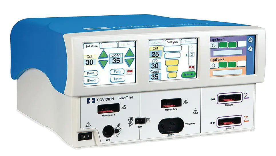
Figure 2.14 The ForceTriad vessel sealing device with standard monopolar radiofrequency on the left, monopolar or bipolar radiofrequency in the center, and vessel sealing on the right side of the control box. All three modalities can be connected and active simultaneously.
( Source: Photo courtesy of Covidien Division of Medtronic's.)

Figure 2.15 A 10 mm diameter 37 cm long “Atlas” vessel sealing instrument for minimally invasive surgery with a round shaft to provide a seal for maintaining insufflation during laparoscopy. This is the workhorse for minimally invasive surgery in small animal practice. Multiple‐length 10 mm instruments and similar 5 mm diameter instruments are available for use with this system.
Читать дальшеИнтервал:
Закладка:
Похожие книги на «Veterinary Endoscopy for the Small Animal Practitioner»
Представляем Вашему вниманию похожие книги на «Veterinary Endoscopy for the Small Animal Practitioner» списком для выбора. Мы отобрали схожую по названию и смыслу литературу в надежде предоставить читателям больше вариантов отыскать новые, интересные, ещё непрочитанные произведения.


Обсуждение, отзывы о книге «Veterinary Endoscopy for the Small Animal Practitioner» и просто собственные мнения читателей. Оставьте ваши комментарии, напишите, что Вы думаете о произведении, его смысле или главных героях. Укажите что конкретно понравилось, а что нет, и почему Вы так считаете.