Veterinary Endoscopy for the Small Animal Practitioner
Здесь есть возможность читать онлайн «Veterinary Endoscopy for the Small Animal Practitioner» — ознакомительный отрывок электронной книги совершенно бесплатно, а после прочтения отрывка купить полную версию. В некоторых случаях можно слушать аудио, скачать через торрент в формате fb2 и присутствует краткое содержание. Жанр: unrecognised, на английском языке. Описание произведения, (предисловие) а так же отзывы посетителей доступны на портале библиотеки ЛибКат.
- Название:Veterinary Endoscopy for the Small Animal Practitioner
- Автор:
- Жанр:
- Год:неизвестен
- ISBN:нет данных
- Рейтинг книги:4 / 5. Голосов: 1
-
Избранное:Добавить в избранное
- Отзывы:
-
Ваша оценка:
Veterinary Endoscopy for the Small Animal Practitioner: краткое содержание, описание и аннотация
Предлагаем к чтению аннотацию, описание, краткое содержание или предисловие (зависит от того, что написал сам автор книги «Veterinary Endoscopy for the Small Animal Practitioner»). Если вы не нашли необходимую информацию о книге — напишите в комментариях, мы постараемся отыскать её.
Covers diagnostic endoscopy, interventional endoscopy, and minimally invasive soft tissue surgery Includes thousands of images to illustrate endoscopy concepts for veterinarians Provides a clinically oriented reference book for using rigid and flexible endoscopy in a small animal practice Supports veterinarians who are seeking to increase their services and enhance their revenue streams Any practitioner who is using or preparing to use endoscopic techniques will find
an essential practice resource.
Veterinary Endoscopy for the Small Animal Practitioner — читать онлайн ознакомительный отрывок
Ниже представлен текст книги, разбитый по страницам. Система сохранения места последней прочитанной страницы, позволяет с удобством читать онлайн бесплатно книгу «Veterinary Endoscopy for the Small Animal Practitioner», без необходимости каждый раз заново искать на чём Вы остановились. Поставьте закладку, и сможете в любой момент перейти на страницу, на которой закончили чтение.
Интервал:
Закладка:
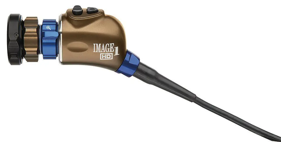
Figure 2.4 A Karl Storz Endoscopy IMAGE 1 FULL HD Three‐Chip Camera Head H3‐Z. This is an example of the small lightweight camera heads designed for endoscopy.
( Source: Photo courtesy of Karl Storz: ©Karl Storz SE & Co KG, Germany.)
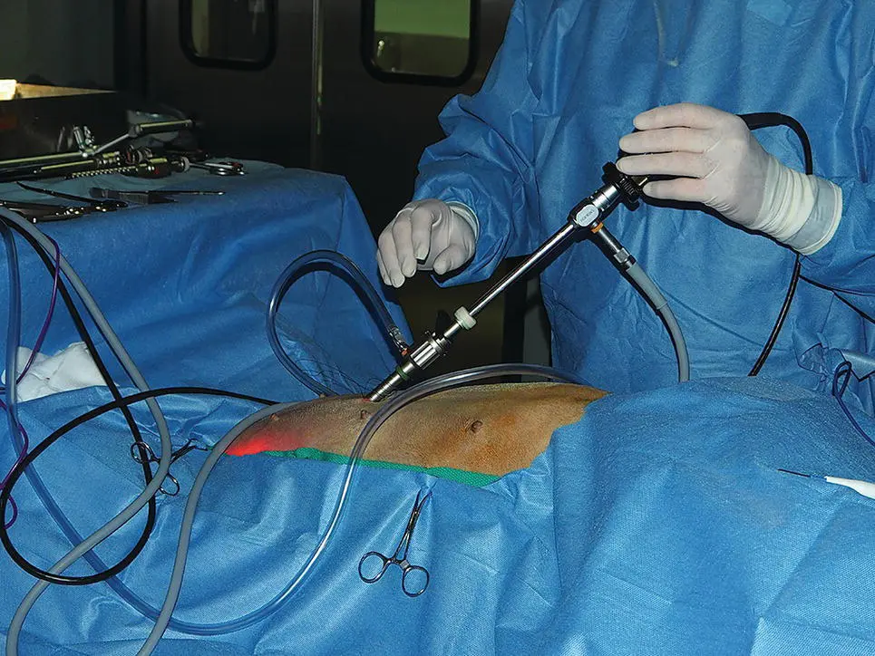
Figure 2.5 An endoscopic video camera head attached securely to a 10 mm laparoscope to create a one‐piece operating system.
An alternative to a video camera system tower for limited space situations and for mobile applications is a Tele Pack ( Figure 2.6). This self‐contained system includes a light source, camera control box, monitor, a capture system, and an air pump for gastrointestinal insufflation in a single small suitcase‐sized unit. This unit uses a standard definition single‐chip camera.
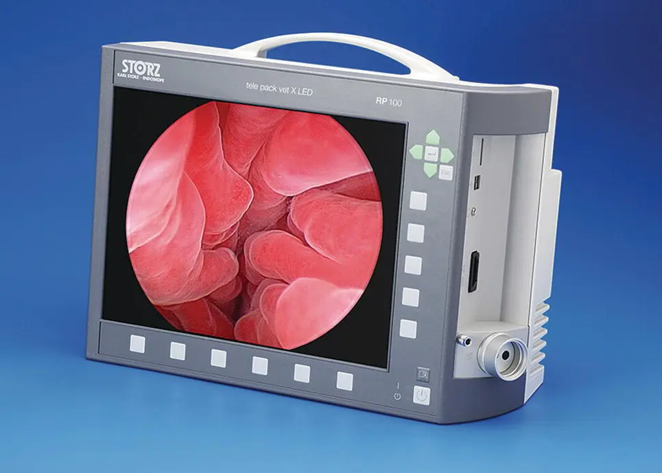
Figure 2.6 A Karl Storz Endoscopy Tele Pack Vet X LED self‐contained endoscopy video system with a camera control box, LED light source, monitor, capture system, and air pump for gastrointestinal insufflation in a single small suitcase‐sized box.
( Source: Photo courtesy of Karl Storz: ©Karl Storz SE & Co KG, Germany.)
A good‐quality autoexposure single‐chip camera is more than adequate for small animal endoscopy and minimally invasive surgery. These cameras are lightweight, compact, connect directly to the telescope, and provide an excellent image. Three‐chip cameras and high‐definition cameras have higher resolution and better color separation but are only necessary for obtaining publication or presentation quality capture images.
A significant recent advance in camera technology is that they are now available as autoclavable versions greatly speeding instrument turnaround. It is critical to determine if the camera that you have is autoclavable or not. If you have a camera that is not autoclavable and it is subjected to the heat and moisture of an autoclave cycle, it will be destroyed. Cameras designated as autoclavable are still soakable and sterilizable with gas or plasma.
2.2.3 Video Monitors ( Table 2.1)
The medical‐grade video monitors previously recommended and considered to be necessary for effective video endoscopy are no longer needed and have been replaced by flat screen monitors ( Figures 2.1– 2.3). The added expense of a medical‐grade monitor is no longer required. The decreased cost for monitors with flat screen technology also allows the use of more than one monitor in the operating room. Currently recommended operating room setup uses two monitors, one on the tower, and one either mounted on a wall in an appropriate location for the operating room or on another movable cart. Wireless transmission systems are readily available to connect the remote monitor to the tower system eliminating the need for wired connections. This arrangement eliminates the problem of monitor placement for complex procedures where more than one monitor position is needed. Wireless transmission is also used to send video to monitors or computers outside the operating room.
2.2.4 Light Source ( Table 2.1)
A good‐quality light source is required for flexible and rigid endoscopy. Halogen was the initial light source used when fiberoptic light transmission was developed for endoscopic use. Xenon light sources ( Figure 2.7) have replaced the original Halogen light sources and have been the preferred technology in endoscopy for many years. Xenon has advantages over Halogen of being much brighter and providing light closer to natural light giving tissues a true color. They are available in 100, 175, and 300‐W sizes. Light sources are available as single function units ( Figure 2.7) or containing an air insufflation pump for gastrointestinal endoscopy ( Figure 2.8). The major disadvantage of Xenon is the cost of bulb replacement and their short bulb life expectancy (approximately 800 hours) compared to other light sources. Xenon is rapidly being replaced with diode light sources ( Figure 2.9). Diode light sources produce light comparable in wavelength to Xenon, use much less energy, produce less heat, and have a bulb life expectancy of 20 000 hours. They are currently more expensive than Xenon light sources, but this initial cost is offset by their other advantages.
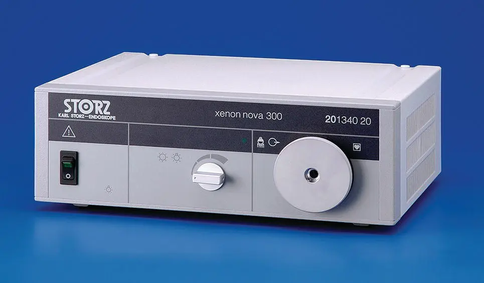
Figure 2.7 A Xenon Nova 300‐W light source.
( Source: Photo courtesy of Karl Storz: ©Karl Storz SE & Co KG, Germany.)
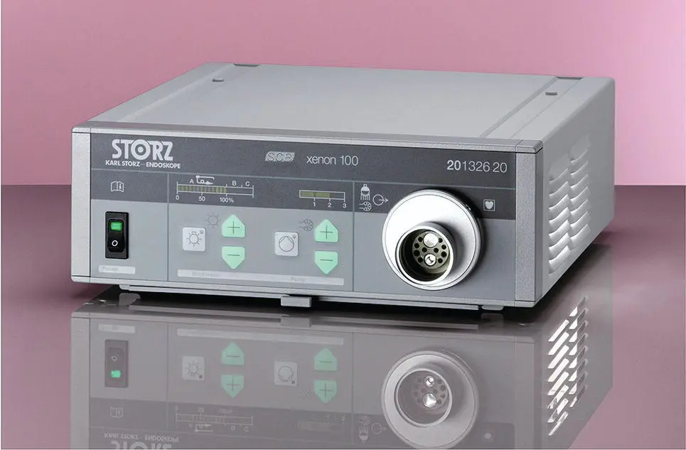
Figure 2.8 A Xenon 100 light source for gastrointestinal endoscopy with integral air insufflation. An adaptor allows this light source to be used with rigid endoscopes.
( Source: Photo courtesy of Karl Storz: ©Karl Storz SE & Co KG, Germany.)
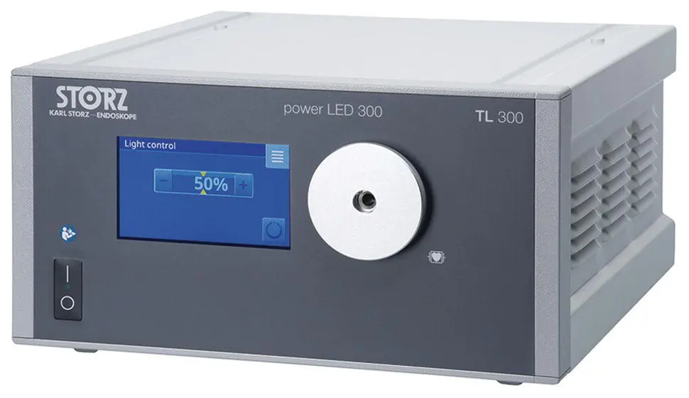
Figure 2.9 A 300‐W LED light source.
( Source: Photo courtesy of Karl Storz: ©Karl Storz SE & Co KG, Germany.)
Light is transmitted from the external light source to the distal tip of flexible endoscopes and rigid telescopes through flexible incoherent fiberoptic light bundles ( Figures 1.3– 1.6and 1.9). In flexible endoscopes, the incoherent fiberoptic light bundle is continuous from the light post at the plug end of the endoscope umbilical cord to the distal tip of the endoscope. For rigid endoscopic telescopes, light transmission is a two‐part system. The first part is from the external light source to the light guidepost of the telescope with an incoherent flexible fiberoptic light transmitting cable ( Figure 1.9). The second part is an incoherent fiberoptic light bundle that goes from the light guidepost through the rigid telescope, usually surrounding the lens system, to the tip of the endoscope. Flexible light guide cables are available in diameters from 2.5 to 4.8 mm and lengths from 180 to 320 cm. Smaller light guide cables are recommended for smaller telescopes and larger light guide cables for larger telescope diameters. Multiple adaptors are available to allow connection to endoscopes and light sources from many manufacturers. Both systems provide light at the working end of the endoscope pointed to illuminate the area visible through the objective lens of the endoscope. Diode technology has also allowed light sources to be placed into endoscopes and into small battery‐operated light sources. The Flex XC flexible video uretero‐cystoscope has this technology and when connected to the video control module produces its own light eliminating the need for an external light source and a light guide cable ( Figure 2.10). Small battery packs with internal LED light sources that connect directly to the light guidepost on rigid telescopes are available with 60‐ and 120‐minute battery life prior to recharging ( Figure 2.11). These battery packs are ideal for use with the video otoscope in the examination room.
Читать дальшеИнтервал:
Закладка:
Похожие книги на «Veterinary Endoscopy for the Small Animal Practitioner»
Представляем Вашему вниманию похожие книги на «Veterinary Endoscopy for the Small Animal Practitioner» списком для выбора. Мы отобрали схожую по названию и смыслу литературу в надежде предоставить читателям больше вариантов отыскать новые, интересные, ещё непрочитанные произведения.


Обсуждение, отзывы о книге «Veterinary Endoscopy for the Small Animal Practitioner» и просто собственные мнения читателей. Оставьте ваши комментарии, напишите, что Вы думаете о произведении, его смысле или главных героях. Укажите что конкретно понравилось, а что нет, и почему Вы так считаете.