Veterinary Endoscopy for the Small Animal Practitioner
Здесь есть возможность читать онлайн «Veterinary Endoscopy for the Small Animal Practitioner» — ознакомительный отрывок электронной книги совершенно бесплатно, а после прочтения отрывка купить полную версию. В некоторых случаях можно слушать аудио, скачать через торрент в формате fb2 и присутствует краткое содержание. Жанр: unrecognised, на английском языке. Описание произведения, (предисловие) а так же отзывы посетителей доступны на портале библиотеки ЛибКат.
- Название:Veterinary Endoscopy for the Small Animal Practitioner
- Автор:
- Жанр:
- Год:неизвестен
- ISBN:нет данных
- Рейтинг книги:4 / 5. Голосов: 1
-
Избранное:Добавить в избранное
- Отзывы:
-
Ваша оценка:
Veterinary Endoscopy for the Small Animal Practitioner: краткое содержание, описание и аннотация
Предлагаем к чтению аннотацию, описание, краткое содержание или предисловие (зависит от того, что написал сам автор книги «Veterinary Endoscopy for the Small Animal Practitioner»). Если вы не нашли необходимую информацию о книге — напишите в комментариях, мы постараемся отыскать её.
Covers diagnostic endoscopy, interventional endoscopy, and minimally invasive soft tissue surgery Includes thousands of images to illustrate endoscopy concepts for veterinarians Provides a clinically oriented reference book for using rigid and flexible endoscopy in a small animal practice Supports veterinarians who are seeking to increase their services and enhance their revenue streams Any practitioner who is using or preparing to use endoscopic techniques will find
an essential practice resource.
Veterinary Endoscopy for the Small Animal Practitioner — читать онлайн ознакомительный отрывок
Ниже представлен текст книги, разбитый по страницам. Система сохранения места последней прочитанной страницы, позволяет с удобством читать онлайн бесплатно книгу «Veterinary Endoscopy for the Small Animal Practitioner», без необходимости каждый раз заново искать на чём Вы остановились. Поставьте закладку, и сможете в любой момент перейти на страницу, на которой закончили чтение.
Интервал:
Закладка:
A manual table that tilts side to side specifically designed for small animal minimally invasive surgery is available that attaches to the top of most currently available small animal surgery tables and works well as an inexpensive alternative ( Figure 2.20). Using this table, patients can be tilted 45° to each side with an intermediate tilt position ( Figure 2.21). This table is more than adequate for the general practice setting primarily interested in performing laparoscopic ovariectomies ( Figure 2.22).
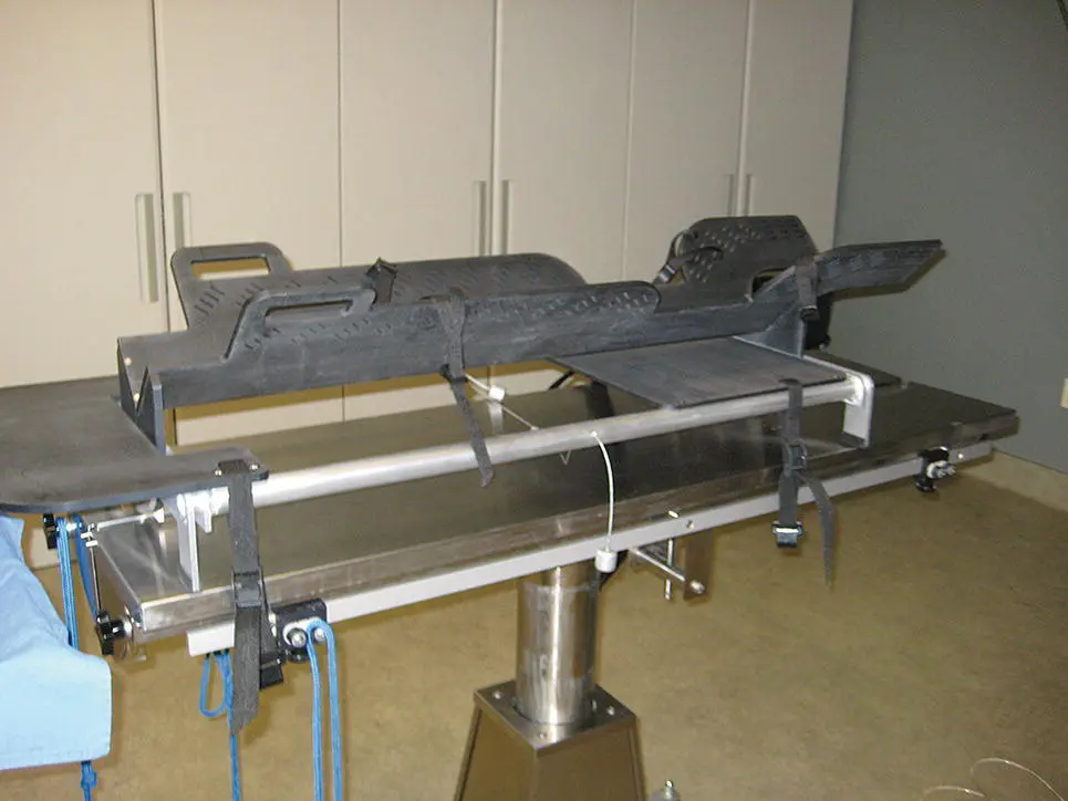
Figure 2.20 A manual Tankersley tilt table (TTT) designed for performing laparoscopic ovariectomies in dogs and cats. This table attaches to the top of most standard‐sized small animal surgery tables.
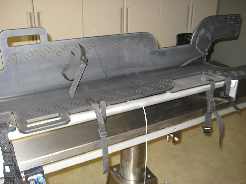
Figure 2.21 The Tankersley table tilted 45° in the position used for laparoscopic ovariectomies. The table tilts to both sides and has an intermediate tilt position for use when less angulation is required.
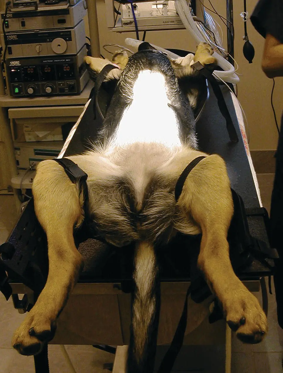
Figure 2.22 A large dog fixed in place on the TTT ready for performing a laparoscopic ovariectomy. The Tankersley tilt table is currently available through Karl Storz.
For the specialty practice planning to advance into complex minimally invasive abdominal and thoracic surgery, a powered operating table with four‐way tilt capability is a needed addition ( Figure 2.23). This table is controlled with a joystick that is accessible and operable through drape material allowing the surgeon to position the patient during surgery and to change positions as needed. The table tilts 45° to the side in both directions ( Figures 2.24and 2.25) and 15° head to tail in both directions ( Figure 2.23).
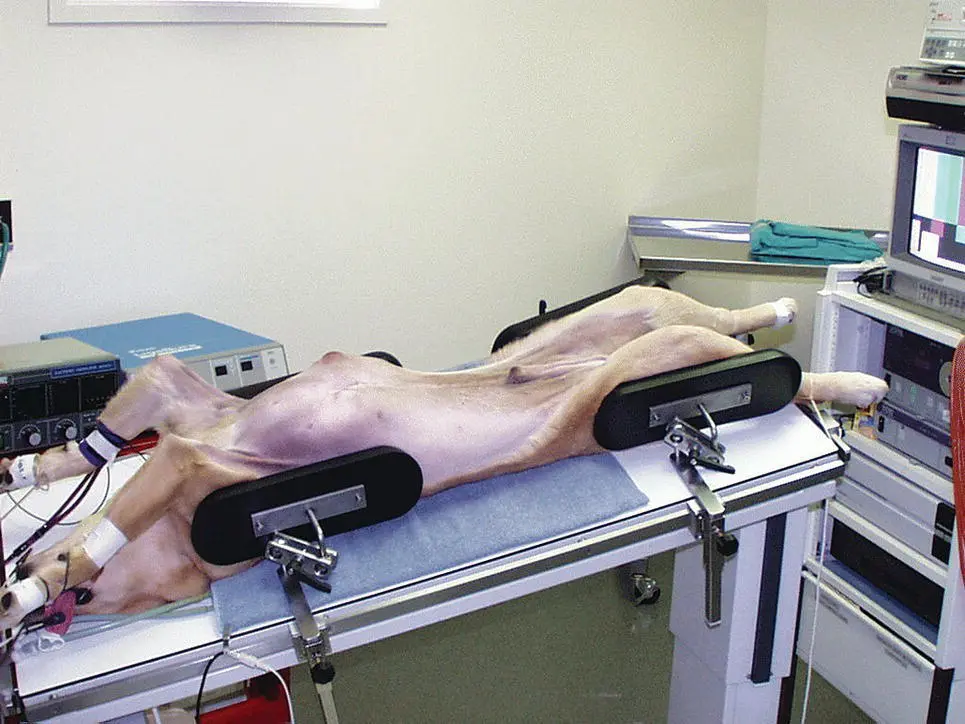
Figure 2.23 A DRE Panomed operating table for small animal use shown with the patient tilted both in a head down position and tilted slightly to the right. The black pads on both ends of the patient and on both sides are positioners to hold the patient in position when the table is tilted to the side. The DRE Panomed table is available through DRE Veterinary ( http://dremed.com) that has changed its name to Avante Medical Surgical ( http://avantehs.com/medical).
Figure 2.24 The DRE Panomed powered operating table for small animal practice with the patient tilted 45° to the left. Note the positioners on both sides of the patient. For a dog of this size when 45° tilting is planned, four of the positioners are needed, one on each side for the thorax and one on each side for the abdomen. Prior to draping, tilting is done to each side to test for patient stability and confirm that the patient will not slide off the table when the table is tilted.
Figure 2.25 The same patient as seen in the Figure 2.24tilted to the right prior to draping completing testing for patient stability. This is easy and quick and can minimize the risk of a disaster.
2.2.9 Rigid Telescopes
Rigid telescopes used for endoscopy are described by the diameter, length, and angle of view. For small animal endoscopy, telescopes range in diameter from 1.0 to 10.0 mm and in length from 6.5 to 31 cm ( Figure 2.26). Angle of view refers to the angle that the center of the image seen through the telescope deviates from the long axis of the telescope and ranges from 0° or straight ahead to 120° ( Figure 2.27). A new rigid telescope design, the “ENDOCAMELEON”, has a variable adjustable angle of view from 0 to 120° for a 10 mm diameter, 32 cm long laparoscope version ( Figure 2.28) and from 15 to 90° for a 4.0 mm diameter, 18 cm long arthroscopy/ENT version. Telescopes in use today are designed with what is termed a Hopkins rod lens system ( Figure 2.29) for image transmission that has dramatically improved quality over previous lens systems. Light transmission into the patient is through a fiberoptic bundle incorporated into the telescope with a light post for attaching a flexible fiberoptic light guide cable that leads to an external remote cold light source ( Figure 1.9).
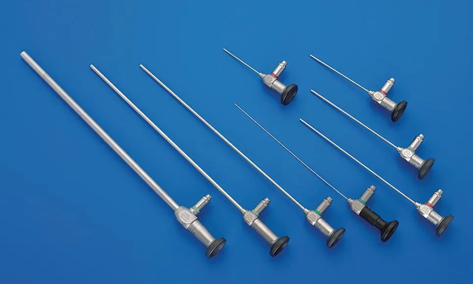
Figure 2.26 Telescopes available for use in small animal practice including from left to right: A 10 mm diameter, 31 cm long, 0° laparoscope; a 5 mm diameter, 29 cm long, 0° laparoscope; a 4.0 mm diameter, 30 cm long, 30° cystoscope; a 1.0 mm diameter, 20 cm long, 0° semirigid needle scope; the 2.7 mm diameter, 18 cm long, 30° multipurpose rigid telescope (MPRT); a 3 mm diameter, 14 cm long, 0° laparoscope; a 2.4 mm diameter, 10 cm long, 30° arthroscope; and a 1.9 mm diameter, 6.5 cm long, 30° arthroscope that is set above the line of telescopes. The degree angles of the telescopes listed are examples and many are available in one to multiple viewing angles.
( Source: Photo courtesy of Karl Storz: ©Karl Storz SE & Co KG, Germany.)
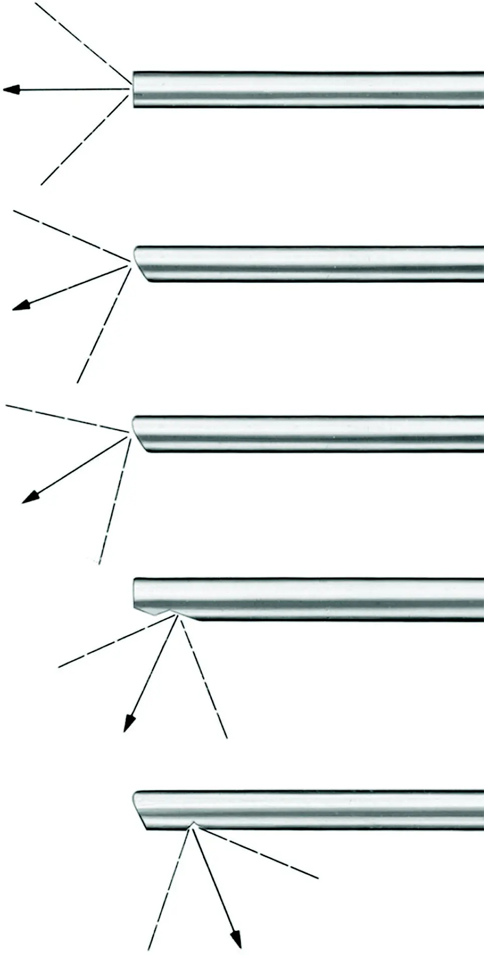
Figure 2.27 A diagram showing the angle of view of rigid telescopes used in small animal practice. Zero and thirty degree angles are the most commonly used angles. Greater angles are occasionally used but increase the difficulty in orientation of the image.
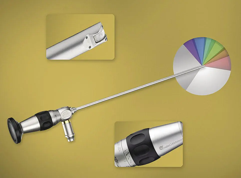
Figure 2.28 The ENDOCAMELEON telescope for laparoscopy with variable angles of view. A 10 mm diameter laparoscope is shown that has a working length of 32 cm and a variable view angle of 0–120°. The tip of the telescope has a curved outer lens housing with an inner lens that rotates through the variable angle range shown in the upper insert. The lens position is adjusted with the black ring behind the light post shown in the lower insert. The angles of the angle of view are shown in the insert at the tip of the endoscope.
( Source: Photo courtesy of Karl Storz: ©Karl Storz SE & Co KG, Germany.)
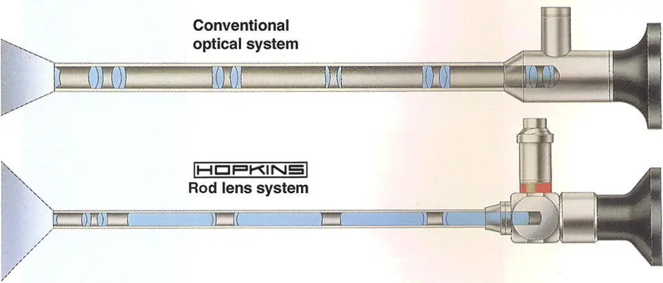
Figure 2.29 A diagram of the Hopkins rod lens system shown in the telescope at the bottom and a conventional lens system in the telescope at the top. Hopkins rod lenses are the standard design for telescopes in current use today having superior image compared to conventional lens telescopes.
Читать дальшеИнтервал:
Закладка:
Похожие книги на «Veterinary Endoscopy for the Small Animal Practitioner»
Представляем Вашему вниманию похожие книги на «Veterinary Endoscopy for the Small Animal Practitioner» списком для выбора. Мы отобрали схожую по названию и смыслу литературу в надежде предоставить читателям больше вариантов отыскать новые, интересные, ещё непрочитанные произведения.


Обсуждение, отзывы о книге «Veterinary Endoscopy for the Small Animal Practitioner» и просто собственные мнения читателей. Оставьте ваши комментарии, напишите, что Вы думаете о произведении, его смысле или главных героях. Укажите что конкретно понравилось, а что нет, и почему Вы так считаете.