Philias R. Garant - Oral Cells and Tissues
Здесь есть возможность читать онлайн «Philias R. Garant - Oral Cells and Tissues» — ознакомительный отрывок электронной книги совершенно бесплатно, а после прочтения отрывка купить полную версию. В некоторых случаях можно слушать аудио, скачать через торрент в формате fb2 и присутствует краткое содержание. Жанр: unrecognised, на английском языке. Описание произведения, (предисловие) а так же отзывы посетителей доступны на портале библиотеки ЛибКат.

- Название:Oral Cells and Tissues
- Автор:
- Жанр:
- Год:неизвестен
- ISBN:нет данных
- Рейтинг книги:3 / 5. Голосов: 1
-
Избранное:Добавить в избранное
- Отзывы:
-
Ваша оценка:
Oral Cells and Tissues: краткое содержание, описание и аннотация
Предлагаем к чтению аннотацию, описание, краткое содержание или предисловие (зависит от того, что написал сам автор книги «Oral Cells and Tissues»). Если вы не нашли необходимую информацию о книге — напишите в комментариях, мы постараемся отыскать её.
Oral Cells and Tissues — читать онлайн ознакомительный отрывок
Ниже представлен текст книги, разбитый по страницам. Система сохранения места последней прочитанной страницы, позволяет с удобством читать онлайн бесплатно книгу «Oral Cells and Tissues», без необходимости каждый раз заново искать на чём Вы остановились. Поставьте закладку, и сможете в любой момент перейти на страницу, на которой закончили чтение.
Интервал:
Закладка:
A second and minor component of circumpulpal dentin, the peritubular dentin, develops around the odontoblastic process (see Fig 2-1). The organic matrix of peritubular dentin is rich in glycosaminoglycans and relatively free of collagen fibrils. Bone sialoprotein and osteonectin have been localized in peritubular dentin. The hydroxyapatite crystals that develop in the peritubular dentin matrix are small in comparison to those that form in the intertubular dentin. Because of its small crystallites (large surface area) and the noncollagenous nature of its organic matrix, peritubular dentin is more susceptible to demineralization and degradation during the caries process.
Transport Across the Odontoblastic Layer
Over the course of several decades, there have been numerous contradictory reports on the transport of calcium and other substances into developing dentin. Some investigators have presented autoradiographic evidence of an intracellular (transcellular) pathway, while others have shown that ions move from pulp to dentin through a paracellular route between odontoblasts.
Increasing evidence supports a direct role for odontoblastic transport of ions into the predentin and dentin. The calcium ion activity in the fluid phase of predentin is two to three times higher than that in the pulp. 135Current physiologic evidence suggests that an ionic gradient exists across the odontoblastic layer and that, under normal conditions, the paracellular pathway is closed. Under these conditions small molecules and ions would be forced to pass through the odontoblast cytoplasm to reach the dentin.
Recent studies have provided indirect support that calcium is transported into the dentin across the odontoblast cell membrane by a calcium ATPase pump, and by Na +-Ca ++antiports. 136 , 137L-type voltage–gated and agonist-gated calcium channels have been localized in odontoblasts by immunocytochemical methods. 24 , 137 , 138Calcium channel inhibitors (nifedipine and neomycin) have been shown to decrease the entry of calcium into the odontoblasts, and eventually to reduce the level of calcium entry into the predentin. Secretory odontoblasts express high levels of inositol 1,4,5-triphosphate–regulated channel proteins. These channel proteins permit calcium flux from intracellular stores into the cytosol. Smutzer et al suggested that 1,4,5-triphosphate receptors might regulate calcium release for dentin mineralization. 139
The presence of ion concentration gradients across the odontoblastic layer appears to be at odds with the morphologic evidence that no zonula occludens is formed at the distal end of the odontoblast cell body. Furthermore, there is free diffusion of intravenously administered tracer molecules into predentin. Additional research is needed to identify the mechanisms responsible for generating and maintaining ion concentration gradients and for regulating solute movement across the odontoblast layer.
Innervation of Dentin and Mechanisms of Pain Sensation
Organization of the nerve supply
The nerve supply to the coronal pulp is especially rich, forming a subodontoblastic plexus of nerve fibers (plexus of Raschkow). Unmyelinated nerves from this plexus penetrate between the odontoblasts and enter the predentin. Although most unmyelinated nerves appear to terminate in the predentin, some enter the dentinal tubules. Nerve endings containing many small vesicles and mitochondria have been identified in close association with the odontoblast cell body and the odontoblastic process in dentin ( Figs 2-7and 2-10). 140 – 143The majority of these nerves are afferent somatosensory pain fibers.
Nerve growth factor and its receptor have been localized in relatively high amounts in the odontoblastic and subodontoblastic layers of the coronal pulp. The presence of nerve growth factor may be responsible for concentrating nerve endings in the vicinity of the odontoblasts. Experimental evidence has shown that nerves grow toward concentrations of nerve growth factor.
Because the cell processes of fibroblasts and odontoblasts can be confused with unmyelinated nerve fibers, special staining techniques are needed to accurately distinguish nerve fibers and their terminals. Protein gene product 9.5, a neuron-specific protein, has been used to identify pulpal nerve fibers at both the light and electron microscopic level (see Figs 2-10ato 2-10d). 140 , 144Fibers containing protein gene product were present in both radicular and coronal predentin. Nerve endings in the predentin have also been identified by immunocytochemistry for calbindin, a calcium-binding protein found in high concentrations in nerve cells. 145Tracer experiments with tritiated proline injected into the brainstem nuclei of the trigeminal nerve have provided convincing proof of a rich supply of sensory nerve terminals in the predentin and dentinal tubules. 146
Figs 2-10a to 2-10dNerve structure of a human premolar. (Human protein gene product [PGP] 9.5 antibody stain. Adapted from Maeda et al 140with permission from Elsevier Science.)
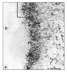
Fig 2-10aPulpodentinal region of human premolar stained with human PGP 9.5 antibody. A dense network of nerve endings can be seen in the predentin (PD). (D) Dentin; (P) pulp. (PGP 9.5 antibody stain. Original magnification × 70.)
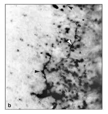
Fig 2-10bInset area in Fig 2-10a at higher magnification. Nerve terminals are beadlike axonal swellings (arrowheads) . (Original magnification × 700.)
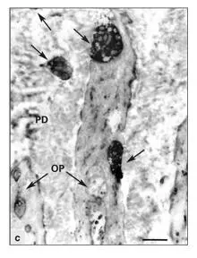
Fig 2-10cElectron immunocytochemistry. Nerve terminals (arrows) stained with PGP 9.5 antibody are juxtaposed to the odontoblastic processes (OP) in the predentin (PD). (Original magnification × 4,000.)
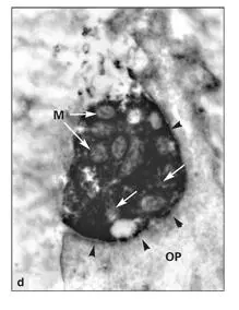
Fig 2-10dElectron immunocytochemistry. The nerve terminals contain mitochondria (M) and many smooth vesicles (arrows) . Although the plasma membranes of the nerve terminals are in close apposition to the plasma membrane of the odontoblastic process (OP) (arrowheads) , no evidence of a synaptic structure is present. (Original magnification × 12,000.)
Vasomotor nerves supply small arteries in the pulp, terminating in close apposition to arteriolar smooth muscle cells. 147The nerve endings contain many dense-cored adrenergic synaptic vesicles.
Theories of dental pain
Although it is well established that most unmyelinated nerve endings in pulp and dentin are nociceptors, the exact mechanisms whereby noxious conditions are converted into dental pain stimuli have yet to be identified. The most widely held theory centers around fluid flow within dentinal tubules. The hydrodynamic theory of dental pain is based on the facts that fluid in the dentinal tubules is constrained by the rigid walls of the peritubular dentin and fluxes in temperature or in osmotic pressure produce rapid expansion and contraction of the dentinal fluid. The bulk flow of the dentinal fluid may distort nerve endings, thereby triggering nerve impulses. Experimental support for this theory is provided by the observation that pain occurs when heat or a substance that changes the osmotic pressure of the dentinal fluid is applied to cut dentin surfaces.
Читать дальшеИнтервал:
Закладка:
Похожие книги на «Oral Cells and Tissues»
Представляем Вашему вниманию похожие книги на «Oral Cells and Tissues» списком для выбора. Мы отобрали схожую по названию и смыслу литературу в надежде предоставить читателям больше вариантов отыскать новые, интересные, ещё непрочитанные произведения.


Обсуждение, отзывы о книге «Oral Cells and Tissues» и просто собственные мнения читателей. Оставьте ваши комментарии, напишите, что Вы думаете о произведении, его смысле или главных героях. Укажите что конкретно понравилось, а что нет, и почему Вы так считаете.