Philias R. Garant - Oral Cells and Tissues
Здесь есть возможность читать онлайн «Philias R. Garant - Oral Cells and Tissues» — ознакомительный отрывок электронной книги совершенно бесплатно, а после прочтения отрывка купить полную версию. В некоторых случаях можно слушать аудио, скачать через торрент в формате fb2 и присутствует краткое содержание. Жанр: unrecognised, на английском языке. Описание произведения, (предисловие) а так же отзывы посетителей доступны на портале библиотеки ЛибКат.

- Название:Oral Cells and Tissues
- Автор:
- Жанр:
- Год:неизвестен
- ISBN:нет данных
- Рейтинг книги:3 / 5. Голосов: 1
-
Избранное:Добавить в избранное
- Отзывы:
-
Ваша оценка:
Oral Cells and Tissues: краткое содержание, описание и аннотация
Предлагаем к чтению аннотацию, описание, краткое содержание или предисловие (зависит от того, что написал сам автор книги «Oral Cells and Tissues»). Если вы не нашли необходимую информацию о книге — напишите в комментариях, мы постараемся отыскать её.
Oral Cells and Tissues — читать онлайн ознакомительный отрывок
Ниже представлен текст книги, разбитый по страницам. Система сохранения места последней прочитанной страницы, позволяет с удобством читать онлайн бесплатно книгу «Oral Cells and Tissues», без необходимости каждый раз заново искать на чём Вы остановились. Поставьте закладку, и сможете в любой момент перейти на страницу, на которой закончили чтение.
Интервал:
Закладка:
Dentin phosphophoryn is the major noncollagenous component of dentin. Immunocytochemical studies indicate that phosphophoryns are localized in small granules distinct from larger collagen-containing secretory granules. The DPPs are secreted from the odontoblastic process at the mineralization front. 49Mineral crystal nucleation is attributed to DPP, a protein rich in aspartic acid and serine residues. Biochemical studies indicate that the DPPs are covalently linked to specific sites on the collagen fibrils of dentin. 69 – 71The serine residues of DPPs are phosphorylated by casein kinase in the extracellular space prior to mineralization. 72 , 73Because of their many phosphate groups, and their capacity to bind calcium, DPPs create a template for calcium and phosphate concentration, and thereby drive crystal nucleation (see Fig 2-4). 74 , 75
An acidic phosphoprotein, DMP1 is localized to mature odontoblasts, cementoblasts, and osteoblasts. 76 – 78It is not expressed in the enamel organ and pulp. The precise role of DMP1 has yet to be identified. The gene coding for DMP1 has been localized to human chromosome 4q21, a region implicated in the autosomal-dominant form of dentinogenesis imperfecta type II. 79Teeth affected by this disease are characterized by discolored and abnormally soft dentin and by fewer and irregular dentinal tubules. 80
Dentin sialoprotein is a sialic acid–rich glycoprotein that is expressed early in tooth development, prior to degradation of the basement membrane. The mRNA for DSP has been detected in preameloblasts and preodontoblasts, suggesting that it may have a signaling role during ameloblast and odontoblast differentiation. 81 , 82
Both DSP and DPP are transcribed from a single mRNA, coded by a gene on human chromosome 4, and coexpressed during tooth development. 83 , 84Both proteins are expressed in preodontoblasts and odontoblasts throughout dentin matrix production. Preameloblasts also express DSP and DPP until enamel secretion, at which point mRNA for DSP and DPP is no longer detected in ameloblasts.
Additional glycoproteins, such as osteocalcin and thrombospondin 1, are found in dentin. Osteocalcin, a glycoprotein rich in glutamic acid, is present in odontoblasts and dentin matrix. 31 , 85In bone, osteocalcin is a chemotactic factor for osteoclasts. Thrombospondin 1 is present in high levels within predentin, especially near the mineralization front. 86Thrombospondin 1 mRNA is localized in odontoblasts but not in the cells of the dental pulp.
Phospholipids
Cytochemical and autoradiographic studies have demonstrated the presence of phospholipids in predentin and dentin matrix. Because they are closely associated with hydroxyapatite crystals at the mineralization front, it has been speculated that they may have a role in mineral nucleation. 87
Growth factors
Bone morphogenetic proteins and TGF-β have been isolated from demineralized dentin matrix. 88It has been suggested that they may trigger the differentiation of new odontoblasts during the induction of reparative dentin. Transforming growth factor β may be retained in the dentin matrix because of its ability to bind to decorin proteoglycan. Odontoblasts express high levels of TGF-β and its receptor. 22 , 89In addition, TGF-β promotes matrix production in most connective tissue cell types. Mutations in TGF-β genes lead to severe pulpal inflammation and attrition of occlusal surfaces. 23
Mineralization of Mantle and circumpulpal Dentin
Two mechanisms for initiating crystal nucleation are responsible for mineralization of the dentin matrix: matrix vesicles in mantle dentin and collagen-phosphophoryn complexes in circumpulpal dentin.
Matrix vesicles
Matrix vesicles in mantle dentin are similar to those first described in mineralizing cartilage. 90 , 91Matrix vesicles in mantle dentin are believed to bud from the tips of odontoblast cytoplasmic processes.
Matrix vesicles initiate mineralization by concentrating calcium and phosphate ions. 92Adenosine triphosphatase (ATPase) activity in matrix vesicle membranes may concentrate ions across the limiting membrane prior to nucleation. 93As the ion concentration increases, hydroxyapatite crystallizes along the inner surface of the matrix vesicle membrane. Calcium-binding phospholipids in the limiting membrane may serve as templates for hydroxyapatite precipitation.
Elemental analysis of freeze-dried matrix vesicles in mantle predentin indicates that earliest mineral deposits appear as dotlike nuclei inside the vesicles. 94Continued crystal growth leads to vesicle rupture, with release of hydroxyapatite crystals into the extracellular matrix. The newly formed crystals act as seeds for continued mineralization of mantle dentin matrix.
After mineralization is initiated, there appears to be no further need for matrix vesicles. Thus matrix vesicles do not participate in mineralization of late mantle dentin and circumpulpal dentin. 95Matrix vesicles are also discussed in chapter 12.
Collagen-phosphophoryn complexes
Mineralization of circumpulpal dentin is initiated by phosphophoryns, independent of matrix vesicle activity. 96Extracellular kinases have been identified in dental matrix. 73These enzymes phosphorylate dentin phosphophoryn. Dentin phosphophoryns, linked to collagen fibrils, act as nucleators of hydroxyapatite crystals in late mantle dentin and circumpulpal dentin. The steric arrangement of negative charges on the phosphophoryns creates a template for hydroxyapatite deposition. 97
Morphologic and histochemical studies of the predentin-dentin border in teeth that have been preserved by rapid pressure freezing and freeze substitution to avoid exposure to aqueous solvents have revealed a 0.5- to 2.0-μm border zone within which mineral deposition occurs gradually. 98In this zone, some proteoglycans are degraded and calcium is bound by phosphophoryn and perhaps by phospholipid as well.
At the mineralization front in the zone of initial mineralization, dotlike mineral nuclei are aligned parallel to and superimposed on collagen fibrils. 99 , 100The mineral nuclei appear positioned over the hole regions of the collagen fibrils, suggesting that the DPPs are bound to collagen at those sites. Additional hydroxyapatite crystals have been located on the surface of the fibrils and in perifibrillar spaces. It has been suggested that dentin sialoproteins and proteoglycans act as nucleating agents for perifibrillar hydroxyapatite crystals. Collagen mineralization is discussed further in chapter 8.
Structure of the Odontoblastic Process and Dentinal Tubules
With progressive deposition of dentin matrix, the odontoblastic process lengthens and becomes embedded in mineralized tissue (Figs 2-2, 2-3, 2-6, and 2-7). The space occupied by the odontoblastic process is known as the dentinal tubule . The dentinal tubule extends from within the mantle dentin to the predentin. 101 , 102
Odontoblastic process
The cytoplasm of the odontoblastic process contains a rich network of microtubules and microfilaments, both of which are oriented parallel to its long axis (see Fig 2-7). 50 , 103 – 105The microtubules and microfilaments form a network that runs the length of the odontoblastic process. Microtubules provide a substratum for granule translocation (discussed in chapter 3). The microfilaments (actin) provide a contractile mechanism that might enable the odontoblastic process to retract toward the pulp. It has been suggested that the odontoblastic process is retracted toward the cell body when exposed to noxious stimuli. This is an interesting idea that should be explored in new research.
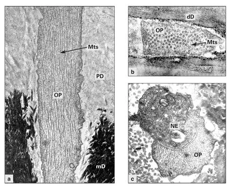
Figs 2-7a to 2-7cElectron micrographs of the odontoblastic process (OP) in longitudinal section (a)and cross section (b, c). The process contains a dense network of microtubules (Mts) and microfilaments, and is free of cytoplasmic organelles. Nerve endings (NE) can be found in close juxtaposition to the OP. (mD) Mineralized dentin; (PD) predentin; (dD) demineralized dentin. (Original magnification × 24,000 [a], × 48,000 [b], and × 30,000 [c].)
Читать дальшеИнтервал:
Закладка:
Похожие книги на «Oral Cells and Tissues»
Представляем Вашему вниманию похожие книги на «Oral Cells and Tissues» списком для выбора. Мы отобрали схожую по названию и смыслу литературу в надежде предоставить читателям больше вариантов отыскать новые, интересные, ещё непрочитанные произведения.


Обсуждение, отзывы о книге «Oral Cells and Tissues» и просто собственные мнения читателей. Оставьте ваши комментарии, напишите, что Вы думаете о произведении, его смысле или главных героях. Укажите что конкретно понравилось, а что нет, и почему Вы так считаете.