Philias R. Garant - Oral Cells and Tissues
Здесь есть возможность читать онлайн «Philias R. Garant - Oral Cells and Tissues» — ознакомительный отрывок электронной книги совершенно бесплатно, а после прочтения отрывка купить полную версию. В некоторых случаях можно слушать аудио, скачать через торрент в формате fb2 и присутствует краткое содержание. Жанр: unrecognised, на английском языке. Описание произведения, (предисловие) а так же отзывы посетителей доступны на портале библиотеки ЛибКат.

- Название:Oral Cells and Tissues
- Автор:
- Жанр:
- Год:неизвестен
- ISBN:нет данных
- Рейтинг книги:3 / 5. Голосов: 1
-
Избранное:Добавить в избранное
- Отзывы:
-
Ваша оценка:
Oral Cells and Tissues: краткое содержание, описание и аннотация
Предлагаем к чтению аннотацию, описание, краткое содержание или предисловие (зависит от того, что написал сам автор книги «Oral Cells and Tissues»). Если вы не нашли необходимую информацию о книге — напишите в комментариях, мы постараемся отыскать её.
Oral Cells and Tissues — читать онлайн ознакомительный отрывок
Ниже представлен текст книги, разбитый по страницам. Система сохранения места последней прочитанной страницы, позволяет с удобством читать онлайн бесплатно книгу «Oral Cells and Tissues», без необходимости каждый раз заново искать на чём Вы остановились. Поставьте закладку, и сможете в любой момент перейти на страницу, на которой закончили чтение.
Интервал:
Закладка:
Differentiation of Odontoblasts
Odontoblast precursors migrate into the developing jaw from the neural crest as part of a large population of ectomesenchymal cells that participate in the formation of many parts of the face and oral cavity. During the cap stage of tooth formation, the preodontoblasts concentrate adjacent to the inner enamel epithelium (IEE) of the enamel organ. Preodontoblasts exit the cell cycle and differentiate before the preameloblasts of the IEE have stopped dividing. 1 , 2
Contact with the IEE basement membrane and/or with other associated extracellular material of epithelial origin has long been held to be a requirement for initial odontoblastic differentiation. 3Recent experiments suggest that a fibronectin-rich substratum is a key requirement. 4Early studies implicating the importance of the basement membrane in odontoblast differentiation were reviewed by Ruch 1 , 2and Ruch et al. 5
Aperiodic fibrils are key structures regulating the differentiation of odontoblasts. They are deposited first at the tip of the future cusp, and then apically toward the cervical border of the developing tooth. Shortly after the first aperiodic fibrils form, the preodontoblasts bind to them through leading-edge cytoplasmic processes (see Fig 1-8). 3 , 6 , 7As leading-edge contacts increase in number, the preodontoblasts are immobilized across the basal lamina from the cells of the IEE. Polarity toward the basal lamina is established at this time. 8 , 9Odontoblast differentiation in organ culture fails when the basement membrane is removed by prior incubation in trypsin. 3
Electron microscopy reveals that aperiodic fibrils, about 15 nm wide and 1.0 to 2.0 μm long, are attached to the basal lamina beneath the IEE. Fluorescent antibodies to collagen types I, III, IV, and VI, tenascin, proteoglycan, and fibronectin bind to basement membrane molecular components in the same location, suggesting that the aperiodic fibrils may consist of one or more of these matrix proteins. 10 – 16Similar patches of extracellular matrix have been observed adjacent to the plasma membrane of preodontoblasts. 12 , 17
Fibronectin receptors (165-kDa protein) are present in the leading-edge plasma membrane of preodontoblasts during differentiation and stabilization. Adherence of a cell surface 165-kDa fibronectin receptor appears to stabilize cytoskeletal elements, promote preodontoblast polarization, and trigger other cytoplasmic processes associated with differentiation. 18 , 19Attachment to fibronectin leads to its uptake and removal at the leading edge of the differentiating odontoblast.
Transforming growth factor β1 (TGF-β1), a growth factor that binds to fibronectin, is a well-known inhibitor of cell proliferation and a promoter of odontoblast differentiation and matrix synthesis. Thus, one important function of fibronectin may be to serve as a reservoir for growth factors that cause preodontoblasts to exit the cell cycle and to undergo differentiation. The importance of fibronectin in dentinogenesis is underscored by the observation that cells of the dental papilla can differentiate into odontoblast-like cells when grown in contact with a supporting surface that is rich in fibronectin and other soluble dentin matrix components. 4 , 20
Odontoblasts sequentially express several members of the TGF-β superfamily of growth factors and their receptors. 21During normal development, TGF-β1 is expressed in the IEE before and during odontoblast polarization. Differentiated odontoblasts express receptors for TGF-β1 and secrete TGF-β1 into the dentin matrix. 22Loss-of-function mutations in the Tgf-β1 gene in mice cause dentin and pulpal pathoses. 23Evidence accumulated over nearly 2 decades suggest that spatial and temporal interactions between cell surface receptors and extracellular matrix molecules and growth factors, such as fibronectin and TGF-β1, provide the necessary information to coordinate odontoblast differentiation.
It has been suggested that the entry of calcium ions might act as a signal for mediating restructuring of the cytoskeleton during the establishment of odontoblast shape and polarity toward the IEE. Cell membrane ligand-gated calcium channels have been localized to the apical pole of the preodontoblasts (nearest the basement membrane). 24
In addition to the potential signaling effects of calcium, fibronectin, and TGF-β, there is evidence to suggest that enamel matrix proteins may serve a similar instructional role during odontoblast differentiation. The expression of enamel proteins in the IEE begins before the cells have acquired the secretory ameloblast phenotype. Electron microscopic studies have identified the presence of enamel matrix protein across the basal lamina in close contact with the apical pole of the developing odontoblasts. 16 , 25The enamel proteins, identified by antiamelogenin antibodies, are endocytosed in coated vesicles at the odontoblast cell surface. 16 , 26The potential instructive role, if any, for these enamel proteins in regulating odontoblast development is unclear.
Secretion of Dentin Matrix
Subsequent to odontoblast differentiation, the basal lamina is degraded. Application of in situ hybridization techniques has shown that preameloblasts and preodontoblasts express matrix metalloproteinase 2, an enzyme that degrades collagen IV and fibronectin, coincident with the removal of the basal lamina. 14Evidence from electron microscopy suggests that the preameloblasts of the IEE phagocytose the partially degraded basal lamina.
After the breakup of the basal lamina, heterotypic cell-to-cell contacts form between cell processes of the newly differentiated odontoblasts and the distal ends of the preameloblasts. Although it was speculated that such contacts might allow the transmission of informational messages needed for differentiation, there has never been any evidence presented that functional gap junctional contacts exist between these two cell types.
In contrast, stable gap junctions and macula adherens–type junctions develop between adjacent odontoblasts during aggregation (see chapter 1). 8 , 27 – 30Coordination of dentin matrix secretion may require communication across gap junctions, permitting ions and small metabolites to cross from odontoblast to odontoblast. Soon after alignment of the odontoblasts, a junctional complex consisting of fascia adherens and fascia occludens forms in the distolateral cell membranes. The fascia adherens is associated with a highly developed terminal web of cytoplasmic filaments. 8The tight junctions of the fascia occludens do not form a zonula occludens. 27
Concomitant with the onset of dentin matrix secretion, odontoblasts grow in length and develop large amounts of rough endoplasmic reticulum (RER). A prominent Golgi complex develops in the supranuclear cytoplasm facing the IEE. In addition to increased expression of messenger ribonucleic acid (mRNA) for collagen type I, developing odontoblasts also express mRNA for osteocalcin, dentin phosphophoryn, and high levels of alkaline phosphatase. 31 , 32As synthesis of type I collagen increases, the expression of type III collagen decreases in odontoblasts. Dentin matrix contains type I collagen and a variety of glycoproteins and glycosaminoglycans. 33 , 34
The earliest layer of dentin to form is called mantle dentin ( Fig 2-1). The collagen fibers of the mantle dentin are thicker than those that form later in circumpulpal dentin. In coronal dentin, the collagen fibers of mantle dentin are polymerized perpendicular to the dentinoenamel junction, while the fibers of the circumpulpal dentin form approximately parallel to the dentinoenamel junction.
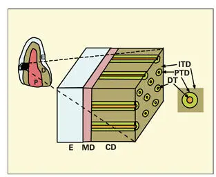
Fig 2-1Components of dentin. The outermost layer of dentin is the mantle dentin (MD). It is deposited during the early stage of odontoblast development. With the appearance of the odontoblastic process, the major portion of dentin, the circumpulpal dentin (CD), is deposited. It consists mainly of intertubular dentin (ITD) and narrow bands of peritubular dentin (PTD) surrounding the dentinal tubule (DT). (E) Enamel; (D) dentin; (P) pulp.
Читать дальшеИнтервал:
Закладка:
Похожие книги на «Oral Cells and Tissues»
Представляем Вашему вниманию похожие книги на «Oral Cells and Tissues» списком для выбора. Мы отобрали схожую по названию и смыслу литературу в надежде предоставить читателям больше вариантов отыскать новые, интересные, ещё непрочитанные произведения.


Обсуждение, отзывы о книге «Oral Cells and Tissues» и просто собственные мнения читателей. Оставьте ваши комментарии, напишите, что Вы думаете о произведении, его смысле или главных героях. Укажите что конкретно понравилось, а что нет, и почему Вы так считаете.