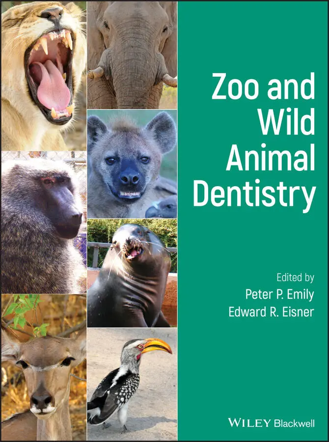
Zoo and Wild Animal Dentistry
Здесь есть возможность читать онлайн «Zoo and Wild Animal Dentistry» — ознакомительный отрывок электронной книги совершенно бесплатно, а после прочтения отрывка купить полную версию. В некоторых случаях можно слушать аудио, скачать через торрент в формате fb2 и присутствует краткое содержание. Жанр: unrecognised, на английском языке. Описание произведения, (предисловие) а так же отзывы посетителей доступны на портале библиотеки ЛибКат.
- Название:Zoo and Wild Animal Dentistry
- Автор:
- Жанр:
- Год:неизвестен
- ISBN:нет данных
- Рейтинг книги:3 / 5. Голосов: 1
-
Избранное:Добавить в избранное
- Отзывы:
-
Ваша оценка:
Zoo and Wild Animal Dentistry: краткое содержание, описание и аннотация
Предлагаем к чтению аннотацию, описание, краткое содержание или предисловие (зависит от того, что написал сам автор книги «Zoo and Wild Animal Dentistry»). Если вы не нашли необходимую информацию о книге — напишите в комментариях, мы постараемся отыскать её.
covers a wide range of zoo and wild species, including cats, bears, primates, dogs, raccoons, weasels, hyenas, marsupials, herbivores, edentates, sea mammals, birds, reptiles, and more. This important resource: Offers a comprehensive reference to oral pathology and dental therapy in captive and wild animals Highlights oral health to promote overall health Includes information on the most recent advances in the field Contains a groundbreaking resource for the dental care of exotic animals Written for zoo and wildlife caretakers and veterinarians, veterinary dentists, veterinary technicians, and veterinary students,
is a practical resource that has information for the dental care of a wide range of animal species that are all too often neglected.
Zoo and Wild Animal Dentistry — читать онлайн ознакомительный отрывок
Ниже представлен текст книги, разбитый по страницам. Система сохранения места последней прочитанной страницы, позволяет с удобством читать онлайн бесплатно книгу «Zoo and Wild Animal Dentistry», без необходимости каждый раз заново искать на чём Вы остановились. Поставьте закладку, и сможете в любой момент перейти на страницу, на которой закончили чтение.
Интервал:
Закладка:
14 Chapter 9AFigure 9A.1 Canal apical foremen, in primates, is approximately 1 mm short o...Figure 9A.2 21‐year‐old chimpanzee. Maxillary incisors are more readily acce...Figure 9A.2a 21‐year‐old chimpanzee. Mandibular incisors are also more readi...Figure 9A.3 Typical primate apical pathology. Notice that the apical canal i...Figure 9A.4 Access to maxillary primate premolars. First PM has two roots. B...Figure 9A.5 Access to primate maxillary molars and three roots.Figure 9A.6 Figure 9A.3a. 21‐year‐old chimpanzee. Maxillary caudal teeth are...Figure 9A.6a Figure 9A.3a. 21‐year‐old chimpanzee. Maxillary molars are acce...Figure 9A.7 21‐year‐old chimpanzee. Mandibular caudal teeth are best accesse...Figure 9A.7a 21‐year‐old chimpanzee. Mandibular second molars are also more ...Figure 9A.8 Postoperative radiograph of root canal therapy on a lower right ...
15 Chapter 9BFigure 9B.1 Cl 1 Lesion (Primate). Class 1 preparations are caries of occlus...Figure 9B.2 Class 2 lesion (Primate). Class 2 cavity preparations are poster...Figure 9B.3 Note: The use of a #330 pear shape bur creates a rounded corner ...Figure 9B.4 Cl 2 preparation (Primate).Figure 9B.5 Class 2 finished preparation with pin retention, shoeing, (reduc...Figure 9B.6 Class 3 lesion (Primate). Class 3 preparations are for interprox...Figure 9B.7 Class 3 preparation with Mylar strip and wedge placement. The re...Figure 9B.8 Class 4 lesion. Class 4 preparations are a continuation of Class...Figure 9B.9a Class 5 Lesion (Shallow). Class 5 preparations are for buccal (...Figure 9B.9b Glass ionomer restoration of the Tiger Class V lesion in Figure...
16 Chapter 10AFigure 10A.1.1 Ibis: Alginate impression being made.Figure 10A.1.2 Ibis: Stone model made from alginate impression.Figure 10A.1.3 Ibis: Acrylic prosthesis construction.Figure 10A.1.4 Ibis: Two #0.030 Markley wires were threaded into caudal lowe...Figure 10A.1.5 Ibis: Insertion of prosthesis onto a Markley wire and secured...Figure 10A.1.6 Ibis: Completed lower beak restoration.Figure 10A.2.1 Goose: Lost upper beak segment.Figure 10A.2.2 Goose: Retention 1 × 4 mm self‐tapping posts are placed into ...Figure 10A.2.3 Goose: Carding red wax matrix in place to establish occlusal ...Figure 10A.2.4 Goose: Composite placed and being shaped.Figure 10A.2.5 Goose: Occlusal check.Figure 10A.2.6 Goose: Postoperative.
17 Chapter 10BFigure 10B.1 Compound TMS pins with elastic chain ligatures; bit plane const...Figure 10B.1.1 Toucan: malocclusion.Figure 10B.1.2 Toucan: TMS Restorative Pins: Drill size 0.20 mm. Self‐thread...Figure 10B.1.3 Dental composite bite plane fabrication.Figure 10B.1.4 Corrected occlusion prior to bite plane removal.Figure 10B.2.1 Great Horned Owl with cross beak.Figure 10B.2.2 1 × 4 mm self‐tapping post for the Great Horned Owl.Figure 10B.2.3 Great Horned Owl. Posts and ligatures in place from bone to b...Figure 10B.3.1 Hornbill: Damaged upper beak.Figure 10B.3.2 Hornbill: Removing necrotic beak section.Figure 10B.3.3 Hornbill: Application of TMS pins.Figure 10B.3.4 Hornbill: TMS pins in place.Figure 10B.3.5 Hornbill: Ligature wire placement across the dorsal void.Figure 10B.3.6 Hornbill: Application of composite hybrid to cover the wire t...Figure 10B.3.7 Hornbill: Completed application of composite, before finishin...Figure 10B.3.8 Hornbill: Prosthetic beak finished and shaped with diamond bu...Figure 10B.3.9 Hornbill: Dye‐stained finish.
18 Chapter 10CFigure 10C.1 Turtle with lost mandibular beak segment.Figure 10C.2 Turtle: Lost beak segment.Figure 10C.3 Turtle: Preparation of lower beak for acrylic prosthesis, with ...Figure 10C.4 Insertion of prosthetic beak segment with Markley wire.Figure 10C.5 Turtle: Acrylic and cyanoacrylate placement.
19 Chapter 10DFigure 10D.1 Anatomical division of the rhamphoteca in the bird.Figure 10D.2 Image obtained by thermography demonstrating the body thermoreg...Figure 10D.3 Macroscopic (A) and microscopic (B) appearance of the trabecula...Figure 10D.4 Radiograph of skull in a chicken showing poor congenital format...Figure 10D.5 CT scan evaluation of a blue and gold macaw skull (Ara araruana...Figure 10D.6 Overgrowth in the blue macaw (Anodorhynchus hyacinthinus). Corr...Figure 10D.7 Scissor beak in Golden Conure (Guaruba guarouba), where the occ...Figure 10D.8 Scheme demonstrating the use of orthopedic pin inside the front...Figure 10D.9 Use of orthopedic screws for fixation of orthodontic elastic fo...Figure 10D.10 Scheme demonstrating the use of the inclined plane technique....Figure 10D.11 Use of inclined plane with acrylic resin for correction of bea...Figure 10D.12 Use of orthodontic buttons for elastic fixation and correction...Figure 10D.13 Fracture of the rhinotheca in cockatoo (Cacatua moluccensis), ...Figure 10D.14 Fracture on the lateral side of the gnathotheca on the blue an...Figure 10D.15 Mandibular symphyseal diastasis in a cockatiel (Nymphicus holl...Figure 10D.16 Surgical correction of symphysis dislocation in a cockatiel (N...Figure 10D.17 Traumatic avulsion of upper and lower beak in green beak touca...Figure 10D.18 Fixation of orthopedic mini‐plates (system 1.5) for fracture c...Figure 10D.19 Rostral fracture of rhinotheca on toco toucan (Ramphastos toco...Figure 10D.20 Taking an impression of a beak fracture in a goose using silic...Figure 10D.21 Toco Toucan (Ramphastos toco), before and after the insertion ...Figure 10D.22 Blue and Gold Macaw (Ara araruana) after the insertion of a he...Figure 10D.23 Rhinoteca metal prosthesis in goose.Figure 10D.24 Use of intra‐dentin pins and orthopedic plaques for the prepar...Figure 10D.25 An example of a 3D scanner and the reference points needed for...Figure 10D.26 Formation of the 3D surface of toucan beak using MeshLab softw...Figure 10D.27 Use of Blender™ software for digital preparation of prosthesis...Figure 10D.28 Virtual model of toucan rhinotheca prosthesis. Identification ...Figure 10D.29 Example of use of 3D‐printed titanium prosthesis in blue and g...Figure 10D.30 Fixation of bipartite prosthesis in green beak toucan (Ramphas...
20 Chapter 11Figure 11.1 Bilateral complicated mandibular canine fracture.Figure 11.2 Dolphin pan bone.Figure 11.3 Radiograph of Dolphin pan bone.Figure 11.4 Palatal inflammation, secondary to adjacent tooth abscess in a C...Figure 11.5 A chart of a Harbor Seal (and most phocid seals) courtesy of Vet...Figure 11.6 A chart of a California Sea Lion (and most otariid seals) courte...Figure 11.7 Maxillary dentition of a California Sea Lion.Figure 11.8 Mandibular dentition of a California Sea Lion. The teeth are ori...Figure 11.9 Sea Lion trained to hold an X‐ray plate. This sea lion has been ...Figure 11.10 Sea Lion target for taking a radiograph. The sea lion touches t...Figure 11.11 Fabricated CR Reusable Radiograph Plate and holder. Pictured is...Figure 11.12 Rostral maxillary radiographic projection. The plate is in a si...Figure 11.13 Caudal maxillary radiographic projection. The plate is placed i...Figure 11.14 Rostral mandibular radiographic projection. The plate is placed...Figure 11.15 Rostral mandibular radiograph.Figure 11.16 Caudal mandibular radiographic projection.Figure 11.17 Maxilla of a Sea Lion: maxilla of an adult California Sea Lion,...Figure 11.18 Maxilla of a juvenile Sea Lion: maxilla of a juvenile Californi...Figure 11.19 Stage 3 Periodontal disease: radiograph of a sea otter showing ...Figure 11.20 Stage IV periodontal disease of all of the mandibular incisors,...Figure 11.21 Sea Lion mandibular fistula from an abscessed left mandibular c...Figure 11.22 Radiograph of Sea Lion showing mandibular fistula and periodont...Figure 11.23 Left mandibular canine periapical lucency. Fractures may be not...Figure 11.24 Bilateral complicated mandibular canine fracture: California Se...Figure 11.25 Complicated crown fracture of a mandibular canine in a Northern...Figure 11.26 Northern Elephant Seal (Mirounga angustirostris), aged four mon...Figure 11.27 Postoperative radiograph of root canal therapy on left upper ca...Figure 11.28 Five sizes of surgical luxators. Luxators are used similarly to...Figure 11.29 XL T Winged Elevators.Figure 11.30 Improper exodontic instrument grip on a winged elevator. The el...Figure 11.31 Proper exodontic instrument grip. The elevator is held in the p...Figure 11.32 Vettome Periotome™. The Powertome Periotome has custom blades t...Figure 11.33 Winged elevators.
Читать дальшеИнтервал:
Закладка:
Похожие книги на «Zoo and Wild Animal Dentistry»
Представляем Вашему вниманию похожие книги на «Zoo and Wild Animal Dentistry» списком для выбора. Мы отобрали схожую по названию и смыслу литературу в надежде предоставить читателям больше вариантов отыскать новые, интересные, ещё непрочитанные произведения.


Обсуждение, отзывы о книге «Zoo and Wild Animal Dentistry» и просто собственные мнения читателей. Оставьте ваши комментарии, напишите, что Вы думаете о произведении, его смысле или главных героях. Укажите что конкретно понравилось, а что нет, и почему Вы так считаете.