Gastrointestinal Pathology
Здесь есть возможность читать онлайн «Gastrointestinal Pathology» — ознакомительный отрывок электронной книги совершенно бесплатно, а после прочтения отрывка купить полную версию. В некоторых случаях можно слушать аудио, скачать через торрент в формате fb2 и присутствует краткое содержание. Жанр: unrecognised, на английском языке. Описание произведения, (предисловие) а так же отзывы посетителей доступны на портале библиотеки ЛибКат.

- Название:Gastrointestinal Pathology
- Автор:
- Жанр:
- Год:неизвестен
- ISBN:нет данных
- Рейтинг книги:3 / 5. Голосов: 1
-
Избранное:Добавить в избранное
- Отзывы:
-
Ваша оценка:
Gastrointestinal Pathology: краткое содержание, описание и аннотация
Предлагаем к чтению аннотацию, описание, краткое содержание или предисловие (зависит от того, что написал сам автор книги «Gastrointestinal Pathology»). Если вы не нашли необходимую информацию о книге — напишите в комментариях, мы постараемся отыскать её.
and assessing biopsies for GI conditions of all kinds
Accurate diagnosis of GI conditions necessarily entails both the careful taking of biopsies and the informed analysis of tissue material. With that being so, gastroenterologists and GI pathologists alike must have a solid understanding of the techniques, handling requirements, and diagnostic characteristics involved if they are to collaborate effectively.
has been designed to provide a clinically focussed and richly illustrated guide to real-world scenarios faced by practicing GI specialists, offering step-by-step instruction and professional advice on the correct diagnosis of all major GI conditions. This essential new book includes:
Full-color illustrations throughout Complete details of biopsy samples required to diagnose specific conditions Reviews of differential diagnoses Clinical management clues based on pathologic findings Featuring information to improve the practice of all gastroenterologists and GI pathologists,
is a practical and every-day resource for the precise diagnosis of a wide range of GI conditions.
Gastrointestinal Pathology — читать онлайн ознакомительный отрывок
Ниже представлен текст книги, разбитый по страницам. Система сохранения места последней прочитанной страницы, позволяет с удобством читать онлайн бесплатно книгу «Gastrointestinal Pathology», без необходимости каждый раз заново искать на чём Вы остановились. Поставьте закладку, и сможете в любой момент перейти на страницу, на которой закончили чтение.
Интервал:
Закладка:
Histologically, pemphigus vulgaris is characterized by the presence of suprabasal clefting and intraepithelial blisters due to prominent acantholysis. The intact basal epithelial cells may appear similar to a row of tombstones at the base of the cleft. The blister may be accompanied by a mild superficial mixed inflammatory infiltrate, which includes eosinophils. Immunofluorescence shows characteristic intercellular deposition of IgG and C3, although the results can be negative in early stages or in disease remission.
Bullous pemphigoid shows subepithelial clefting, often containing a mixture of fibrin, red cells, neutrophils, and eosinophils. Immunofluorescence studies may demonstrate the deposition of complement components (typically C3), and linear deposits of IgG at the level of the basement membrane, although these studies may be of limited value if the mucosa has been extensively eroded.
In epidermolysis bullosa, the lesions are thought to be similar to those found in the skin, with the esophageal mucosa exhibiting a subepithelial cleavage plane, with little or no associated inflammatory infiltrate.
Differential Diagnosis
The differential diagnosis of lichen planus includes other disorders that may show intraepithelial lymphocytic infiltration, such as drug injury, reflux, infections, Crohn’s disease and lymphocytic esophagitis. Clinical correlation is necessary, with particular attention to other mucocutaneous findings, medication or IBD history, location of injury, and response to therapy.
Differentiating bullous diseases from sloughing esophagitis requires clinical and/or IF correlation.
Prognosis, Evolution, and Clinical Management
Esophageal lichen planus has a tendency to cause persistent dysphagia and stricture formation. Systemic treatment is usually required to prevent stricturing disease. Injection of corticosteroids and a response to oral fluticasone have been reported. Squamous cell dysplasia and carcinoma can develop and long‐term follow‐up is advised.
Mucosal pemphigus vulgaris may remain localized to the mucous membranes or progress to skin involvement. Corticosteroids and immunosuppressive agents are considered first‐line treatments.
In epidermolysis bullosa, stricturing disease is common and treatment is symptomatic. Although these patients are at increased risk of skin cancer, esophageal carcinoma is rarely reported.
Scleroderma‐associated chronic GERD can lead to Barrett's esophagus with an increased risk of esophageal adenocarcinoma.
Esophagitis and Inflammatory Bowel Disease
Definition, General Features, Predisposing Factors
Isolated esophageal Crohn’s disease is rare, and usually associated with established extraesophageal disease. Endoscopic studies report a prevalence of esophageal involvement between 0.2 and 11% in adults with Crohn’s disease. However, in children up to 43% may show histological esophageal involvement on mucosal biopsy.
Esophageal inflammation has been documented in patients with ulcerative colitis, although there have been no documented patterns that occur more commonly in ulcerative colitis than in controls.
Clinical and Endoscopic Characteristics
The most common clinical presentation in Crohn’s disease is dysphagia. However, esophageal symptoms may be absent or not correlate well with the endoscopic or biopsy findings. In fact, upper gastrointestinal symptoms related to involvement of other organs may predominate.
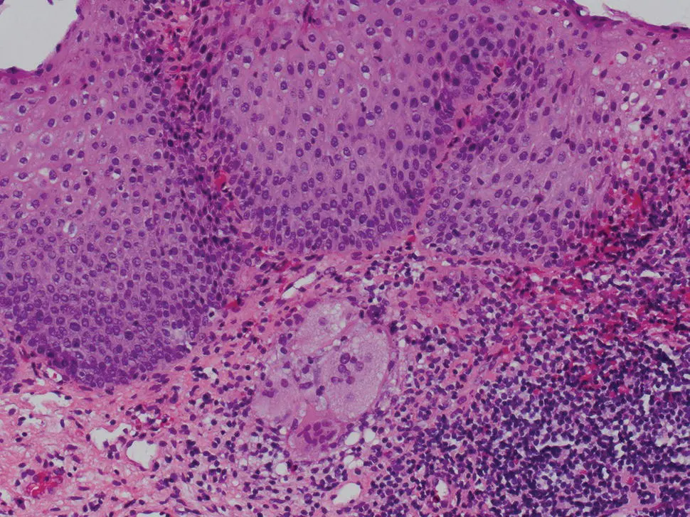
Figure 2.23 Esophageal granuloma in a previously diagnosed patient with Crohn's disease.
Endoscopically, the mid‐ to distal esophagus is most commonly involved. Findings may include erythema, erythematous nodules, single or multiple erosions, and aphthous or deep punched‐out ulcers, which may mimic pill‐induced esophagitis, but are usually differentiated by the extensive mucosal involvement. Strictures and fistulas may occur.
Microscopic Features
Histologic features can be found in asymptomatic patients (particularly children) and those with endoscopically normal appearing mucosa. Therefore biopsies are an important diagnostic tool. The findings include focal active mucosal infiltration with neutrophils and eosinophils, erosions, and ulceration. Erosions and ulcers are associated with an inflammatory infiltrate of the lamina propria and submucosa, which may contain histiocytes or small, poorly formed granulomas ( Figure 2.23). Epithelioid granulomas are documented in up to 39% of esophageal biopsies, and in the proper clinical setting the presence of granulomas is considered diagnostic of Crohn’s esophagitis. Transmural inflammation has been documented in surgical specimens.
Crohn’s disease has also been associated with lymphocytic and eosinophilic esophagitis patterns of mucosal inflammation. The lymphocytic pattern is entirely nonspecific. Crohn’s disease‐related eosinophilic inflammation may be similar to allergic eosinophilic esophagitis, although the underlying immunologic mechanisms appear to differ.
Differential Diagnosis
The histological differential diagnosis includes lymphocytic esophagitis, eosinophilic esophagitis, certain infections (fungal, mycobacterial), and pill esophagitis. The clinical context as well as corroborating histological abnormalities in other mucosal biopsies is a key consideration. Special stains may be indicated to exclude infectious agents.
Prognosis, Evolution, and Clinical Management
Esophageal Crohn’s disease is unlikely to progress or lead to recurrent esophageal lesions or symptoms during subsequent disease exacerbations. Esophageal symptoms, when present, typically improve with therapy. Some patients may require dilatation for strictures, or surgery for complications such as fistulae or abscesses.
Further Reading
1 Abid, S., Mumtaz, K., Jafri, W. et al. (2005). Pill‐induced esophageal injury: endoscopic features and clinical outcomes. Endoscopy 37: 740–744.
2 Abraham, S.C., Yardley, J.H., and Wu, T. (1999). Erosive injury to the upper gastrointestinal tract in patients receiving iron medication: an underrecognized entitiy. Am. J. Surg. Pathol. 23: 1241–1247.
3 Abraham, S.C., Cruz‐Correa, M., Lee, L. et al. (1999). Alendronate‐associated esophageal injury: pathologic and endoscopic features. Mod. Pathol. 12: 1152–1157.
4 Abraham, S.C., Ravish, W.J., Anhalt, G.J. et al. (2000). Esophageal lichen planus: case report and review of the literature. Am. J. Surg. Pathol. 24: 1678–1682.
5 Abraham, S.C., Bhagavan, B., Lee, L. et al. (2001). Upper gastrointestinal tract injury in patients receiving Kayexalate (sodium polystyrene sulfonate) in sorbitol. Am. J. Surg. Pathol. 25: 637–644.
6 Aceves, S.S., Newbury, R.O., Dohil, R. et al. (2007). Distinguishing eosinophilic esophagitis in pediatric patients: clinical, endoscopic, and histologic features of an emerging disorder. J. Clin. Gastroenterol. 41: 252–256.
7 Adler, D.G., Farraye, F.A., and Crawford, J.M. (2015). Gastrointestinal tract endoscopic and tissue processing techniques and normal histology. In: Surgical Pathology of the GI Tract, Liver Biliary Tract, and Pancreas, 3e (eds. R.D. Odze and J.R. Goldblum), 4–33. Philadelphia: Saunders.
8 Almashat, S.J., Duan, L., and Goldsmith, J.D. (2014). Non‐reflux esophagitis: a review of inflammatory diseases of the esophagus exclusive of reflux esophagitis. Semin. Diagn. Pathol. 31: 89–99.
9 Armstrong, D., Bennett, J.R., Blum, A.L. et al. (1996). The endoscopic assessment of esophagitis: a progress report on observer agreement. Gastroenterology 111: 85–92.
Читать дальшеИнтервал:
Закладка:
Похожие книги на «Gastrointestinal Pathology»
Представляем Вашему вниманию похожие книги на «Gastrointestinal Pathology» списком для выбора. Мы отобрали схожую по названию и смыслу литературу в надежде предоставить читателям больше вариантов отыскать новые, интересные, ещё непрочитанные произведения.


Обсуждение, отзывы о книге «Gastrointestinal Pathology» и просто собственные мнения читателей. Оставьте ваши комментарии, напишите, что Вы думаете о произведении, его смысле или главных героях. Укажите что конкретно понравилось, а что нет, и почему Вы так считаете.