Clinical and Endoscopic Characteristics
Patients with acute esophageal radiation toxicity (which develops within two to three weeks of exposure) complain of dysphagia, odynophagia, and/or substernal chest pain. Late manifestations (≥3 months) include stricture formation, chronic ulcerations, and fistula formation. Endoscopic abnormalities, depending on the time course, include mucositis, ulceration, strictures, or fistulas.
Macrocytic changes and cytologic atypia of squamous epithelial cells and stromal cells are common, often associated with multinucleation and bizarre hyperchromatic nuclei. Notably, the cells maintain an abundant cytoplasm and mitosis is rare. Submucosal fibrosis and obliterative vasculitis may also be evident in deeper biopsy specimens.
Immunohistochemical Studies and Molecular Features
Immunohistochemical stains may be useful in the differential diagnosis of viral esophagitis.
The squamous epithelial cytologic atypia may mimic carcinoma, which can be of particular concern for recurrent or persistent tumor in patients being treated for primary esophageal carcinomas, as radiation changes and neoplasia can coexist. Review of the prior tumor histology, if available, and assessment for evidence of invasion can be helpful.
The nuclear changes may also resemble viral infection, particularly Herpes simplex , which can be excluded by appropriate immunohistochemical stains for HSV.
Prognosis, Evolution and Clinical Management
The outcome is dependent on the radiation dose and the overall severity of injury. The lesion may heal completely, although severe injury may lead to chronic ulcers, strictures, and fistulas. Oral glutamine therapy may have a protective effect during therapy. Radiation‐induced esophageal carcinoma may also be a late complication of therapy for extraesophageal thoracic tumors. Endoscopic dilation and stenting may be required to maintain luminal patency.
Dermatological Diseases Involving the Esophagus
Definition, General Features, Predisposing Factors
The esophagus may be involved in multiple primary dermatologic conditions ( Table 2.8). Esophageal manifestation may develop in the presence or absence of skin lesions. As these disorders are generally not well recognized by both gastroenterologists and pathologists, they may be overlooked for some time and the diagnosis delayed with potentially important consequences.
Table 2.8 Dermatologic diseases involving the esophagus.
| Lichen planus |
| Bullous diseases |
| Pemphigus vulgaris |
| Bullous pemphigoid |
| Epidermolysis bullosa acquisita |
| Behcet’s syndrome |
| Toxic epidermal necrolysis/Stevens–Johnson syndrome |
| Scleroderma |
Lichen planus involving the esophagus is rare and can occur in conjunction with other mucosal (or cutaneous) lesions or as the sole manifestation. It is predominantly seen in middle‐aged women. Oral involvement coexists with skin lesions in approximately 30–50% of patients. Lichenoid esophagitis refers to a subset of patients with an identical lichen planus pattern of histologic injury without oral or cutaneous lesions, negative immunofluorescence studies and some distinct demographics, such as polypharmacy and an association with viral hepatitis or HIV.
The bullous diseases are also difficult to diagnose in the esophagus and correlation of the findings with the clinical manifestations, separate biopsies of the skin lesions, and ancillary immunofluorescence studies can help secure the diagnosis. Pemphigus vulgaris is the most common of the autoimmune mucocutaneous diseases with bulla formation. Men and women are affected equally, with a predominance of middle‐aged to elderly individuals, although often asymptomatic, esophageal involvement can be demonstrated in the majority of patients if sought by endoscopy and biopsy. Pemphigus vulgaris is caused by loss of integrity of the normal intercellular attachments within the mucosal epithelium resulting in intramucosal bullae of various sizes. Although esophageal involvement may be a frequent occurrence in active mucocutaneous disease, it may also rarely occur as the sole or initial manifestation.
Bullous pemphigoid is a chronic, immune‐mediated, subepidermal blistering skin disease that rarely involves mucosal surfaces and affects the esophagus less commonly than pemphigus vulgaris. It primarily affects patients in the fifth through seventh decades.
Epidermolysis bullosa is a diverse group of inherited skin disorders caused by mutations of genes that encode for structural proteins located at the dermal‐epidermal junction. The patients characteristically develop blisters after minor trauma, with a range of clinical phenotypes and severity. Minor trauma from a food bolus may elicit similar esophageal mucosal injury.
There are also rare reports of Behcet’s disease involving the esophagus, ranging from asymptomatic to severe stricturing disease. In scleroderma, dysmotility‐associated esophageal inflammatory injury is due primarily to reflux and/or candidiasis.
Clinical and Endoscopic Characteristics
Endoscopic evaluation often plays a central role in the evaluation of patients with suspected esophageal manifestations of these dermatologic diseases, although in some cases the procedure must be approached with caution, particularly in the bullous or blistering disorders.
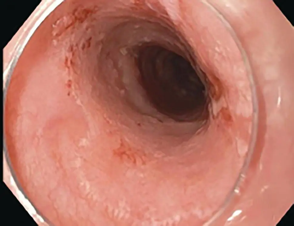
Figure 2.22 Endoscopic appearance of esophageal lichen planus with diffuse mucosal plaques.
In lichen planus, dysphagia and esophageal strictures may clinically suggest GERD. Magnification chromoendoscopy was used in one study showing a high prevalence of esophageal disease. Mucosal stripping, hyperemia, and submucosal plaques ( Figure 2.22) are typically limited to the upper and mid‐esophagus.
Odynophagia and dysphagia are the usual symptoms of esophageal pemphigus vulgaris, as well as the other mucosal blistering diseases. Endoscopic diagnosis involves both evaluation of the appearance of the lesions and tissue sampling. The mucosa may initially appear normal, with the subsequent appearance of erosions, diffuse longitudinal erythematous lines, or mucosal sloughing. Induction of bullae in normal mucosa by the endoscope (Nikolsky's sign) may be observed.
The esophagus is among the most common sites of GI involvement in epidermolysis bullosa, manifested by symptoms of dysphagia and odynophagia. Clinical phenotypes range from mild to severe with bullae and blister formation, ulceration, scarring, webs, and strictures, most commonly involving the proximal esophagus. The trauma of a food bolus, or the endoscope itself, may precipitate of exacerbate bullae and ulcer formation.
Microscopic and Immunofluorescence Features
In addition to hematoxylin and eosin stained sections, immunofluorescence (IF) studies can be of diagnostic value for the diagnosis of lichen planus and vesiculobullous disorders ( Table 2.9). If suspected, additional biopsy samples should be submitted fresh or in an appropriate IF transport medium (e.g. Michel's or Zeus)
Table 2.9 Histologic features of bullous diseases involving the esophagus.
|
Cleft |
Inflammation |
Ancillary IF |
| Pemphigus vulgaris |
Suprabasal with acantholysis |
Mixed, with eosinophils |
Intracellular IgG and C3 |
| Bullous pemphigoid |
Subepithelial |
Neutrophils, eosinophils, fibrin, red blood cells |
Linear basement membrane IgG and C3 |
| Epidermolysis bullosa |
Subepithelial |
Minimal or absent |
N/A |
In lichen planus of the esophagus, the histologic features more closely resemble those seen in oral, rather than cutaneous disease. The characteristic histologic features include a prominent band‐like lymphocytic infiltrate involving the epithelium and lamina propria, acanthotic or atrophic mucosal changes, and scattered degenerated or apoptotic keratinocytes (Civatte bodies). The latter are an important diagnostic feature of lichen planus and a distinguishing characteristic from lymphocytic esophagitis. Immunofluorescence studies showing globular IgM deposits at the epithelial‐subepithelial junction along with complement staining in apoptotic squamous cells, similar to cutaneous findings, may be useful in establishing the diagnosis.
Читать дальше
