Euclid Seeram - A Comprehensive Guide to Radiographic Sciences and Technology
Здесь есть возможность читать онлайн «Euclid Seeram - A Comprehensive Guide to Radiographic Sciences and Technology» — ознакомительный отрывок электронной книги совершенно бесплатно, а после прочтения отрывка купить полную версию. В некоторых случаях можно слушать аудио, скачать через торрент в формате fb2 и присутствует краткое содержание. Жанр: unrecognised, на английском языке. Описание произведения, (предисловие) а так же отзывы посетителей доступны на портале библиотеки ЛибКат.
- Название:A Comprehensive Guide to Radiographic Sciences and Technology
- Автор:
- Жанр:
- Год:неизвестен
- ISBN:нет данных
- Рейтинг книги:4 / 5. Голосов: 1
-
Избранное:Добавить в избранное
- Отзывы:
-
Ваша оценка:
A Comprehensive Guide to Radiographic Sciences and Technology: краткое содержание, описание и аннотация
Предлагаем к чтению аннотацию, описание, краткое содержание или предисловие (зависит от того, что написал сам автор книги «A Comprehensive Guide to Radiographic Sciences and Technology»). Если вы не нашли необходимую информацию о книге — напишите в комментариях, мы постараемся отыскать её.
Designed to support students preparing to sit certification and board examinations, including the American Registry for Radiologic Technologists (ARRT) and other global radiography certification examinations. Addresses the core radiographic science components of the ASRT curriculum, including digital imaging production and equipment, radiation protection and safety, and the principles of Computed Tomography. Useful for students and practitioners in diagnostic medical radiation technology, radiography and medical radiation sciences, as well as in biomedical engineering technology.
A Comprehensive Guide to Radiographic Sciences and Technology — читать онлайн ознакомительный отрывок
Ниже представлен текст книги, разбитый по страницам. Система сохранения места последней прочитанной страницы, позволяет с удобством читать онлайн бесплатно книгу «A Comprehensive Guide to Radiographic Sciences and Technology», без необходимости каждый раз заново искать на чём Вы остановились. Поставьте закладку, и сможете в любой момент перейти на страницу, на которой закончили чтение.
Интервал:
Закладка:
A significant limitation of FSR that has been overcome by digital radiography (DR) relates to the narrow exposure latitude which means that exposure technique selection in FSR must be accurate to achieve an image with acceptable density and contrast. This poses a challenge as stated in point 8. For technologists, DR detectors “have wide exposure latitude, a variable speed class of operation, and image postprocessing capabilities that provide consistent image appearance even with underexposed or overexposed radiographs” [1]. This is illustrated in Figure 2.3. It is clear that DR images taken with low and high exposures appear visually the same on the viewing monitor (due to the image processing of DR systems). Note, however, while low exposures produce images with more noise (grainy image), high exposures produce images with less noise but at the expense of increased dose to the patient. The result is that technologists face a difficult task of recognizing underexposed and overexposed images. If overexposed images cannot be determined, the patient receives an unnecessary dose. Overexposures 5–10 times a normal exposure will appear acceptable to the technologist. Subsequently, this will lead to what has been popularly referred to as exposure creep or dose creep [1].
In FSR, the radiation exposure used for an examination is determined by the exposure technique factors selected on the radiographic control console by the technologist. These include the kilovoltage (kV), the milliamperage (mA), and the time of exposure in seconds (s). Consoles may allow the selection of mAs (mA × s). While the kV determines the x‐ray beam quality (beam penetration), the mA determines the quantity of photons falling on the patient per unit time. The length of time of the exposure is influenced by the exposure time. Today, automatic exposure control (AEC) is often used to ensure that correct exposure factors are used for examinations, and to reduce errors made by manual technique selection. The basic principle of an AEC timer is such when a preset quantity of radiation reaching the film detector (acceptable image density) is measured, the exposure is automatically terminated. The result is almost always perfect exposures.
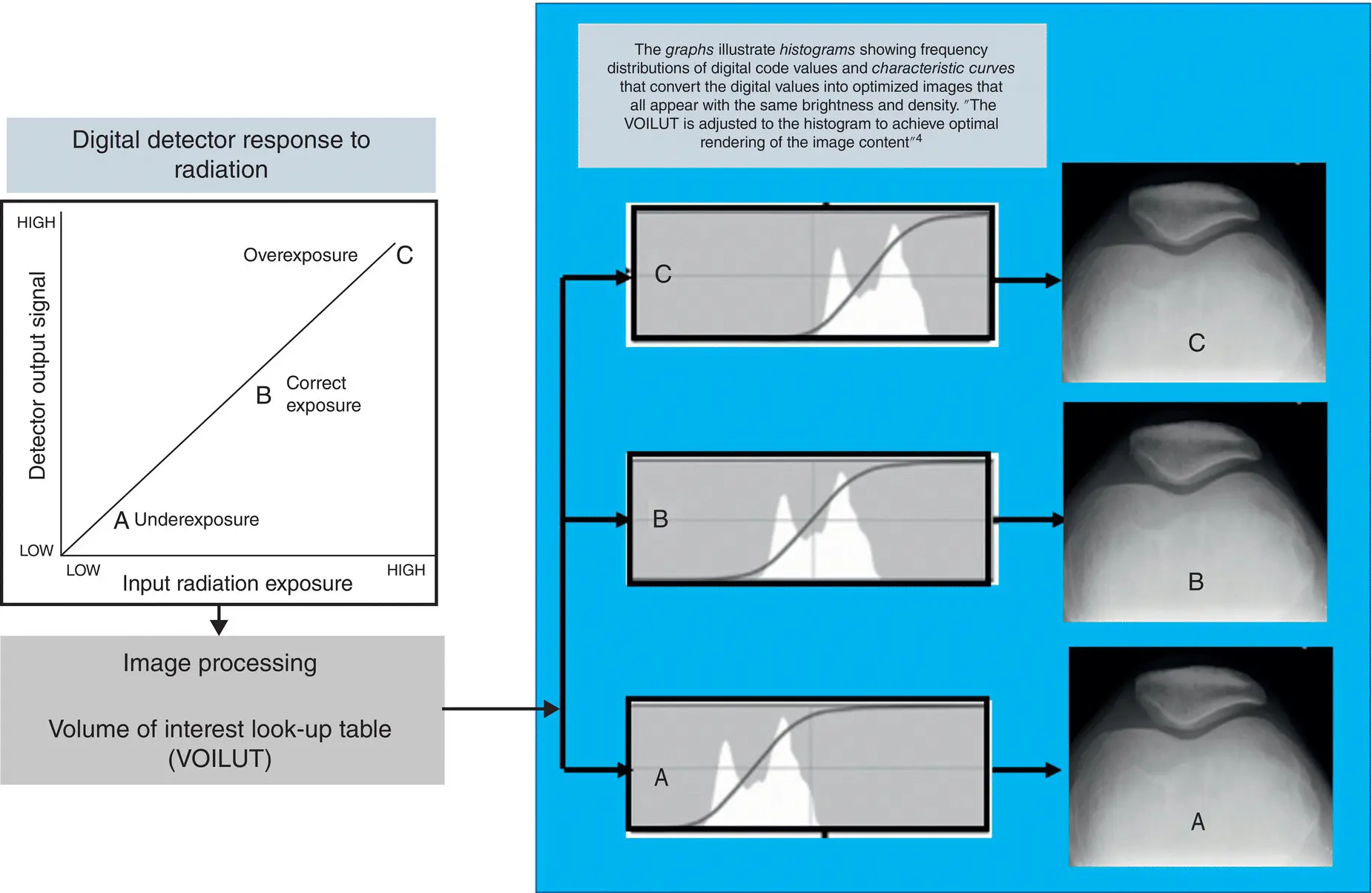
Figure 2.3 DR detectors have wide‐exposure latitude and image postprocessing provides consistent image appearance even with underexposed or overexposed radiographs. See text for further explanation.
Another essential concept in FSR is image quality . The quality of a film‐based image can be described by several technical factors including resolution, contrast, noise, distortion, and artifacts. Only the first three will be reviewed in this chapter. Resolution includes two types, namely, spatial resolution and contrast resolution.
1 Spatial resolution refers to the detail or sharpness of the image, and is measured in line pairs/mm (lp/mm). The higher the number of lp/mm, the greater the sharpness of the image. FSR has the highest spatial resolution ranging from 5 to 15 lp/mm compared to all other imaging modalities [2]. As noted by Bushong [3], “Detail is affected by several factors such as the focal spot size, motion of the patient, and the image receptor design characteristics. Detail is optimum when small focal spots are used, when the patient does not move during the exposure, and when detail cassettes are used.”
2 Contrast resolution on the other hand describes the differences in tissue contrast that the film can show. As a radiation detector, film‐screen cannot show differences in tissue contrast less than 10%. This means that film‐based imaging is limited in its contrast resolution. For example, while the contrast resolution (mm at 0.5% difference) for FSR is 10, it is 20 for nuclear medicine, 10 for ultrasound, 4 for computed tomography (CT), and 1 for magnetic resonance imaging [2]. “The contrast of a radiographic film image, including the object, energy of the beam, scattered radiation, grids, and the film. The main controlling factor for image contrast, however, is kV. Optimum contrast is produced when low kV techniques are used. A grid improves radiographic contrast by absorbing scattered radiation before it gets to the film” [3].
3 Noise is seen on an image as having a grainy appearance. This occurs if few photons (quantity) are used to create the image. Noise can be reduced if more photons are used by using higher mAs settings; however, this will result in more dose to the patient. Less noise is produced when higher kV techniques are used for the same mA settings. The goal of the technologist is to use the lowest possible radiation dose and not compromise image quality. This is an important consideration in observing and working within the as low as reasonably achievable (ALARA) philosophy established by the ICRP.
DIGITAL RADIOGRAPHY MODALITIES: MAJOR SYSTEM COMPONENTS
Digital radiographic imaging systems generally referred to as DR has replaced the workhorse of diagnostic radiography, FSR. DR is defined by the American Association of Physicists in Medicine (AAPM) [4] as “radiographic imaging technology producing digital projection images such as those using photostimulable storage phosphor (computed radiography or CR), amorphous selenium, amorphous silicon, charge‐coupled device (CCD), and metal oxide semiconductor‐field effect transistor (MOSFET) technology.” DR includes computed radiography (CR) and flat‐panel digital radiography (FPDR); digital fluoroscopy (DF); digital mammography (DM); digital radiographic tomosynthesis (DRT) and digital breast tomosynthesis (DBT); and CT. These modalities are described in detail by Seeram [5].
The overall major system components of any DR modality are illustrated in Figure 2.4, and includes an x‐ray generator, an x‐ray tube, a digital detector, a computer, an image display monitor, and finally a digital communications system.
1 The x‐ray generator provides the electrical power to the x‐ray tube to provide the appropriate radiation exposure for the examination under investigation.
2 The patient is exposed and a latent image is created on the digital detector.
3 The latent image is processed by the computer and subsequently displayed on a monitor for viewing and interpretation.
4 The image can be stored and communicated to remote sites for viewing.
Each of the DR modalities listed above will be reviewed below with respect to major imaging system components only. The details of each of these modalities work will be described in later chapters.
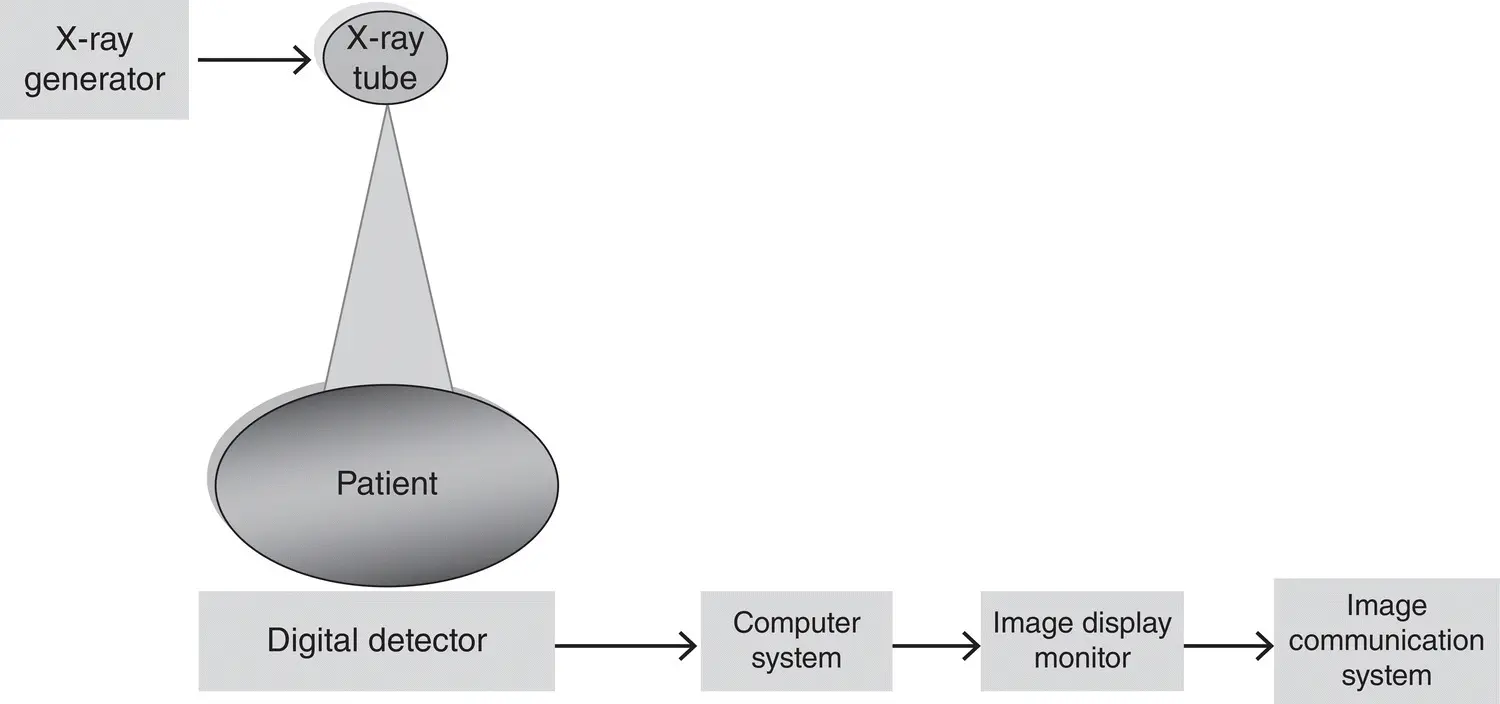
Figure 2.4 The overall major system components of any DR modality includes an x‐ray generator, an x‐ray tube, a digital detector, a computer, an image display monitor, and finally a digital communications system.
Computed radiography
The major components of a CR system are shown in Figure 2.5. These components include the imaging plate (IP), the IP processor, IP erasure, and image display monitor. After the IP is exposed as shown, a latent image is created on the IP. Subsequently, the IP is placed in the processor where it is scanned by a laser beam to render the latent image visible which is then displayed for viewing and interpretation. The IP is exposed to a bright light to erase any residual image, so that it can be used again.
Flat‐panel digital radiography
Интервал:
Закладка:
Похожие книги на «A Comprehensive Guide to Radiographic Sciences and Technology»
Представляем Вашему вниманию похожие книги на «A Comprehensive Guide to Radiographic Sciences and Technology» списком для выбора. Мы отобрали схожую по названию и смыслу литературу в надежде предоставить читателям больше вариантов отыскать новые, интересные, ещё непрочитанные произведения.


Обсуждение, отзывы о книге «A Comprehensive Guide to Radiographic Sciences and Technology» и просто собственные мнения читателей. Оставьте ваши комментарии, напишите, что Вы думаете о произведении, его смысле или главных героях. Укажите что конкретно понравилось, а что нет, и почему Вы так считаете.