Euclid Seeram - A Comprehensive Guide to Radiographic Sciences and Technology
Здесь есть возможность читать онлайн «Euclid Seeram - A Comprehensive Guide to Radiographic Sciences and Technology» — ознакомительный отрывок электронной книги совершенно бесплатно, а после прочтения отрывка купить полную версию. В некоторых случаях можно слушать аудио, скачать через торрент в формате fb2 и присутствует краткое содержание. Жанр: unrecognised, на английском языке. Описание произведения, (предисловие) а так же отзывы посетителей доступны на портале библиотеки ЛибКат.
- Название:A Comprehensive Guide to Radiographic Sciences and Technology
- Автор:
- Жанр:
- Год:неизвестен
- ISBN:нет данных
- Рейтинг книги:4 / 5. Голосов: 1
-
Избранное:Добавить в избранное
- Отзывы:
-
Ваша оценка:
A Comprehensive Guide to Radiographic Sciences and Technology: краткое содержание, описание и аннотация
Предлагаем к чтению аннотацию, описание, краткое содержание или предисловие (зависит от того, что написал сам автор книги «A Comprehensive Guide to Radiographic Sciences and Technology»). Если вы не нашли необходимую информацию о книге — напишите в комментариях, мы постараемся отыскать её.
Designed to support students preparing to sit certification and board examinations, including the American Registry for Radiologic Technologists (ARRT) and other global radiography certification examinations. Addresses the core radiographic science components of the ASRT curriculum, including digital imaging production and equipment, radiation protection and safety, and the principles of Computed Tomography. Useful for students and practitioners in diagnostic medical radiation technology, radiography and medical radiation sciences, as well as in biomedical engineering technology.
A Comprehensive Guide to Radiographic Sciences and Technology — читать онлайн ознакомительный отрывок
Ниже представлен текст книги, разбитый по страницам. Система сохранения места последней прочитанной страницы, позволяет с удобством читать онлайн бесплатно книгу «A Comprehensive Guide to Radiographic Sciences and Technology», без необходимости каждый раз заново искать на чём Вы остановились. Поставьте закладку, и сможете в любой момент перейти на страницу, на которой закончили чтение.
Интервал:
Закладка:
The journal Radiation Protection Dosimetry dedicated a special issue to optimization strategies in medical imaging for fluoroscopy, radiography, mammography, and CT. Several studies identified at least four important requirements for dose and image‐quality optimization research. First, patient safety must be a priority in any study. Second, the level of image quality needed for a particular diagnostic task must be determined. The third requirement involves acquiring images at various exposure levels from high to low and in such a manner that accurate diagnosis can still be made, and finally, use reliable and valid methodologies for the dosimetry, image acquisition, and evaluation of image quality using human observers, keeping in mind the nature of the detection task.
An interesting study utilizing these elements of dose optimization is one by Seeram and colleagues published in Radiologic Technology in 2016. The purpose was to investigate a technique for optimizing radiation dose and image quality for a CR system. The researchers measure the entrance skin doses for phantom models of the pelvis and lumbar spine imaged using the vendor's recommended exposure settings (i.e. the reference doses) as well as doses above and below the vendor's recommended settings for both body parts. Images were assessed using visual grading analysis (VGA). The phantom dosimetry results revealed strong positive linear relationships between dose and milliampere seconds (mAs), mAs and inverse EI, and dose and inverse EI for both body parts. The VGA showed that optimized values of 16 mAs/EI = 136 for the anteroposterior (AP) pelvis and 32 mAs/EI = 139 for the AP lumbar spine did not compromise image quality. Selecting optimized mAs reduced dose by 36% compared with the vendor's recommended mAs (dose) values. The study concluded that optimizing the mAs and associated EIs can be an ED management strategy.
Bibliography
1 1. American Society of Radiologic Technologists (2016). Radiography Curriculum. Albuquerque, NM: American Society of Radiologic Technologists.
2 2. Bushong, S. (2017). Radiologic Science for Technologists. St Louis, MO: Elsevier.
3 3. Bushberg, J.T., Seibert, J.A., Leidholdt, E.M. Jr., and Boone, J.M. (2012). The Essential Physics of Medical Imaging, 3e. Philadelphia, PA: Wolters Kluwer/Lippincott Williams & Wilkins.
4 4. Seeram, E. and Brennan, P. (2017). Radiation Protection in Diagnostic X‐Ray Imaging. Burlington, MA: Jones and Bartlett Learning.
5 5. Seeram, E. (2016). Computed Tomography: Physical Principles, Clinical Applications, and Quality Control. St Louis, MO: Elsevier.
6 6. Wolbarst, A.B., Capasso, P., and Wyant, A. (2013). Medical Imaging: Essentials for Physicians. Hoboken, NJ: Wiley.
7 7. Seeram, E. (2020). Rad Tech's Guide to Radiation Protection, 2e. Hoboken, NJ: Wiley.
8 8. Seeram, E. (2019). Digital Radiography: Physical Principles and Quality Control. Singapore: Springer.
2 Digital radiographic imaging systems: major components
FILM‐SCREEN RADIOGRAPHY: A SHORT REVIEW OF PRINCIPLES
DIGITAL RADIOGRAPHY MODALITIES: MAJOR SYSTEM COMPONENTS
Computed radiography
Flat‐panel digital radiography
Digital fluoroscopy
Digital mammography
Computed tomography
IMAGE COMMUNICATION SYSTEMS
Picture archiving and communication system
References
The purpose of this chapter is to present a general overview of the digital radiographic imaging systems used in diagnostic radiology, by describing briefly, the general system components of each of the modalities in an effort to lay the foundations needed for a good understanding of how each component works to create images and protect not only patients but technologists as well. The more technical details will be reviewed in later chapters on each of the modalities. First, it is important to review the essential principles of film‐screen radiography (FSR) in order to fully understand the rationale for the emergence of digital imaging modalities.
FILM‐SCREEN RADIOGRAPHY: A SHORT REVIEW OF PRINCIPLES
The overall system components of FSR are shown in Figure 2.1. These include the x‐ray generator, the x‐ray tube, a radiographic table, the image receptor, a chemical processing unit, and a light view‐box. A film‐based image is created as follows:
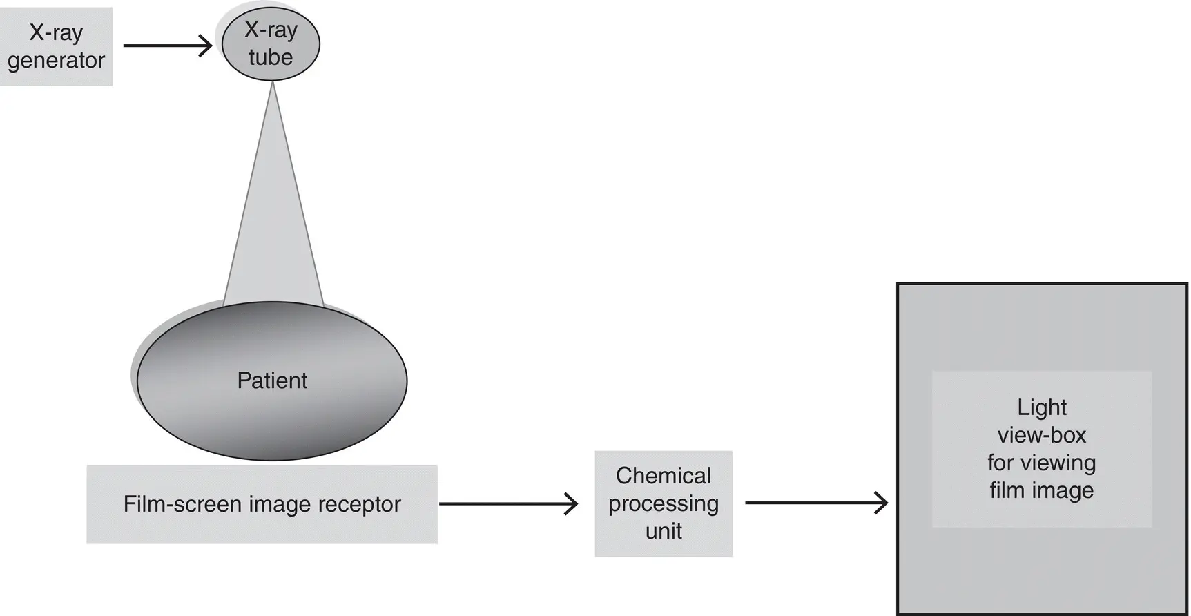
Figure 2.1 The overall system components of film screen radiography (FSR).These include the x‐ray generator, the x‐ray tube, the image receptor, a chemical processing unit, and a light view‐box, for viewing the film image.
1 X‐rays pass through the patient and fall upon the film to form a latent image.
2 The latent image is then rendered visible using chemical processing.
3 The visible image on the film is displayed on a light view‐box for viewing and interpretation by a radiologist. This image consists of varying degrees of blackening as a result of the amount of exposure transmitted by different parts of the anatomy. While the blackening is referred to as the film density, the difference in densities in the image is referred to as the film contrast. The film converts the radiation transmitted through the various types of tissues (tissue contrast) into film contrast.
4 The light from the view‐box is transmitted through the film and can be measured using a densitometer and is referred to as the optical density (OD), which is defined as the log of the ratio of the intensity of the view‐box (original intensity) to the intensity of the transmitted light. The OD is used to describe the degree of film blackening as a result of radiation exposure.
5 A plot of the OD as a function of the log of the relative radiation exposure is described by the well‐known characteristic curve (Hurter–Driffield Curve), which provides information about the film response to the exposure ( Figure 2.2). There are three parts of the curve: the toe region, the slope, and the shoulder region. Exposures that fall in the toe and shoulder region of the curve will result in images that are light (underexposed) and images that are dark (overexposed), respectively. Acceptable image contrast is obtained when the exposure that falls within the slope of the curve. This slope defines the exposure latitude as well as the film contrast characteristics (the steeper the gradient, the higher the contrast).
6 The density of the image is hence used as an exposure indicator that provides immediate feedback to the technologist that the correct exposure technique factors have been used for the examination. This curve also shows that FSR has a fixed film speed (sensitivity) and a fixed‐dose requirement.
7 Furthermore, the characteristic curve can be used to describe the film speed, average gradient, the film gamma, and the film latitude. Only film speed and film latitude will be reviewed here. The interested reader should refer to any standard radiography physics text for a further description of the other terms. While film speed refers to the sensitivity of the film to radiation and it is inversely proportional to the exposure, film latitude describes the range of exposures that would produce useful densities (contrast). Figure 2.2 A plot of the OD as a function of the log of the relative radiation exposure is described by the well‐known characteristic curve (Hurter–Driffield Curve), which provides information about the film response to the exposure. See text for further explanation.
8 High‐speed films (fast films) require less exposure than films with low speeds (slow films). On the other hand, while wide‐exposure latitude films respond to a wide range of exposures, films with narrow exposure latitude can respond only to a small range of exposures. In the latter situation, the technologist has to be very precise in the selection of the exposure technique factors for examination.
Читать дальшеИнтервал:
Закладка:
Похожие книги на «A Comprehensive Guide to Radiographic Sciences and Technology»
Представляем Вашему вниманию похожие книги на «A Comprehensive Guide to Radiographic Sciences and Technology» списком для выбора. Мы отобрали схожую по названию и смыслу литературу в надежде предоставить читателям больше вариантов отыскать новые, интересные, ещё непрочитанные произведения.


Обсуждение, отзывы о книге «A Comprehensive Guide to Radiographic Sciences and Technology» и просто собственные мнения читателей. Оставьте ваши комментарии, напишите, что Вы думаете о произведении, его смысле или главных героях. Укажите что конкретно понравилось, а что нет, и почему Вы так считаете.