Euclid Seeram - A Comprehensive Guide to Radiographic Sciences and Technology
Здесь есть возможность читать онлайн «Euclid Seeram - A Comprehensive Guide to Radiographic Sciences and Technology» — ознакомительный отрывок электронной книги совершенно бесплатно, а после прочтения отрывка купить полную версию. В некоторых случаях можно слушать аудио, скачать через торрент в формате fb2 и присутствует краткое содержание. Жанр: unrecognised, на английском языке. Описание произведения, (предисловие) а так же отзывы посетителей доступны на портале библиотеки ЛибКат.
- Название:A Comprehensive Guide to Radiographic Sciences and Technology
- Автор:
- Жанр:
- Год:неизвестен
- ISBN:нет данных
- Рейтинг книги:4 / 5. Голосов: 1
-
Избранное:Добавить в избранное
- Отзывы:
-
Ваша оценка:
A Comprehensive Guide to Radiographic Sciences and Technology: краткое содержание, описание и аннотация
Предлагаем к чтению аннотацию, описание, краткое содержание или предисловие (зависит от того, что написал сам автор книги «A Comprehensive Guide to Radiographic Sciences and Technology»). Если вы не нашли необходимую информацию о книге — напишите в комментариях, мы постараемся отыскать её.
Designed to support students preparing to sit certification and board examinations, including the American Registry for Radiologic Technologists (ARRT) and other global radiography certification examinations. Addresses the core radiographic science components of the ASRT curriculum, including digital imaging production and equipment, radiation protection and safety, and the principles of Computed Tomography. Useful for students and practitioners in diagnostic medical radiation technology, radiography and medical radiation sciences, as well as in biomedical engineering technology.
A Comprehensive Guide to Radiographic Sciences and Technology — читать онлайн ознакомительный отрывок
Ниже представлен текст книги, разбитый по страницам. Система сохранения места последней прочитанной страницы, позволяет с удобством читать онлайн бесплатно книгу «A Comprehensive Guide to Radiographic Sciences and Technology», без необходимости каждый раз заново искать на чём Вы остановились. Поставьте закладку, и сможете в любой момент перейти на страницу, на которой закончили чтение.
Интервал:
Закладка:
Image quality considerations
Image quality is a significant goal of radiographic imaging modalities. The attenuated radiation data from the patient are used to create images that are used for diagnostic interpretation by a human observer. There are at least five important descriptors of digital image quality and these include spatial resolution, contrast resolution, noise, detective quantum efficiency (DQE), and image artifacts. While spatial resolution addresses the sharpness of images, and is related to the size of the pixels (picture elements) in an image, contrast resolution or density resolution deals with the ability of the imaging system to demonstrate differences in tissue contra, and is linked to the bit depth, that is the range of gray levels per pixel. Noise, on the other hand, depends on the number of x‐ray photons used to create the image. While fewer photons (low exposure technique factors) will result in more noise (grainy appearance), more photons (higher exposure technique factors) will create a better image (less noisy image), but at the expense of dose. Another digital image quality descriptor is the DQE, which is a measure of the efficiency and fidelity with which the detector can convert an input exposure into a useful output image. Finally, digital images are not free of artifacts. These are features seen on the image that are not present in the patient, and can pose challenges for the human observer in detecting fact from artifact.
Computed tomography – physics and instrumentation
This section will present a broad overview of the essential elements of the Physics and Instrumentation of Computed Tomography . One of the major advantages of CT is that it provides improved contrast resolution compared to radiography and for this reason, it has proven to be worthy of further developments in imaging soft tissues of the human body. It is important to note, however, that magnetic resonance imaging (MRI) has superior contrast resolution compared to all other imaging modalities, such as radiography, nuclear medicine, and diagnostic medical sonography.
CT is a sectional imaging technique that produces direct cross‐sectional digital images referred to as transverse axial images which has been referred to as planar sections that are perpendicular to the long axis of the patient. The word “computed” implies that a computer is used to process and reconstruct x‐ray transmission data collected from the patient. The CT scanner has evolved from single‐slice CT scanners (SSCT) to multi‐slice CT scanners (MSCT). State‐of‐the‐art CT scanners are now MSCT scanners capable of a wide range of applications. The increasing use is that CT in clinical practice has led to increasing doses to the patient and a well‐documented fact is that CT delivered the highest collective dose in the United States compared to other medical imaging modalities.
Two individuals shared the Nobel Prize in Medicine and Physiology in 1979 for their development of the CT scanner. These include Godfrey Newbold Hounsfield in the United Kingdom (UK) who invented the first clinically useful scanner, and Allan Cormack, a physicist at Tufts University in Massachusetts.
CT is a multidisciplinary technology and has its roots in physics, mathematics, engineering, and computer science. The CT process consists of at least three major system components that are used to produce the CT image; the data acquisition system; the computer system; and the image display, storage, and communication systems.
Data acquisition means that radiation attenuation data are collected from the patient during the scanning. In this respect, an x‐ray tube coupled to special electronic detectors rotate around the patient to collect and measure attenuation readings as the x‐ray beam passes through the patient.
The attenuation is according to Beer–Lambert's law:

where I is the transmitted x‐ray beam intensity, I ois the original x‐ray beam intensity, e represents Euler's constant, μ is the linear attenuation coefficient, and Δx is the finite thickness of the section. In CT, the system calculates all μs for all structures seen on the image. Special detectors and detector electronics are used to calculate the attenuation data and convert them into integers (0, a positive number, or a negative number) referred to as CT numbers using an image reconstruction algorithm to build up the image in numerical format. The CT numbers (numerical image format) are converted into a gray‐scale image for display on a monitor for the observer to interpret.
The CT numbers are calculated using the following relationship:
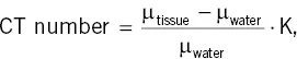
where K represents a scaling factor. In general, K is equal to 1000. When Hounsfield invented the scanner, K was equal to 500.
The technology aspects of CT are complex and are responsible for using the attenuation values collected around the patient for 360° to build up an image of the internal anatomy of the patient, and displays such image for interpretation by radiologists. The technology addressing the collection of these values includes the x‐ray tube which is coupled to special electronic detectors and detector electronics. Another major technology component in CT is the computer system which captures the raw data from the detectors and uses sophisticated image reconstruction algorithms for creating the image from the raw data.
Present‐day CT scanners are MSCT scanners. One characteristic feature of MSCT is the two‐dimensional detector array, compared to a one‐dimensional detector array of SSCT. This means that there will be additional specific technical factors that affect the dose in CT. One such notable factor is the pitch (P), which is defined by the International Electrotechnical Commission (IEC) as the distance the table travels per rotation (D) divided by the total collimation (W). This can be expressed algebraically as:

The increasing use of CT has led to widespread concerns about high patient radiation doses from CT examinations relative to other radiography examinations. The distribution of the dose to the patient in CT is significantly different than the distribution of the dose in radiography. These differences require additional CT‐specific dose metrics. There are essential four CT‐specific dose metrics: the computed tomography dose index (CTDI), the dose length product (DLP), the size‐specific dose estimate (SSDE), and the ED. These and other elements of CT physical principles will be described further in Chapter 7.
Quality control
QC is an essential activity of all medical imaging departments and it is part of a QA program. QA deals with people and includes the administrative aspects of patient care and quality outcomes. QC addresses the technical aspects of equipment performance used to image patients. QA and QC programs have evolved into what is now referred to as Continuous Quality Improvement (CQI) which includes Total Quality Management (TQM). CQI was introduced by the Joint Committee on Accreditation of Healthcare organizations (JCAHO) to stress the importance that all employees play an active role in ensuring a quality product. The purpose of the procedures and techniques of CQI, QA, and QC is threefold: to ensure optimum image quality for the purpose of enhancing diagnosis, to optimize the radiation dose to patients and reduce the dose to personnel, and to reduce costs to the healthcare facility.
Читать дальшеИнтервал:
Закладка:
Похожие книги на «A Comprehensive Guide to Radiographic Sciences and Technology»
Представляем Вашему вниманию похожие книги на «A Comprehensive Guide to Radiographic Sciences and Technology» списком для выбора. Мы отобрали схожую по названию и смыслу литературу в надежде предоставить читателям больше вариантов отыскать новые, интересные, ещё непрочитанные произведения.


Обсуждение, отзывы о книге «A Comprehensive Guide to Radiographic Sciences and Technology» и просто собственные мнения читателей. Оставьте ваши комментарии, напишите, что Вы думаете о произведении, его смысле или главных героях. Укажите что конкретно понравилось, а что нет, и почему Вы так считаете.