Javid A. Parray - Nano-Technological Intervention in Agricultural Productivity
Здесь есть возможность читать онлайн «Javid A. Parray - Nano-Technological Intervention in Agricultural Productivity» — ознакомительный отрывок электронной книги совершенно бесплатно, а после прочтения отрывка купить полную версию. В некоторых случаях можно слушать аудио, скачать через торрент в формате fb2 и присутствует краткое содержание. Жанр: unrecognised, на английском языке. Описание произведения, (предисловие) а так же отзывы посетителей доступны на портале библиотеки ЛибКат.
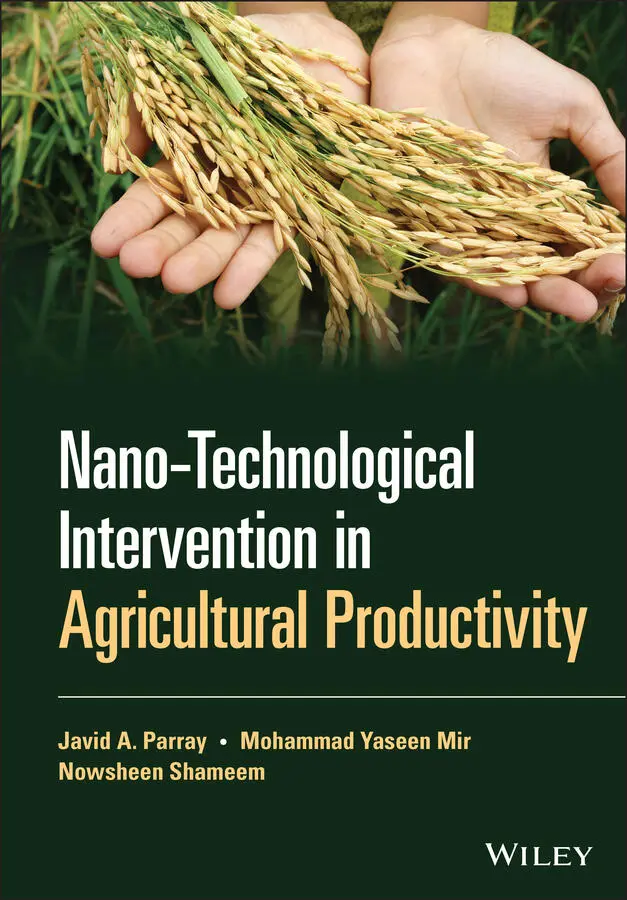
- Название:Nano-Technological Intervention in Agricultural Productivity
- Автор:
- Жанр:
- Год:неизвестен
- ISBN:нет данных
- Рейтинг книги:3 / 5. Голосов: 1
-
Избранное:Добавить в избранное
- Отзывы:
-
Ваша оценка:
Nano-Technological Intervention in Agricultural Productivity: краткое содержание, описание и аннотация
Предлагаем к чтению аннотацию, описание, краткое содержание или предисловие (зависит от того, что написал сам автор книги «Nano-Technological Intervention in Agricultural Productivity»). Если вы не нашли необходимую информацию о книге — напишите в комментариях, мы постараемся отыскать её.
Explores biotechnological advances in the development of sophisticated green technologies for waste minimization and waste control Emphasizes the use of microbes for degradation and removal of various xenobiotic substances Discusses bioremediation approaches in relation to the impact of increased urbanization and industrialization on the environment Covers a variety of applications of nanotechnology in agriculture, including nano-fertilizers, nano-biosensors, nano-pesticides, and nanoparticle protection in plants
is a valuable resource for students in plant biotechnology and agricultural science and engineering, as well as an important reference for researchers in plant biotechnology and agricultural sciences, particularly those with interest in the use of nanomaterials for pollution remediation and sustainable development.
Nano-Technological Intervention in Agricultural Productivity — читать онлайн ознакомительный отрывок
Ниже представлен текст книги, разбитый по страницам. Система сохранения места последней прочитанной страницы, позволяет с удобством читать онлайн бесплатно книгу «Nano-Technological Intervention in Agricultural Productivity», без необходимости каждый раз заново искать на чём Вы остановились. Поставьте закладку, и сможете в любой момент перейти на страницу, на которой закончили чтение.
Интервал:
Закладка:
27 27 Gujrati, M., Malamas, A., Shin, T. et al. (2014). Multifunctional cationic lipid‐based nanoparticles facilitate endosomal escape and reduction‐triggered cytosolic siRNA release. Mol. Pharmaceutics 11: 2734–2744. https://doi.org/10.1021/mp400787s.
28 28 Wang, Y. and Xia, Y. (2004). Bottom‐up and top‐down approaches to the synthesis of monodispersed spherical colloids of low melting‐point metals. Nano Lett. 4: 2047–2050. https://doi.org/10.1021/nl048689j.
29 29 Iravani, S. (2011). Green synthesis of metal nanoparticles using plants. Green Chem. 13: 2638. https://doi.org/10.1039/c1gc15386b.
30 30 Bello, S.A., Agunsoye, J.O., and Hassan, S.B. (2015). Synthesis of coconut shell nanoparticles via a top‐down approach: assessment of milling duration on the particle sizes and morphologies of coconut shell nanoparticles. Mater. Lett. https://doi.org/10.1016/j.matlet.2015.07.063.
31 31 Priyadarshana, G., Kottegoda, N., Senaratne, A. et al. (2015). Synthesis of magnetite nanoparticles by top‐down approach from a high purity ore. J. Nanomater.: 1–8. https://doi.org/10.1155/2015/317312.
32 32 Garrigue, P., Delville, M.‐H., Labruge're, C. et al. (2004). Top‐down approach for the preparation of colloidal carbon nanoparticles. Chem. Mater. 16: 2984–2986. https://doi.org/10.1021/cm049685i.
33 33 Zhang, X., Lai, Z., Liu, Z. et al. (2015). A facile and universal top‐down method for preparation of monodisperse transition‐metal dichalcogenide nanodots. Angew. Chem. Int. Ed. 54: 5425–5428. https://doi.org/10.1002/anie.201501071.
34 34 Zhou, Y., Dong, C.K., Han, L. et al. (2016). Top‐down preparation of active cobalt oxide catalyst. ACS Catal. 6: 6699–6703. https://doi.org/10.1021/acscatal.6b02416.
35 35 Mogilevsky, G., Hartman, O., Emmons, E.D. et al. (2014). Bottom‐up synthesis of anatase nanoparticles with graphene domains. ACS Appl. Mater. Interfaces 6: 10638–10648. https://doi.org/10.1021/am502322y.
36 36 Liu, D., Li, C., Zhou, F. et al. (2015). Rapid synthesis of monodisperse Au nanospheres through a laser irradiation‐induced shape conversion, self‐assembly and their electromagnetic coupling SERS enhancement. Sci. Rep. 5: 7686. https://doi.org/10.1038/srep07686.
37 37 Liu, J., Liu, Y., Liu, N. et al. (2015). Metal‐free efficient photocatalyst for stable visible water splitting via a two‐electron pathway. Science 80 (347): 970–974. https://doi.org/10.1126/science.aaa3145.
38 38 Needham, D., Arslanagic, A., Glud, K. et al. (2016). Bottom‐up design of nanoparticles for anti‐cancer diapeutics: “put the drug in the cancer's food”. J. Drug Targeting: 1–21. https://doi.org/10.1080/1061186X.2016.1238092.
39 39 Wang, Y. and Xia, Y. (2004). Bottom‐up and top‐down approaches to synthesizing monodispersed spherical colloids of low melting‐point metals. Nano Lett. 4: 2047–2050. https://doi.org/10.1021/nl048689j.
40 40 Parveen, K., Banse, V., and Ledwani, L. (2016). Green synthesis of nanoparticles: their advantages and disadvantages. Acta Naturae: 20048. https://doi.org/10.1063/1.4945168.
41 41 Ahmed, S., Annu, S., and Yudha, S.S. (2016). Biosynthesis of gold nanoparticles: a green approach. J. Photochem. Photobiol., B 161: 141–153. https://doi.org/10.1016/j.jphotobiol.2016.04.034.
42 42 Biswas, A., Bayer, I.S., Biris, A.S. et al. (2012). Advances in top‐down and bottom‐up surface nanofabrication: techniques, applications and prospects. Adv. Colloid Interface Sci. 170: 2–27. https://doi.org/10.1016/j.cis.2011.11.001.
43 43 Mirzadeh, E. and Akhbari, K. (2016). Synthesis of nanomaterials with desirable morphologies from metal–organic frameworks for various applications. CrystEngComm 18: 7410–7424. https://doi.org/10.1039/C6CE01076H.
44 44 Khlebtsov, N. and Dykman, L. (2011). Biodistribution and toxicity of engineered gold nanoparticles: a review of in vitro and in vivo studies. Chem. Soc. Rev. 40: 1647–1671. https://doi.org/10.1039/C0CS00018C.
45 45 Wang, J., Yang, N., Tang, H. et al. (2013). Accurate control of multishelled Co3O4 hollow microspheres as high‐performance anode materials in lithium‐ion batteries. Angew. Chem. Int. Ed. 52: 6417–6420. https://doi.org/10.1002/anie.201301622.
46 46 Emery, A.A., Saal, J.E., Kirklin, S. et al. (2016). High‐throughput computational screening of perovskites for thermochemical water splitting applications. Chem. Mater. 28 https://doi.org/10.1021/acs.chemmater.6b01182.
47 47 Ingham, B. (2015). X‐ray scattering characterization of nanoparticles. Crystallogr. Rev. 21: 229–303. https://doi.org/10.1080/0889311X.2015.1024114.
48 48 Avasare, V., Zhang, Z., Avasare, D. et al. (2015). Room‐temperature synthesis of TiO2 nanospheres and their solar‐driven photoelectrochemical hydrogen production. Int. J. Energy Res. 39: 1714–1719. https://doi.org/10.1002/er.3372.
49 49 Khan, I., Ali, S., Mansha, M., and Qurashi, A. (2017). Sonochemical assisted hydrothermal synthesis of pseudo‐flower shaped Bismuth vanadate (BiVO4) and their solar‐driven water splitting application. Ultrason. Sonochem. 36: 386–392. https://doi.org/10.1016/j.ultsonch.2016.12.014.
50 50 Mansha, M., Qurashi, A., Ullah, N. et al. (2016). Synthesis of In2O3/graphene heterostructure and their hydrogen gas sensing properties. Ceram. Int. 42: 11490–11495. https://doi.org/10.1016/j.ceramint.2016.04.035.
51 51 Lykhach, Y., Kozlov, S.M., Skála, T. et al. (2015). Counting electrons on supported nanoparticles. Nat. Mater. https://doi.org/10.1038/nmat4500.
52 52 Oprea, B., Martínez, L., Román, E. et al. (2015). Dispersion and functionalization of nanoparticles synthesized by gas aggregation source: opening new routes toward the fabrication of nanoparticles for biomedicine. Langmuir 31: 13813–13820. https://doi.org/10.1021/acs.langmuir.5b03399.
53 53 Wang, Y.C., Engelhard, M.H., Baer, D.R., and Castner, D.G. (2016). Quantifying the impact of nanoparticle coatings and nonuniformities on XPS analysis: gold/silver core‐shell nanoparticles. Anal. Chem. 88: 3917–3925. https://doi.org/10.1021/acs.analchem.6b00100.
54 54 Dablemont, C., Lang, P., Mangeney, C. et al. (2008). FTIR and XPS study of Pt nanoparticle functionalization and interaction with alumina. Langmuir 24: 5832–5841. https://doi.org/10.1021/la7028643.
55 55 Pokhrel, M., Wahid, K., and Mao, Y. (2016). Systematic studies on RE2‐Hf2O7:5%Eu3+ (RE = Y, La, Pr, Gd, Er, and Lu) nanoparticles: effects of the A‐site RE3+ cation and calcination on structure and photoluminescence. J. Phys. Chem. C 120: 14828–14839. https://doi.org/10.1021/acs.jpcc.6b04798.
56 56 Muehlethaler, C., Leona, M., and Lombardi, J.R. (2016). Review of surface‐enhanced Raman scattering applications in forensic science. Anal. Chem. 88: 152–169. https://doi.org/10.1021/acs.analchem.5b04131.
57 57 Ma, S., Livingstone, R., Zhao, B., and Lombardi, J.R. (2011). Enhanced Raman spectroscopy of nanostructured semiconductor phonon modes. J. Phys. Chem. Lett. 2: 671–674. https://doi.org/10.1021/jz2001562.
58 58 Sikora, A., Shard, A.G., and Minelli, C. (2016). Size and ζ‐potential measurement of silica nanoparticles in serum using tunable resistive pulse sensing. Langmuir 32: 2216–2224. https://doi.org/10.1021/acs.Langmuir.5b04160.
59 59 Filipe, V., Hawe, A., and Jiskoot, W. (2010). Critical evaluation of nanoparticle tracking analysis (NTA) by insight to measure nanoparticles and protein aggregates. Pharm. Res. 27: 796–810. https://doi.org/10.1007/s11095-010-0073-2.
60 60 Gross, J., Sayle, S., Karow, A.R. et al. (2016). Nanoparticle tracking analysis of particle size and concentration detection in suspensions of polymer and protein samples: influence of experimental and data evaluation parameters. Eur. J. Pharm. Biopharm. 104: 30–41. https://doi.org/10.1016/j.ejpb.2016.04.013.
Читать дальшеИнтервал:
Закладка:
Похожие книги на «Nano-Technological Intervention in Agricultural Productivity»
Представляем Вашему вниманию похожие книги на «Nano-Technological Intervention in Agricultural Productivity» списком для выбора. Мы отобрали схожую по названию и смыслу литературу в надежде предоставить читателям больше вариантов отыскать новые, интересные, ещё непрочитанные произведения.


![Chade-Meng Tan - Search Inside Yourself - Increase Productivity, Creativity and Happiness [ePub edition]](/books/703803/chade-thumb.webp)

Обсуждение, отзывы о книге «Nano-Technological Intervention in Agricultural Productivity» и просто собственные мнения читателей. Оставьте ваши комментарии, напишите, что Вы думаете о произведении, его смысле или главных героях. Укажите что конкретно понравилось, а что нет, и почему Вы так считаете.