Robert E. Blankenship - Molecular Mechanisms of Photosynthesis
Здесь есть возможность читать онлайн «Robert E. Blankenship - Molecular Mechanisms of Photosynthesis» — ознакомительный отрывок электронной книги совершенно бесплатно, а после прочтения отрывка купить полную версию. В некоторых случаях можно слушать аудио, скачать через торрент в формате fb2 и присутствует краткое содержание. Жанр: unrecognised, на английском языке. Описание произведения, (предисловие) а так же отзывы посетителей доступны на портале библиотеки ЛибКат.
- Название:Molecular Mechanisms of Photosynthesis
- Автор:
- Жанр:
- Год:неизвестен
- ISBN:нет данных
- Рейтинг книги:3 / 5. Голосов: 1
-
Избранное:Добавить в избранное
- Отзывы:
-
Ваша оценка:
Molecular Mechanisms of Photosynthesis: краткое содержание, описание и аннотация
Предлагаем к чтению аннотацию, описание, краткое содержание или предисловие (зависит от того, что написал сам автор книги «Molecular Mechanisms of Photosynthesis»). Если вы не нашли необходимую информацию о книге — напишите в комментариях, мы постараемся отыскать её.
Molecular Mechanisms of Photosynthesis — читать онлайн ознакомительный отрывок
Ниже представлен текст книги, разбитый по страницам. Система сохранения места последней прочитанной страницы, позволяет с удобством читать онлайн бесплатно книгу «Molecular Mechanisms of Photosynthesis», без необходимости каждый раз заново искать на чём Вы остановились. Поставьте закладку, и сможете в любой момент перейти на страницу, на которой закончили чтение.
Интервал:
Закладка:
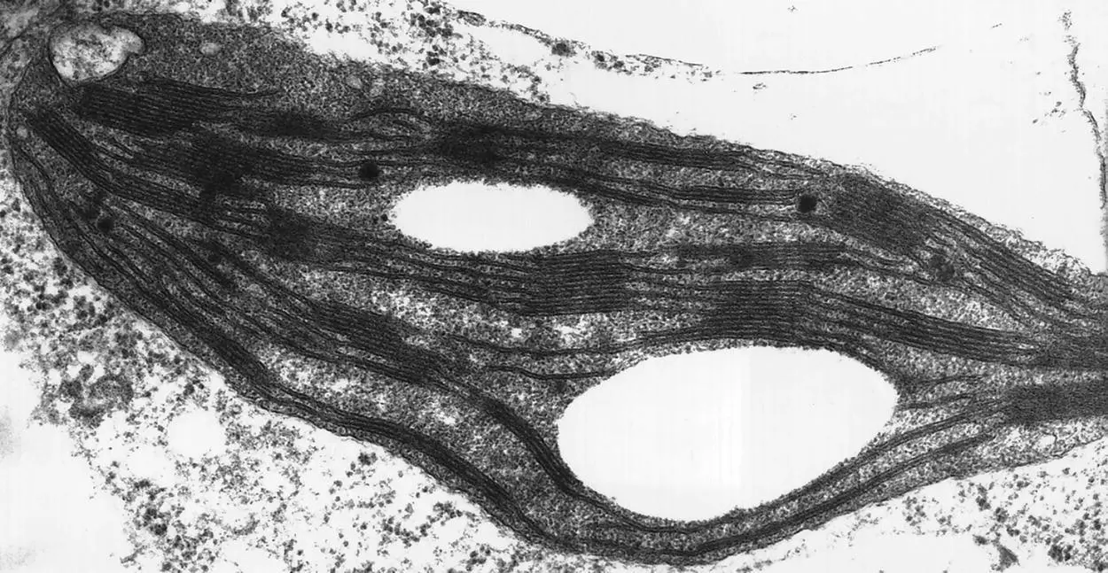
Figure 2.5 Electron micrograph of chloroplast from tobacco.
Source: Courtesy of Kenneth Hoober.
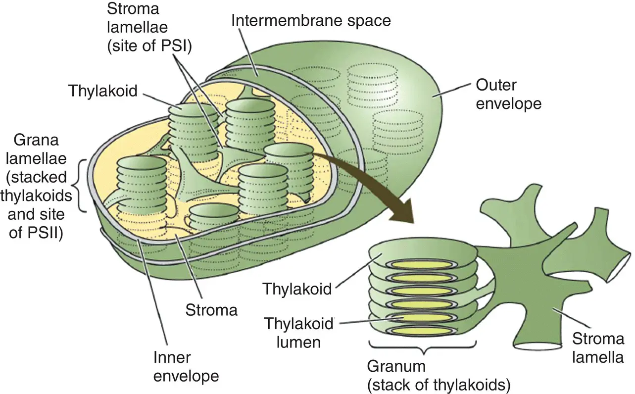
Figure 2.6 Schematic diagram of a chloroplast, showing the inner and outer envelope membranes, the thylakoid membranes – which are divided into grana and stroma lamellae – and the nonmembraneous stroma, containing soluble enzymes.
Source: Taiz et al . (2018)/Oxford University Press.
2.6.1 Algae
Algaeare a large group of eukaryotic organisms (Graham et al ., 2008). Most of them are pigmented and carry out oxygenic photosynthesis. They are either unicellular, and therefore usually microscopic in size, or colonial, containing many cells. The colonial algae are macroscopic in size and can sometimes form huge structures that may look like plants but are quite distinct. There are many different groups of algae, which are usually distinguished by their pigment compositions and morphological features. In aquatic habitats, algae are the dominant photosynthetic life forms, although they are also found on land, including habitats as seemingly unlikely as the surface of snowfields and the hairs of polar bears. Many competing systems of algal classification are in use. We will not attempt to enumerate all the myriad types, but will instead just list some of the most commonly studied types in terms of their photosynthetic properties. In general, the algae all have rather similar electron transport chains, but widely variable antenna complexes from one group to another.
The green algae( chlorophytes) are the most widely studied, because their properties are the closest to higher plants. They contain both chlorophyll a and chlorophyll b as photopigments. They are certainly the evolutionary precursors to plants. The red algae( rhodophytes) are mostly marine organisms that contain chlorophyll a and phycobilisomes, antenna complexes similar to those found in most cyanobacteria. They often have a complex life cycle. The green and red algae, plus one other group (the glaucophytes), are thought to be primary endosymbionts, in that they arose from a single endosymbiotic event. All other algal groups are the result of additional endosymbiotic events, in which a eukaryotic alga was itself incorporated into an organism to form a new type of chimeric cell that in many cases retained the photosynthetic capability of the endosymbiont. Most of these secondary and in some cases tertiary endosymbiotic events involved the red algal line and the complex history of this group includes a dizzying array of gain, loss, and regain of photosynthesis. Many of these organisms contain chlorophyll c as an accessory pigment. The chromoalveolatehypothesis proposes that most of the non‐green eukaryotic algae have been derived from secondary endosymbiosis of red algae and subsequent events (Cavalier‐Smith, 1999).
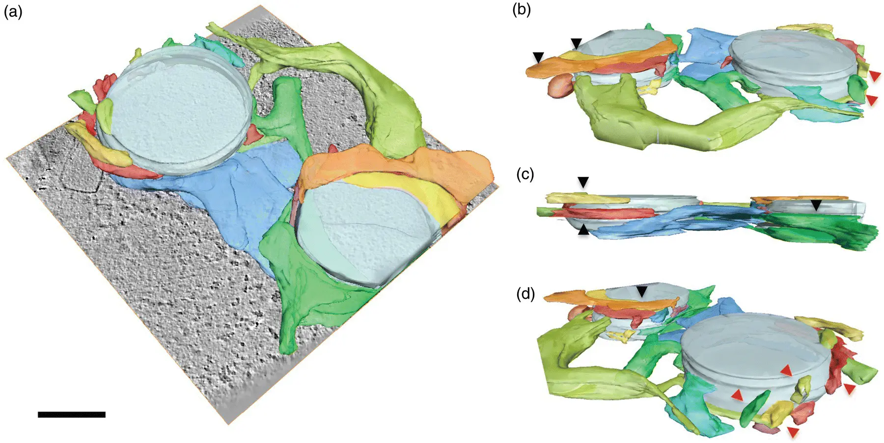
Figure 2.7 Electron microscopic tomographic surface representation of the thylakoid network within a ruptured chloroplast. The different views are of the same thylakoid network from different angles.
Source: Daum and Kühlbrandt (2011). Reproduced with permission of Oxford University Press.
2.6.2 Plants
Plantsare the most complex of all photosynthetic organisms (Taiz et al ., 2018). The simplest plants are the bryophytes, including the mosses, liverworts, and hornworts. They are in many ways like algae and do not have true roots or leaves, or a vascular (liquid‐transporting) system. They do not produce hard tissues for support. The vascular plantsinclude the fernsand the seed plants. The ferns reproduce by means of spores, and have roots, leaves, and vascular tissues, as well as woody tissues for mechanical support. The seed plants reproduce by means of seeds and also contain roots and leaves, as well as vascular and woody tissues. The seed plants are divided into two groups: gymnospermsand angiosperms. Gymnosperms are the more primitive group and include coniferous trees. Angiosperms, also known as flowering plants, make up the vast majority of the species of plants around us.
Remarkably, the basic structure of the photosynthetic membrane and the mechanism of photosynthesis are generally similar in all oxygenic photosynthetic organisms. Some cells include novel antenna complexes, and certain regulatory mechanisms are clearly more sophisticated as one moves from cyanobacteria to algae to higher plants. However, the same basic principles and complexes are found throughout this wide range of organisms. It seems that nature perfected the ability to carry out photosynthesis several billion years ago and has not made major alterations since then. Even the anoxygenic photosynthetic bacteria, while clearly much more primitive than oxygenic forms, carry out photosynthesis using the same basic physical principles. As we proceed, we will examine each of these groups, comparing and contrasting them, trying to find common principles, and pointing out significant differences where they occur.
References
1 Alberts, B., Johnson, A. D., Lewis, J., Morgan, D., Raff, M., Roberts, K., and Walter, P. (2014) The Molecular Biology of the Cell, 6th Edn. New York: W.W. Norton.
2 Beatty, J. T., Overmann, J., Lince, M. T., Manske, A. K., Lang, A. S., Blankenship, R. E., Van Dover, C. L., Martinson, T. A., and Plumley, F. G. (2005) An obligately photosynthetic bacterial anaerobe from a deep‐sea hydrothermal vent. Proceedings of the National Academy of Sciences USA 102: 9306–9310.
3 Blankenship, R. E. (2010) Early evolution of photosynthesis. Plant Physiology 154: 434–438.
4 Blankenship, R. E., Madigan, M. T., and Bauer, C. E., (eds.) (1995) Anoxygenic Photosynthetic Bacteria. Dordrecht: Kluwer Academic Press.
5 Bryant, D. A., (ed.) (1994) The Molecular Biology of Cyanobacteria. Dordrecht: Kluwer Academic Press.
6 Bryant, D. A., Costas, A. M., Maresca, J. A., Chew, A. G. M., Klatt, C., Bateson, M. M., Tallon, L. J., Hostetler, J., Nelson, W. C., Heidelberg, J. F., and Ward, D. M. (2007) Candidatus Chloracidobacterium thermophilum: An aerobic phototrophic acidobacterium. Science 317: 523–526.
7 Cavalier‐Smith, T. (1999) Principles of protein and lipid targeting in secondary symbiogenesis: Euglenoid, dinoflagellate, and sporozoan plastid origins and the eukaryote family tree. Journal of Eukaryotic Microbiology 46: 347–366.
8 Chen, M., Schliep, M., Willows, R. D., Cai, Z.‐L., Neilan, B. A., and Scheer, H. (2010) A red‐shifted chlorophyll. Science 329: 1318–1319.
9 Daum, B. and Kühlbrandt, W. (2011) Electron tomography of plant thylakoid membranes. Journal of Experimenatal Botany 62: 2393–2402.
10 Doolittle, W. F. (1999) Phylogenetic classification and the universal tree. Science 284: 2124–2128.
11 Fleischman, D. and Kramer, D. (1998) Photosynthetic rhizobia. Biochimica et Biophysica Acta 1364: 17–36.
Читать дальшеИнтервал:
Закладка:
Похожие книги на «Molecular Mechanisms of Photosynthesis»
Представляем Вашему вниманию похожие книги на «Molecular Mechanisms of Photosynthesis» списком для выбора. Мы отобрали схожую по названию и смыслу литературу в надежде предоставить читателям больше вариантов отыскать новые, интересные, ещё непрочитанные произведения.


Обсуждение, отзывы о книге «Molecular Mechanisms of Photosynthesis» и просто собственные мнения читателей. Оставьте ваши комментарии, напишите, что Вы думаете о произведении, его смысле или главных героях. Укажите что конкретно понравилось, а что нет, и почему Вы так считаете.