Ron Ben-Amotz - Complications in Canine Cranial Cruciate Ligament Surgery
Здесь есть возможность читать онлайн «Ron Ben-Amotz - Complications in Canine Cranial Cruciate Ligament Surgery» — ознакомительный отрывок электронной книги совершенно бесплатно, а после прочтения отрывка купить полную версию. В некоторых случаях можно слушать аудио, скачать через торрент в формате fb2 и присутствует краткое содержание. Жанр: unrecognised, на английском языке. Описание произведения, (предисловие) а так же отзывы посетителей доступны на портале библиотеки ЛибКат.
- Название:Complications in Canine Cranial Cruciate Ligament Surgery
- Автор:
- Жанр:
- Год:неизвестен
- ISBN:нет данных
- Рейтинг книги:4 / 5. Голосов: 1
-
Избранное:Добавить в избранное
- Отзывы:
-
Ваша оценка:
Complications in Canine Cranial Cruciate Ligament Surgery: краткое содержание, описание и аннотация
Предлагаем к чтению аннотацию, описание, краткое содержание или предисловие (зависит от того, что написал сам автор книги «Complications in Canine Cranial Cruciate Ligament Surgery»). Если вы не нашли необходимую информацию о книге — напишите в комментариях, мы постараемся отыскать её.
provides revision strategies for correcting the complications associated with surgical repair techniques for cranial cruciate ligament rupture, one of the most common causes of a hind limb lameness in dogs. Presenting step-by-step instructions for numerous surgical correction techniques, this practical guide covers articular, extra-articular and osteotomy repair techniques as well as non-surgical management, physical rehabilitation, clinical decision making, and more.
The book begins with an overview of cranial cruciate ligament tear, diagnosis, and treatment goals, followed by a discussion of methods for minimizing surgical site infection and complications. Subsequent chapters describe the potential complications of a particular technique and explain how to identify, evaluate, and correct the complication. Throughout the book, hundreds of high-quality clinical photographs show the appearance of complications and demonstrate each step of the corrective procedure. This authoritative guide:
Provides step-by-step techniques for surgical corrections of common complications Emphasizes surgical decision making and specific strategies for surgical correction Contains revision strategies for identification of intra-operative complications Covers evaluation and identification of post-operative complications Features more than 400 photographs and clinical images Part of the
state-of-the-art
series,
is an invaluable resource for surgical residents, veterinary surgeons, and general practice veterinarians alike.
Complications in Canine Cranial Cruciate Ligament Surgery — читать онлайн ознакомительный отрывок
Ниже представлен текст книги, разбитый по страницам. Система сохранения места последней прочитанной страницы, позволяет с удобством читать онлайн бесплатно книгу «Complications in Canine Cranial Cruciate Ligament Surgery», без необходимости каждый раз заново искать на чём Вы остановились. Поставьте закладку, и сможете в любой момент перейти на страницу, на которой закончили чтение.
Интервал:
Закладка:
1.2 Diagnosis
The diagnosis of CCL pathology is made based on signalment, history and clinical signs, orthopedic examination findings, and diagnostic imaging. While the old saying “a hindlimb lameness in a mature dog is cruciate disease until proven otherwise” holds true, it is important to ensure that the lameness is due to CCL pathology and not another underlying pathological condition of the stifle or another anatomical structure of the pelvic limb.
Signalment information in canines with CCL pathology varies, with most affected dogs presenting for evaluation between 2 and 10 years of age. However, the authors have seen dogs as young as 12 weeks ( Figure 1.2) and as old as 16 years present for CCL pathology. The authors (DD and RBA) have also observed a high incidence of 9–13‐month‐old American Pit Bull Terriers and American Staffordshire Terriers presenting with bilateral CCL pathology. The reason for this is unknown but could be due to demographics and possible breeding practices. Virtually any breed can be affected with CCL pathology; one study revealed the highest prevalence in the Rottweiler, Newfoundland, and Staffordshire Terrier [18]. Another study revealed that breeds at risk for suffering CCL rupture before 2 years of age included the Neapolitan Mastiff, Mastiff, St Bernard, Rottweiler, Akita, Newfoundland, Chesapeake Bay Retriever, Labrador Retriever, and American Staffordshire Terrier [19]. Female dogs have an increased prevalence of CCL pathology (similar to human female athletes), as do neutered canines compared to sexually intact canines [19].
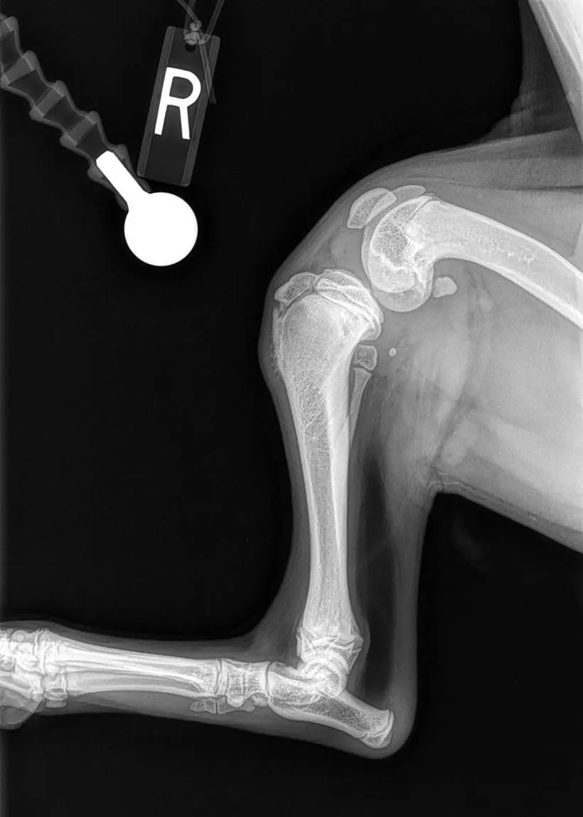
Figure 1.2 Radiograph of a 12‐week‐old dog that suffered a traumatic CCL injury.
Historical findings of patients with CCL pathology may include an acute or chronic onset of hindlimb lameness that may range in severity from mild to nonweight bearing. Some patients may only exhibit stiffness when rising or after heavy activity or may offload the affected limb at a stance ( Figure 1.3). When questioning owners, it is important to obtain a timeline of abnormalities they have noticed even when the lameness appears acute. The authors have found that when questioning owners, many patients have a longer standing history of subtle clinical signs before an acute lameness develops. This further emphasizes the degeneration of the CCL. The common scenario following complete rupture of the CCL is a nonweight‐bearing status for several days. As the inflammatory phase starts to subside, patients will begin to toe touch and use the limb with a noticeable weight‐bearing lameness. Many owners perceive this as their dog improving, when in reality this is consistent with the normal progression of CCL pathology.
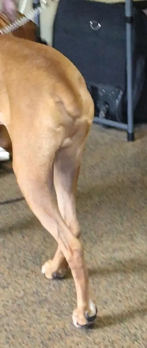
Figure 1.3 Representation of offloading the left pelvic limb at a stance. This patient was diagnosed with a left CCL rupture. Notice when the dog is standing full weight is not applied to the left pelvic limb.
The orthopedic examination is aimed at demonstrating stifle instability; however, other aspects of the stifle and pelvic limb should be evaluated. Pending the severity of the condition as well as the timeframe, there may be pain upon flexion and extension of the stifle, in particular with hyperflexion and hyperextension. In chronic cases of CCL pathology, there may be thickening of the proximal medial tibial or the so‐called “medial buttress” which is development of periarticular fibrosis. The periarticular fibrosis can limit range of motion in the stifle, which may ultimately result in loss of active range of motion. A loss of active range of motion can translate into loss of limb function [20]. In cases with more advanced OA, crepitus may be noted as range of motion is evaluated, and in cases in which meniscal pathology is present, there may be either a consistent or intermittent clicking (“meniscal click”) during range of motion of the stifle. A recent study concluded that a meniscal click in CCL‐deficient stifles carries a high specificity for a bucket handle tear of the meniscus. In addition, the presence of a meniscal click during examination is strongly indicative of a meniscal tear being diagnosed at surgery. However, a lack of meniscal click carries a low sensitivity in diagnosing the absence of meniscal tear [21, 22]. This emphasizes the need for meniscal evaluation in every joint undergoing CCL stabilization.
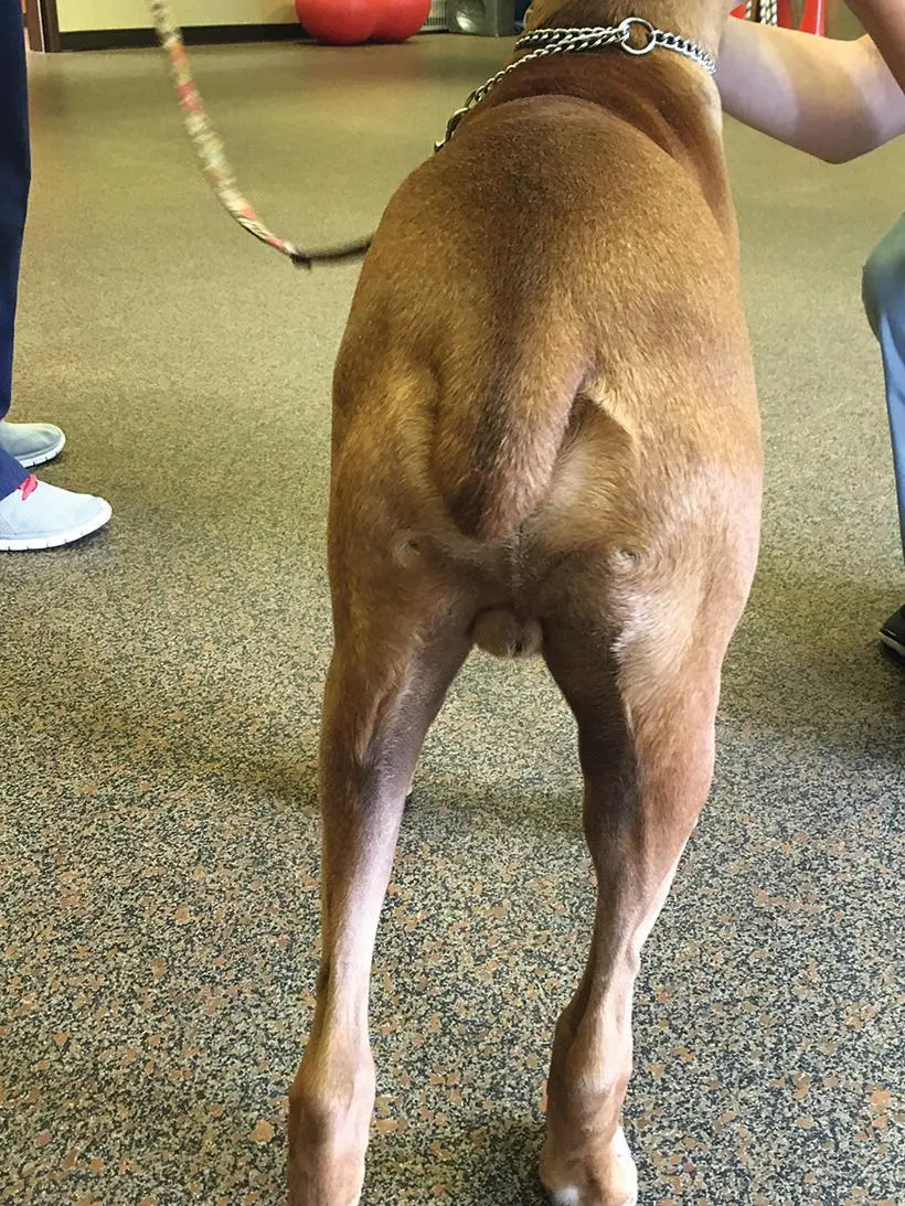
Figure 1.4 A patient with instability of the left pelvic limb secondary to a CCL rupture. Notice the degree of muscle loss on the left hindlimb compared to the right.
Loss of function of a limb will ultimately lead to loss of muscle strength. Unfortunately, in the canine, loss of muscle strength has never been reported but loss of muscle mass has [23] ( Figure 1.4). Therefore, it is important to measure mass at the time of the initial consultation as well as at each follow‐up visit. Additional information on measurement of hindlimb muscle mass can be found in Chapter 15. In addition, measurement of joint motion of the stifle should be performed [24] to determine the flexion and extension of both the affected and unaffected limbs not only at the initial consultation but at all follow‐up visits. Additional information on measurement of joint motion can be found in Chapter 15.
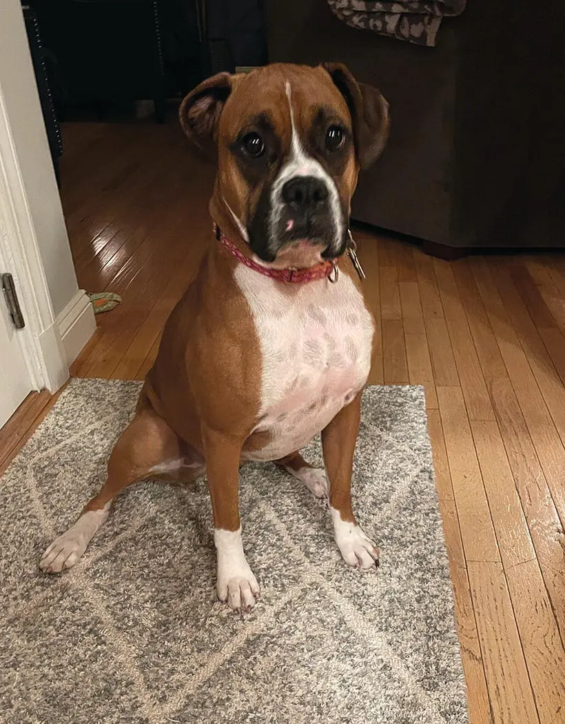
Figure 1.5 An example of a positive sit test in a patient with a right CCL tear. Notice how the right hindlimb is projected outward and the patient is not sitting squarely.
One question that every clinician should ask themselves is: how do you know your patients are truly improving and how can you detect subtle complications? Gathering as much objective information as possible is vital; muscle mass measurements as well as measurement of joint motion of both the affected and unaffected limbs should be part of the objective information gathered (more information on this can be found in Chapter 15). With CCL pathology, there may be an abnormal sit, sometimes referred to as a “positive sit test.” This is characterized by patients sitting with the affected limb projecting out to the side or tucked under them ( Figure 1.5) rather than sitting square ( Figure 1.6). Alternatively, patients may sit with weight shifted off the affected limb ( Figure 1.5). The abnormal sitting posture is thought to be due to discomfort associated with hyperflexion of the stifle when forced to sit squarely. Unfortunately, some dogs may still exhibit an abnormal sit test following CCL stabilization. The reason for this is unknown but it could be due to continued stifle discomfort upon full flexion.
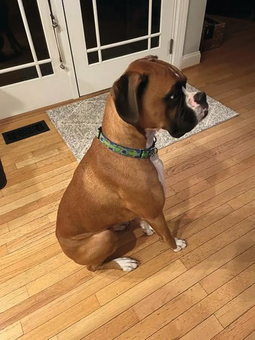
Figure 1.6 An example of a square sit in a patient with no CCL pathology. Notice how both stifles are fully flexed and the patient is sitting square.
Instability of the stifle is commonly demonstrated through the cranial drawer test and tibial compression test. The cranial drawer test ( Figure 1.7) is performed most commonly and tends to be the mainstay of testing for stifle instability by general veterinarians. It is performed by applying a force to the tibia while holding the femur stable, thereby creating craniocaudal translation of the tibia. The operator may stand either behind (caudal) the patient or behind and slightly to the side (caudal and lateral). One hand is placed on the distal femur with the thumb on the lateral fabella and the index finger on the patella. The other hand is placed on the proximal tibia with the thumb on the fibular head and the index finger on the tibial tuberosity. The goal is to move the hand on the proximal tibia cranially while holding the hand on the distal femur stable. Commonly, mistakes made by the inexperienced operator stem from trying to move both hands simultaneously or trying to grab the tissues (both soft and bony) too firmly. Forcing cranial drawer or grasping the tissues too firmly will cause the patient's muscles to tense, making interpretation difficult. Cranial drawer should first be checked in extension, and if positive is likely indicative of a complete tear (typically greater than 75% tearing of the CCL as subjectively noted by one author, DD). If negative, it should then be checked in flexion. A positive cranial drawer in flexion but negative in extension typically indicates an incompetent (unstable) partial CCL tear (usually 50–75% tearing of the CCL as subjectively noted by one author, DD). If cranial drawer is negative in both extension and flexion, then the stifle should be placed in hyperextension to evaluate for discomfort. Discomfort with joint effusion and negative cranial drawer may indicate a competent (stable) partial CCL tear (usually less than 50% tearing of the CCL as subjectively noted by one author, DD).
Читать дальшеИнтервал:
Закладка:
Похожие книги на «Complications in Canine Cranial Cruciate Ligament Surgery»
Представляем Вашему вниманию похожие книги на «Complications in Canine Cranial Cruciate Ligament Surgery» списком для выбора. Мы отобрали схожую по названию и смыслу литературу в надежде предоставить читателям больше вариантов отыскать новые, интересные, ещё непрочитанные произведения.


Обсуждение, отзывы о книге «Complications in Canine Cranial Cruciate Ligament Surgery» и просто собственные мнения читателей. Оставьте ваши комментарии, напишите, что Вы думаете о произведении, его смысле или главных героях. Укажите что конкретно понравилось, а что нет, и почему Вы так считаете.