Ron Ben-Amotz - Complications in Canine Cranial Cruciate Ligament Surgery
Здесь есть возможность читать онлайн «Ron Ben-Amotz - Complications in Canine Cranial Cruciate Ligament Surgery» — ознакомительный отрывок электронной книги совершенно бесплатно, а после прочтения отрывка купить полную версию. В некоторых случаях можно слушать аудио, скачать через торрент в формате fb2 и присутствует краткое содержание. Жанр: unrecognised, на английском языке. Описание произведения, (предисловие) а так же отзывы посетителей доступны на портале библиотеки ЛибКат.
- Название:Complications in Canine Cranial Cruciate Ligament Surgery
- Автор:
- Жанр:
- Год:неизвестен
- ISBN:нет данных
- Рейтинг книги:4 / 5. Голосов: 1
-
Избранное:Добавить в избранное
- Отзывы:
-
Ваша оценка:
Complications in Canine Cranial Cruciate Ligament Surgery: краткое содержание, описание и аннотация
Предлагаем к чтению аннотацию, описание, краткое содержание или предисловие (зависит от того, что написал сам автор книги «Complications in Canine Cranial Cruciate Ligament Surgery»). Если вы не нашли необходимую информацию о книге — напишите в комментариях, мы постараемся отыскать её.
provides revision strategies for correcting the complications associated with surgical repair techniques for cranial cruciate ligament rupture, one of the most common causes of a hind limb lameness in dogs. Presenting step-by-step instructions for numerous surgical correction techniques, this practical guide covers articular, extra-articular and osteotomy repair techniques as well as non-surgical management, physical rehabilitation, clinical decision making, and more.
The book begins with an overview of cranial cruciate ligament tear, diagnosis, and treatment goals, followed by a discussion of methods for minimizing surgical site infection and complications. Subsequent chapters describe the potential complications of a particular technique and explain how to identify, evaluate, and correct the complication. Throughout the book, hundreds of high-quality clinical photographs show the appearance of complications and demonstrate each step of the corrective procedure. This authoritative guide:
Provides step-by-step techniques for surgical corrections of common complications Emphasizes surgical decision making and specific strategies for surgical correction Contains revision strategies for identification of intra-operative complications Covers evaluation and identification of post-operative complications Features more than 400 photographs and clinical images Part of the
state-of-the-art
series,
is an invaluable resource for surgical residents, veterinary surgeons, and general practice veterinarians alike.
Complications in Canine Cranial Cruciate Ligament Surgery — читать онлайн ознакомительный отрывок
Ниже представлен текст книги, разбитый по страницам. Система сохранения места последней прочитанной страницы, позволяет с удобством читать онлайн бесплатно книгу «Complications in Canine Cranial Cruciate Ligament Surgery», без необходимости каждый раз заново искать на чём Вы остановились. Поставьте закладку, и сможете в любой момент перейти на страницу, на которой закончили чтение.
Интервал:
Закладка:
Finally, no veterinarian would be able to do what we do without our patients and their owners. Recognizing the bond that animals bring to our lives helps inspire all veterinary professionals to do our best. Animals bring out the best in all of us and we certainly don't deserve them, so being able to help them in a time of need is one of the most humbling and wonderful experiences.
David L. Dycus
Disclosures
Matthew D. Barnhart is a paid lecturer for Securos and Everost (Steris) and receives royalties from the sales of some of their products.
Matt Corse is a paid consultant for Arthrex Vet Systems.
David L. Dycus is a paid lecturer for Veterinary Orthopedic Implants.
Ian G. Holsworth is a paid consultant and receives royalties from IMEX Veterinary Inc., Veterinary Instrumentation~Covetrus, Sontec Instruments, VetSurg Surgical instrumentation, and Arthrex Vet Systems.
1 Pathology, Diagnosis, and Treatment Goals of Cranial Cruciate Ligament Rupture and Defining Complications
David L. Dycus, Jeffery Biskup, Michael G. Conzemius and Ron Ben‐Amotz
1.1 Introduction
The cranial cruciate ligament (CCL) is a robust intraarticular, yet extrasynovial structure responsible for preventing stifle hyperextension, excessive internal rotation, and cranial subluxation of the tibia in relation to the femur. It originates from the caudomedial aspect of the lateral femoral condyle, and travels in a craniomedial direction to insert on the cranial intercondyloid region of the tibia [1]. It is composed of two distinct bands: the craniomedial and caudolateral portions. During stifle extension, both the craniomedial and caudolateral bands are taut, while during flexion the craniomedial band remains taut while the caudolateral band becomes lax [2].
Cranial cruciate ligament disease is considered to be the most common cause of pelvic limb lameness in the canine, affecting approximately 2.55% of the population [3, 4]. It is a broad term that encompasses a variety of different pathological disorders that may affect the ligament. For example, in skeletally immature canines, avulsion of the CCL may occur secondary to a traumatic event. In the skeletally mature canine, traumatic rupture occurs less commonly, although it may still be induced by forced hyperextension with internal tibial rotation [5]. The most likely etiology for rupture of the CCL in mature dogs is progressive degeneration of the ligament. This degenerative process likely explains the high incidence of bilateral and contralateral CCL pathology. Contralateral CCL pathology has been documented in over 50% of canines [6]. Complicating the matter is the fact that the exact cause of degeneration is poorly understood. Various factors have been investigated in the etiology and pathogenesis of CCL degeneration. Tibial plateau angle [7], genetics, age, bony confirmation, body weight, gait, and vascularity of the ligament [8–10] have all been identified as risk factors for CCL rupture. To date, none of these factors has proved definitive and as such, the degeneration of the CCL is considered multifactorial.
Regardless of the cause of CCL damage, rupture results in a cascade of events that causes altered kinetics and kinematics of the stifle. It is these altered dynamics that likely lead to progressive osteoarthritis (OA) and increased likelihood of meniscal pathology. Kinematic analysis of the CCL‐deficient stifle has shown that the joint remains in a more flexed position throughout the gait cycle. As a result of the increased stifle flexion, the hip and tarsocrural joints compensate by maintaining a greater degree of extension during the stance phase [11]. Kinetic analysis has revealed decreases in peak vertical force (PVF), vertical impulse, braking, and propulsion impulses in dogs with CCL pathology [12]. For example, the PVF in the normal canine pelvic limb was 70% of static body weight; however, when the CCL was transected, the PVF decreased to 25% of static body weight. This was only improved to 32% and 37% at 6 and 12 weeks post transection respectively [13]. Therefore, this demonstrates continued loss of PVF even after periarticular fibrosis has developed. It could be argued that because of this lack of improvement in kinetic function, surgical stabilization of the cruciate‐deficient stifle should be considered.
Along with altered weight bearing and joint flexion/extension, there is also evidence to suggest that the CCL‐deficient stifle exhibits increased cranial subluxation of the tibia in relation to the femur. Up to 8–12 mm of subluxation has been noted during the stance phase of the gait [14].
Long‐term changes have been evaluated in the canine following CCL transection. Following initial transection of the CCL, there was 10 mm more tibial subluxation compared to patients with an intact CCL. Two months following transection, tibial subluxation was noted only during the stance phase and not during the late swing/early stance phase (paw strike). Two years following transection, 5 mm of tibial thrust was noted at the end of the swing phase. It was suggested that the reason for these changes was the presence of an intact medial meniscus. It was theorized that in the CCL‐deficient stifle, the meniscus minimizes tibial subluxation by elastically deforming during tibial subluxation and aiding in reduction of the subluxation once the swing phase begins (alternatively, as the load from the stance phase is removed) [15]. Interestingly, tibial subluxation as the result of instability has been called into question as it demonstrated that instability following CCL rupture is actually a caudal slippage of the femur at the beginning of the stance phase. As such, documented continuation of instability in some stifles exists following common osteotomy procedures such as the tibial plateau leveling osteotomy (TPLO) and tibial tuberosity advancement (TTA) [16].
In addition to controlling sagittal joint motion of the stifle, the CCL also aids in limiting internal rotation. To date, no osteotomy modifying procedures effectively eliminate excessive internal rotation. Initially, this was thought to play a minimal role in the CCL‐deficient stifle. While significant changes in internal rotation were not noted immediately following transection of the CCL, 2 months following transection the range of abduction and adduction in the stifle was increased. These rotary changes remained increased 2 years following tran section [15]. Recognition of the importance of preventing excessive internal rotation has led to the idea and development of augmentation to prevent excessive internal rotation following certain osteotomy procedures such as the TPLO ( Figure 1.1).
Interestingly, a population of CCL‐deficient canines will exhibit persistent internal rotation following surgical stabilization. This persistent internal rotation likely falls under the umbrella term of “pivot shift.” Unfortunately, to date, it is not possible to predict which population of patients will exhibit persistent internal rotation following surgical stabilization. In the authors' opinion, patients with acute CCL rupture, large medial meniscal bucket handle tears, and lack of periarticular fibrosis tend to have persistent internal rotation following certain surgical procedures such as the TPLO. Recently, it was shown that the use of a lateral fabellotibial suture in combination with a TPLO was effective for managing CCL instability in patients with excessive internal rotation identified either preoperatively or intraoperatively [17]. Therefore, the authors recommend the use of adjunct lateral fabellotibial suture in identified patients with excessive internal rotation or “pivot shift” ( Figure 1.1).
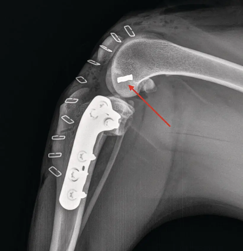
Figure 1.1 An immediate postoperative radiograph of stifle stabilization via a TPLO. Intraoperatively persistent excessive internal rotation was noted so a lateral fabellotibial suture was placed. Note the metallic crimp (red arrow) that has been added.
Читать дальшеИнтервал:
Закладка:
Похожие книги на «Complications in Canine Cranial Cruciate Ligament Surgery»
Представляем Вашему вниманию похожие книги на «Complications in Canine Cranial Cruciate Ligament Surgery» списком для выбора. Мы отобрали схожую по названию и смыслу литературу в надежде предоставить читателям больше вариантов отыскать новые, интересные, ещё непрочитанные произведения.


Обсуждение, отзывы о книге «Complications in Canine Cranial Cruciate Ligament Surgery» и просто собственные мнения читателей. Оставьте ваши комментарии, напишите, что Вы думаете о произведении, его смысле или главных героях. Укажите что конкретно понравилось, а что нет, и почему Вы так считаете.