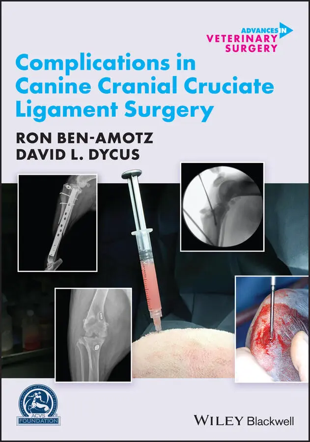
Ron Ben-Amotz - Complications in Canine Cranial Cruciate Ligament Surgery
Здесь есть возможность читать онлайн «Ron Ben-Amotz - Complications in Canine Cranial Cruciate Ligament Surgery» — ознакомительный отрывок электронной книги совершенно бесплатно, а после прочтения отрывка купить полную версию. В некоторых случаях можно слушать аудио, скачать через торрент в формате fb2 и присутствует краткое содержание. Жанр: unrecognised, на английском языке. Описание произведения, (предисловие) а так же отзывы посетителей доступны на портале библиотеки ЛибКат.
- Название:Complications in Canine Cranial Cruciate Ligament Surgery
- Автор:
- Жанр:
- Год:неизвестен
- ISBN:нет данных
- Рейтинг книги:4 / 5. Голосов: 1
-
Избранное:Добавить в избранное
- Отзывы:
-
Ваша оценка:
Complications in Canine Cranial Cruciate Ligament Surgery: краткое содержание, описание и аннотация
Предлагаем к чтению аннотацию, описание, краткое содержание или предисловие (зависит от того, что написал сам автор книги «Complications in Canine Cranial Cruciate Ligament Surgery»). Если вы не нашли необходимую информацию о книге — напишите в комментариях, мы постараемся отыскать её.
provides revision strategies for correcting the complications associated with surgical repair techniques for cranial cruciate ligament rupture, one of the most common causes of a hind limb lameness in dogs. Presenting step-by-step instructions for numerous surgical correction techniques, this practical guide covers articular, extra-articular and osteotomy repair techniques as well as non-surgical management, physical rehabilitation, clinical decision making, and more.
The book begins with an overview of cranial cruciate ligament tear, diagnosis, and treatment goals, followed by a discussion of methods for minimizing surgical site infection and complications. Subsequent chapters describe the potential complications of a particular technique and explain how to identify, evaluate, and correct the complication. Throughout the book, hundreds of high-quality clinical photographs show the appearance of complications and demonstrate each step of the corrective procedure. This authoritative guide:
Provides step-by-step techniques for surgical corrections of common complications Emphasizes surgical decision making and specific strategies for surgical correction Contains revision strategies for identification of intra-operative complications Covers evaluation and identification of post-operative complications Features more than 400 photographs and clinical images Part of the
state-of-the-art
series,
is an invaluable resource for surgical residents, veterinary surgeons, and general practice veterinarians alike.
Complications in Canine Cranial Cruciate Ligament Surgery — читать онлайн ознакомительный отрывок
Ниже представлен текст книги, разбитый по страницам. Система сохранения места последней прочитанной страницы, позволяет с удобством читать онлайн бесплатно книгу «Complications in Canine Cranial Cruciate Ligament Surgery», без необходимости каждый раз заново искать на чём Вы остановились. Поставьте закладку, и сможете в любой момент перейти на страницу, на которой закончили чтение.
Интервал:
Закладка:
Katie L. Hoddinott, J. Scott Weese, and Ameet Singh
2.1 Introduction
The reported incidence of surgical site infections (SSIs) in companion animal veterinary medicine ranges from 3% to 18.1%, with increasing risk of SSI development associated with increasing classification of the surgical procedure ( Table 2.1) [1–7]. Orthopedic surgeries are most commonly classified as clean procedures; however, SSI rates are often higher in clean orthopedic surgeries (3.54–12.9%) when compared to other clean surgical procedures (2.5–4.8%) [5,7–9]. Furthermore, SSI rates vary amongst surgical stabilization techniques for treatment of cranial cruciate ligament ruptures. Extracapsular repair techniques, including the lateral fabellotibial suture and Arthrex Canine Cruciate Ligament Repair Anchor System™, have reported SSI rates ranging from 3.9% to 21% [6,10–12] while proximal tibial osteotomy procedures, including tibial plateau leveling osteotomy (TPLO), tibial tuberosity advancement (TTA), triple tibial osteotomy, and cranial closing wedge osteotomies, have reported SSI rates ranging from 4.7% to 25.9% [13–26].
Many risk factors have been associated with development of SSIs following surgical stabilization of the cranial cruciate‐deficient stifle. These risk factors may be associated with the host, the environment, details surrounding the surgical procedure and the use of antimicrobials. Host factors include breed, sex, body weight, American Society of Anesthesiologists (ASA) status, methicillin‐resistant Staphylococcus pseudintermedius (MRSP) carrier status, and skin microbiome. Environmental factors include the number of personnel in the surgical suite and potential for bacterial contamination from surrounding surfaces. Surgical procedure factors include the method of stifle stabilization, length of general anesthesia, length of surgical procedure, surgeon preparation, intraoperative contamination, adherence to Halstead's principles ( Table 2.2), and choice of implant and wound closure materials. Finally, the use of antimicrobials in the preoperative, perioperative, and postoperative periods has been documented as a risk factor for SSI.
2.2 Host Factors
2.2.1 Breed, Sex, and Body Weight
Several host factors identified as risk factors for SSI development are beyond the control of the veterinary team, such as breed, sex, and body weight. Bulldogs and German Shepherds have been identified as having an increased risk for development of SSI following TPLO surgery [21, 27]. Labrador Retrievers and mixed‐breed dogs have been identified as having a lower SSI development risk following TPLO surgery [4, 3]. Both Fitzpatrick and Solano and Nicholson et al. identified intact male dogs to be at higher risk of SSI; however, sex was not identified as a risk factor in other studies [3, 4, 17, 28]. Increased body weight has been significantly associated with an increased SSI risk in several studies [2, 4, 10, 15,28–30]. In one study, each 1 kg increase in body weight resulted in 1.03 times increased odds of developing an SSI, while a second study noted that for each 1 kg increase in body weight, the odds of developing an SSI increased by 4.7% [28, 29]. As these factors are inherent to the patient and cannot be specifically controlled, this information can be utilized to distinguish those animals at higher risk of developing an SSI and thus requiring greater preventive measures.
Table 2.1 Surgical procedure classification.
Source: Based on Turk et al. [1], Eugster et al. [2], Nicholson et al. [3], Fitzpatrick and Solano [4], Beal et al. [5], Frey et al. [6], and Vasseur et al. [7].
| Clean | No infection No break in aseptic technique Nontraumatic |
| Clean‐contaminated | Controlled access to a hollow viscus Minor break in aseptic technique |
| Contaminated | Entry through nonseptic, yet inflamed tissues Spillage from a hollow viscus – localized, controlled Major break in aseptic technique Fresh, traumatic wounds (<4 h) |
| Dirty | Perforated hollow viscus Septic purulent discharge encountered Chronic, traumatic wounds (>4 h) |
Table 2.2 Halstead's principles.
| Gentle tissue handling Meticulous hemostasis Strict aseptic technique Preservation of blood supply Elimination of dead space Accurate apposition of tissues while minimizing tension |
2.2.2 ASA Status and Endocrinopathies
Preoperative ASA score ( Table 2.3) has been correlated with risk for developing an SSI, such that the risk for SSI increases with each increment in ASA score [2]. As ASA scores take into consideration the overall health status of a patient, the higher the ASA score, the more systemically ill the patient. Animals with endocrinopathies have also been identified to be 8.2 times more likely to develop an SSI, likely due to alterations in immune function [3]. Logically, if an animal has a chronic illness, such as an unmanaged or poorly controlled endocrinopathy, postponement of elective orthopedic procedures until these illnesses are adequately addressed should be considered, when possible.
2.2.3 MRSP Carrier Status
As Staphylococcus spp. have been identified as one of the most common bacteria contributing to the development of SSIs following TPLO, and MRSP SSIs are increasing in frequency, monitoring for carriers of MRSP has been investigated [4, 13, 21,31–34]. In one study, 4.4% of animals were identified to be MRSP carriers preoperatively and carriers were 6.72 times more likely to develop an SSI. While screening for MRSP may help to determine those animals at higher risk for SSI, current screening tests are time‐consuming and not practical for routine implementation as this would result in delaying surgery [21]. Additionally, in human medicine, the goals of methicillin‐resistant Staphylococcus aureus (MRSA) screening are to allow for decolonization preoperatively [35–38]. In veterinary medicine, preoperative decolonization of MRSP is a challenge, due to not only the limited number of effective antimicrobials against MRSP, but also the inherent challenges of topical treatment of the sites most commonly colonized – the nasal passages, pharynx, and rectum [21].
Table 2.3 American society of anesthesiologists (ASA) scores.
Source: Based on Eugster et al. [2].
| ASA I | Normal, healthy patients |
| ASA II | Patients with mild systemic disease |
| ASA III | Patients with severe systemic disease |
| ASA IV | Patients with severe systemic disease that is life‐threatening |
| ASA V | Patients that will not survive 24 h without surgical intervention |
While decolonization may not be feasible, it is reasonable to alter perioperative antimicrobial prophylaxis in MRSP carriers undergoing higher risk procedures like TPLO. This may include measures such as adding a single dose of amikacin preoperatively to the typical (e.g., cefazolin) antimicrobial regime, assuming renal health has been evaluated.
2.2.4 Dermatitis, Clipping, and Skin Preparation
As mentioned, Staphylococcus spp. are amongst the most common bacteria causing SSIs. S. pseudintermedius is a commensal bacteria within the normal microbiome of dogs [39]. Assessment of the animal's skin for evidence of local or distant dermatitis is recommended, to reduce the risk of contamination of the surgical site ( Figure 2.1). Despite this logical recommendation, 17.5% of all animals undergoing TPLO in one study had evidence of active local or distant dermatitis. Of those with local dermatitis, 16.7% developed an SSI and 10.2% with distant dermatitis developed an SSI [27]. As there was no significant difference identified between local and distant dermatitis resulting in SSI, the risk for SSI development should not be considered lower for animals with dermatitis not affecting the direct surgical site, as anecdotally thought, and postponement of elective orthopedic surgeries with local or distant dermatitis should be considered [27].
Читать дальшеИнтервал:
Закладка:
Похожие книги на «Complications in Canine Cranial Cruciate Ligament Surgery»
Представляем Вашему вниманию похожие книги на «Complications in Canine Cranial Cruciate Ligament Surgery» списком для выбора. Мы отобрали схожую по названию и смыслу литературу в надежде предоставить читателям больше вариантов отыскать новые, интересные, ещё непрочитанные произведения.


Обсуждение, отзывы о книге «Complications in Canine Cranial Cruciate Ligament Surgery» и просто собственные мнения читателей. Оставьте ваши комментарии, напишите, что Вы думаете о произведении, его смысле или главных героях. Укажите что конкретно понравилось, а что нет, и почему Вы так считаете.