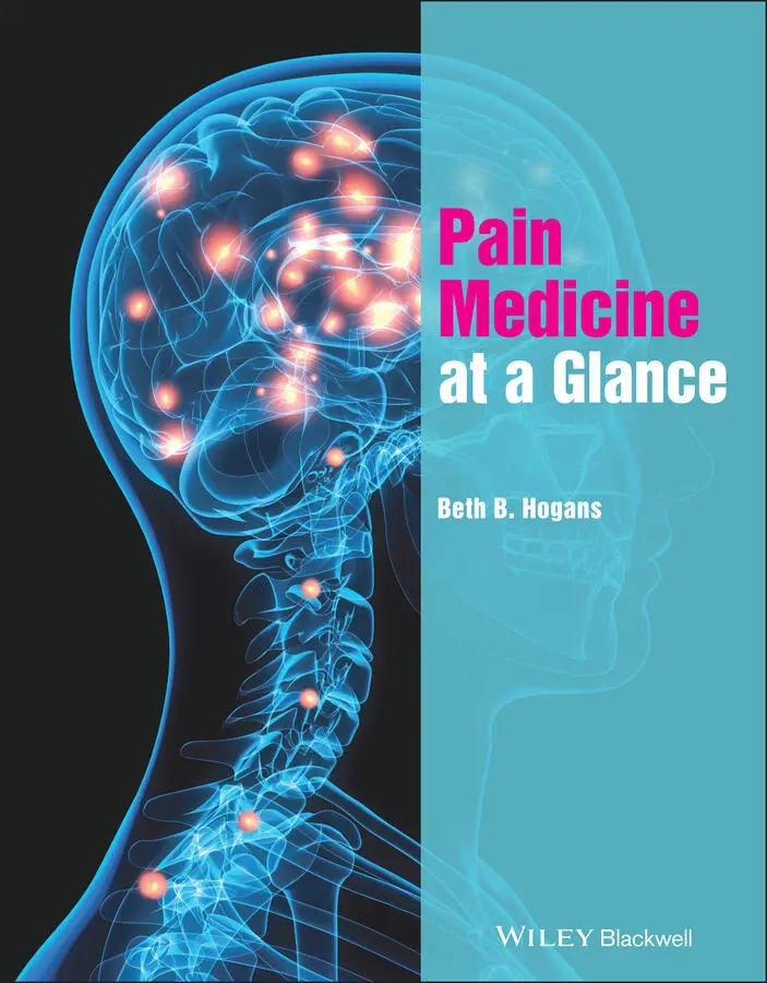
Beth B. Hogans - Pain Medicine at a Glance
Здесь есть возможность читать онлайн «Beth B. Hogans - Pain Medicine at a Glance» — ознакомительный отрывок электронной книги совершенно бесплатно, а после прочтения отрывка купить полную версию. В некоторых случаях можно слушать аудио, скачать через торрент в формате fb2 и присутствует краткое содержание. Жанр: unrecognised, на английском языке. Описание произведения, (предисловие) а так же отзывы посетителей доступны на портале библиотеки ЛибКат.
- Название:Pain Medicine at a Glance
- Автор:
- Жанр:
- Год:неизвестен
- ISBN:нет данных
- Рейтинг книги:4 / 5. Голосов: 1
-
Избранное:Добавить в избранное
- Отзывы:
-
Ваша оценка:
Pain Medicine at a Glance: краткое содержание, описание и аннотация
Предлагаем к чтению аннотацию, описание, краткое содержание или предисловие (зависит от того, что написал сам автор книги «Pain Medicine at a Glance»). Если вы не нашли необходимую информацию о книге — напишите в комментариях, мы постараемся отыскать её.
Pain Medicine at a Glance — читать онлайн ознакомительный отрывок
Ниже представлен текст книги, разбитый по страницам. Система сохранения места последней прочитанной страницы, позволяет с удобством читать онлайн бесплатно книгу «Pain Medicine at a Glance», без необходимости каждый раз заново искать на чём Вы остановились. Поставьте закладку, и сможете в любой момент перейти на страницу, на которой закончили чтение.
Интервал:
Закладка:
19 Chapter 53Table 53.1 Eclampsia: symptoms and signs.Table 53.2 Peripartum headache differential diagnosis.
List of Illustrations
1 Chapter 1 Figure 1.1 Pain has sensory‐discriminative and emotional‐motivational compon... Figure 1.2 Interindividual variability in pain showing tremendous variabilit... Figure 1.3 Standard pain assessment: the pain ‘Alphabet’. Figure 1.4 The numerical rating scale of pain severity (intensity). Figure 1.5 Pain interferes with function in multiple domains of daily functi...
2 Chapter 2 Figure 2.1 Simplified overview of nociceptive processing in the nervous syst... Figure 2.2 Transmission and modulation events in the spinal dorsal horn. Inf...
3 Chapter 3 Figure 3.1 The basic mechanisms of pain. Figure 3.2 How basic pain mechanisms interact. Figure 3.3 Stimulus response curve: normal and abnormal pain perception.
4 Chapter 4 Figure 4.1 (a) Pain is highly prevalent, present in about 38% of the populat... Figure 4.2 Access to pain‐relieving medication varies widely with location. ...
5 Chapter 5 Figure 5.1 Healthcare ethics rests on the “four pillars.” Figure 5.2 Each of the four pillars has distinctive aspects that shape ethic...
6 Chapter 6 Figure 6.1 Pain patterns, examples. Pain can present with many different pat... Figure 6.2 Qualities of pain, examples.
7 Chapter 8 Figure 8.1 Parallel pathway model. Diagnosis and initiation of treatment pro... Figure 8.3 Balancing knowledge of disease with patient‐centered understandin... Figure 8.4 (a) Normal functioning demonstrating processes of eudynia; (b) Am... Figure 8.2 Mechanism‐based classification of pain overview: rationale for de...
8 Chapter 9 Figure 9.1 The numerical rating scale. Figure 9.2 In the effective patient‐provider relationship, there are many fo...
9 Chapter 10 Figure 10.1 Affect and cognition are both communicated in the pain‐focused c... Figure 10.2 Recognizing and modulating the emotional range of a pain‐focused...
10 Chapter 11Figure 11.1 Aspects of emotional development.
11 Chapter 12Figure 12.1 Anatomical posterior view of the torso.Figure 12.2 Dermatomes on male figure. Developed by the author (BBH) from pu...
12 Chapter 13Figure 13.1 Epstein and Hundert model of professional competence is based on...Figure 13.2 Dimensions of emotional competence: applied to pain care.
13 Chapter 14Figure 14.1 Action items for each stage of stages of change model.
14 Chapter 16Figure 16.1 Comprehensive pain management involves incorporation and coordin...Figure 16.2 Exploring therapy options:providing patients with prompts and r...
15 Chapter 17Figure 17.1 Schematic for rational selection of agents for pain based on bas...Figure 17.2 Treatment according to pain type, summary notes.Figure 17.3 Efficacy (NNT) of selected agents.
16 Chapter 18Figure 18.1 Structure and pharmacokinetics of Acetaminophen and Ibuprofen.
17 Chapter 19Figure 19.1 Structure of selected neuromodulating agents.
18 Chapter 20Figure 20.1 Representations of opioids in antiquity.Figure 20.2 Consumption of opioids in the U.S. compared with other global re...Figure 20.3 Opioid precautions.
19 Chapter 21Figure 21.1 Preclinical studies show that there can be very rapid tolerance ...
20 Chapter 22Figure 22.1 Drawing of the neuroaxis showing potential sites of opioid deliv...
21 Chapter 24Figure 24.1 Spinal column views with (a) herniated disc compressing nerve ro...Figure 24.2 Spinal cord stimulator illustration.Figure 24.3 Spinal cord indwelling pump illustration.
22 Chapter 25Figure 25.1 Estimated relative pain Score impacts for chronic pain.Figure 25.2 Examples of activating therapies.
23 Chapter 26Figure 26.1 CBT: a situation leads to thoughts which produce feelings that l...Figure 26.2 The six “pivots” of ACT, adapted from Stephen Hayes. In ACT, the...
24 Chapter 27Figure 27.1 Massage therapy – trigger points, X indicates location of trigge...Figure 27.2 Example of acupressure points in forehead.Figure 27.3 Lunge stretch illustration, stretching routines should be person...Figure 27.4 Inversion table illustration, please observe appropriate clinica...
25 Chapter 28Figure 28.1 Meditation – evidence supports the use of mindfulness‐based stre...Figure 28.2 Vocational engagement – engaging in meaningful and purposeful ac...Figure 28.3 Video games and virtual may help reduce stress and pain, however...Figure 28.4 Connecting with nature is very therapeutic for some.
26 Chapter 29Figure 29.1 Chronic vs. Persistent pain: conditions and associated recovery ...Figure 29.2 In chronic pain, allodynia and hyperalgesia are more prevalent a...Figure 29.3 Acute vs. Chronic pain: how pain types shift in importance betwe...
27 Chapter 30Figure 30.1 Flow diagram for enhanced recovery after surgery protocols. Adap...Figure 30.2 Prevalent factors that impact outcomes.
28 Chapter 31Figure 31.1 Chronic vs. Persistent pain: conditions and associated recovery ...Figure 31.2 Selected nonpharmacological therapies for musculoskeletal pain c...
29 Chapter 32Figure 32.1 Segmental (dermatomal) innervation of face and head, lateral vie...Figure 32.2 Innervation of the tongue.Figure 32.3 Pulpitis typically resolves rapidly and completely with denervat...Figure 32.4 Pain of TMD can involve large areas of the face.
30 Chapter 33Figure 33.1 Sagittal view of the neck, illustrating structures of anterior n...Figure 33.2 Anatomy of cervical spine with a level showing some features of ...Figure 33.3 Cross‐sectional view of cervical spinal column, muscles removed....
31 Chapter 34Figure 34.1 Diagram of arm with major nerves: median, ulnar, and radial; ill...Figure 34.2 Diagram of shoulder with muscles and tendons of rotator cuff.Figure 34.3 Diagram of wrist and hand illustrating location of carpal tunnel...Figure 34.4 Medial elbow, illustrating location of ulnar nerve compressive n...Figure 34.5 Comparison of pain patterns in hand.
32 Chapter 35Figure 35.1 Pain patterns often associated with common causes of low back pa...Figure 35.2 Lumbosacral spine sagittal MRI. Degenerated, desiccated disc is ...Figure 35.3 Schematic diagram illustrating changes associated with disc dege...Figure 35.4 Diagram illustrating SI joint in posterior pelvis/lateral low ba...
33 Chapter 36Figure 36.1 Comparison of Type I and Type II red flags for low back pain eme...Figure 36.2 Imaging modalities and spinal abnormalities. (a) CT of lumbosacr...
34 Chapter 37Figure 37.1 Diagnostic prevalence of radiculopathy in older adults by spinal...Figure 37.2 Patterns of common lumbar radicular pain, L3 (least common), L4,...Figure 37.3 Illustration of sciatic nerve passes between piriformis and geme...Figure 37.4 Illustration of meralgia paresthetica (MAYO – need substitute)....
35 Chapter 38Figure 38.1 Two views of the knee, anterior anatomical view with muscles, me...Figure 38.2 Interior view of the knee from above with femur remove, illustra...Figure 38.3 Common knee injuries with provoking activities and examination n...
36 Chapter 39Figure 39.1 Bones and ligaments of the foot and ankle, with (a) anterior, (b...Figure 39.2 Diagram illustrating selected common foot ailments. Kiel J (2020...
37 Chapter 40Figure 40.1 Classic CSF appearance, varies with diagnosis, may visualize at ...Figure 40.2 Subarachnoid blood in SAH.
38 Chapter 41Figure 41.1 Headache calendars are essential for gathering sufficiently deta...Figure 41.2 Headaches are principally subjective; patient‐reported features ...
39 Chapter 42Figure 42.1 Comprehensive pain management is highly effective for chronic he...Figure 42.2 Chronic headaches can be very impairing: perturbing focus or in ...
40 Chapter 43Figure 43.1 Visceral pain often manifests with “referred pain,” pain perceiv...Figure 43.2 Peripheral and central structures may contribute to the phenomen...
Читать дальшеИнтервал:
Закладка:
Похожие книги на «Pain Medicine at a Glance»
Представляем Вашему вниманию похожие книги на «Pain Medicine at a Glance» списком для выбора. Мы отобрали схожую по названию и смыслу литературу в надежде предоставить читателям больше вариантов отыскать новые, интересные, ещё непрочитанные произведения.


Обсуждение, отзывы о книге «Pain Medicine at a Glance» и просто собственные мнения читателей. Оставьте ваши комментарии, напишите, что Вы думаете о произведении, его смысле или главных героях. Укажите что конкретно понравилось, а что нет, и почему Вы так считаете.