Louie Al-Faraje - Clinical Anatomy for Oral Implantology
Здесь есть возможность читать онлайн «Louie Al-Faraje - Clinical Anatomy for Oral Implantology» — ознакомительный отрывок электронной книги совершенно бесплатно, а после прочтения отрывка купить полную версию. В некоторых случаях можно слушать аудио, скачать через торрент в формате fb2 и присутствует краткое содержание. Жанр: unrecognised, на английском языке. Описание произведения, (предисловие) а так же отзывы посетителей доступны на портале библиотеки ЛибКат.
- Название:Clinical Anatomy for Oral Implantology
- Автор:
- Жанр:
- Год:неизвестен
- ISBN:нет данных
- Рейтинг книги:3 / 5. Голосов: 1
-
Избранное:Добавить в избранное
- Отзывы:
-
Ваша оценка:
Clinical Anatomy for Oral Implantology: краткое содержание, описание и аннотация
Предлагаем к чтению аннотацию, описание, краткое содержание или предисловие (зависит от того, что написал сам автор книги «Clinical Anatomy for Oral Implantology»). Если вы не нашли необходимую информацию о книге — напишите в комментариях, мы постараемся отыскать её.
Clinical Anatomy for Oral Implantology — читать онлайн ознакомительный отрывок
Ниже представлен текст книги, разбитый по страницам. Система сохранения места последней прочитанной страницы, позволяет с удобством читать онлайн бесплатно книгу «Clinical Anatomy for Oral Implantology», без необходимости каждый раз заново искать на чём Вы остановились. Поставьте закладку, и сможете в любой момент перейти на страницу, на которой закончили чтение.
Интервал:
Закладка:
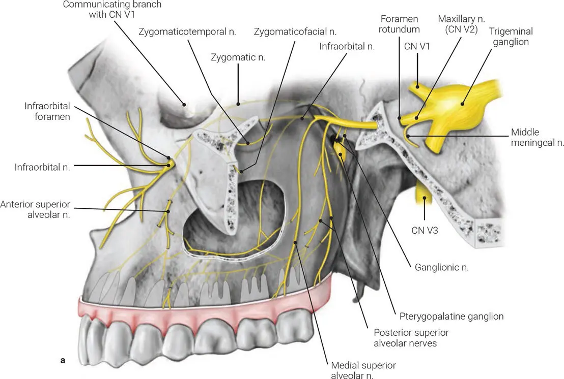
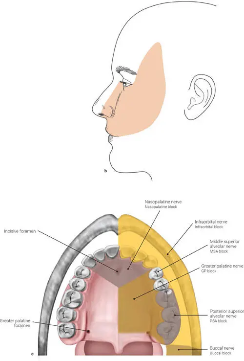
FIG 1-11 (a) The maxillary nerve. n.—nerve; CN V1—ophthalmic nerve; CN V3—mandibular nerve. (b) Region of skin supplied by the maxillary nerve. (c) Innervation of the maxilla along the recommended anesthesia technique per area.
The zygomatic nerve passes through the inferior orbital fissure and gives branches of sensory fibers to the lacrimal nerve, then divides into the zygomaticotemporal branch (temple) and the zygomaticofacial branch (for the skin over the zygomatic arch).
The ganglionic branches are nasal branches (nasopalatine branches) that pass through the sphenopalatine foramen into the nasal cavity, the palatine nerves (greater and lesser) for the soft and hard palates, and the pharyngeal nerve, which provides sensory supply to the upper pharynx.
The infraorbital nerve enters the orbit through the inferior orbital fissure (after branching off into the posterior superior alveolar nerves to the molars and the medial superior alveolar nerves); it traverses the infraorbital groove and canal in the floor of the orbit, where it branches off into the anterior superior alveolar nerve, and appears on the face at the infraorbital foramen. Here it is referred to as the infraorbital nerve, a terminal branch. At its termination, the nerve lies beneath the quadratus labii superioris and divides into several branches that innervate the side of the nose, the lower eyelid (inferior palpebral nerve), and the upper lip (the superior labial nerve), joining with filaments of the facial nerve.
Mandibular nerve (CN V3)
The mandibular nerve (Fig 1-12a) is the third branch of the trigeminal nerve, arising from the trigeminal ganglion. Unlike the other two branches (the maxillary and the ophthalmic nerves, both entirely sensory), the mandibular nerve has both sensory and motor divisions.
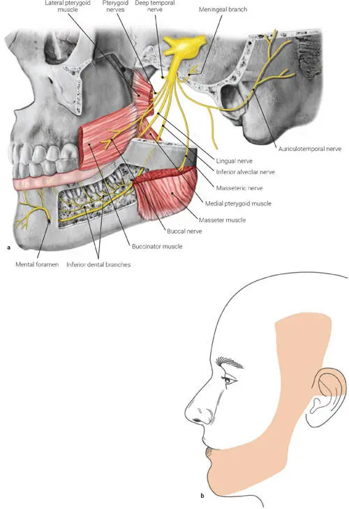
FIG 1-12 (a) The mandibular nerve. (b) Region of skin supplied by the mandibular nerve.
After passing through the foramen ovale and branching off into a meningeal branch in the infratemporal fossa, the nerve divides into the sensory branches—the auriculotemporal, lingual, inferior alveolar, and buccal nerves to the skin over the mandible, lower lip, temporal region, and much of the oral cavity (Fig 1-12b)—and the motor branches that innervate the muscles of mastication (masseteric, deep temporal, and pterygoid nerves).
The inferior alveolar nerve carries motor fibers for the mylohyoid muscle and the anterior belly of the digastric muscle and sensory fibers that enter the canal through the mandibular foramen; it gives branches to the mandibular teeth and exits through the mental foramen under the mental nerve (see chapter 7). Damaging the inferior alveolar nerve will alter the sensation to areas supplied by it and by the mental nerve. Branches of the trigeminal nerve are also frequently used to distribute fibers derived from other cranial nerves.
References
1. Choi J, Park HS. The clinical anatomy of the maxillary artery in the pterygopalatine fossa. J Oral Maxillofac Surg 2003;61:72–78.
2. Li J, Xu X, Wang J, Jing X, Guo Q, Qiu Y. Endoscopic study for the pterygopalatine fossa anatomy: Via the middle nasal meatus-sphenopalatine foramen approach. J Craniofac Surg 2009;20:944–947.
3. Osawa S, Rhoton AL Jr, Seker A, Shimizu S, Fujii K, Kassam AB. Microsurgical and endoscopic anatomy of the vidian canal. Neurosurgery 2009;64(5 suppl 2):385–411.
2
MUSCLES OF FACIAL EXPRESSION AND MASTICATION
This chapter describes the muscles of facial expression and the muscles of mastication and their relevance to implant-related oral surgical procedures.
Muscles of Facial Expression
The muscles of facial expression are paired muscles in the superficial fascia of the facial tissues ( Table 2-1and Figs 2-1 to 2-4). Almost all of them originate from bone (rarely the fascia) and insert on skin tissue, and all of them are innervated by the facial nerve (CN VII).
TABLE 2-1 Muscles of facial expression
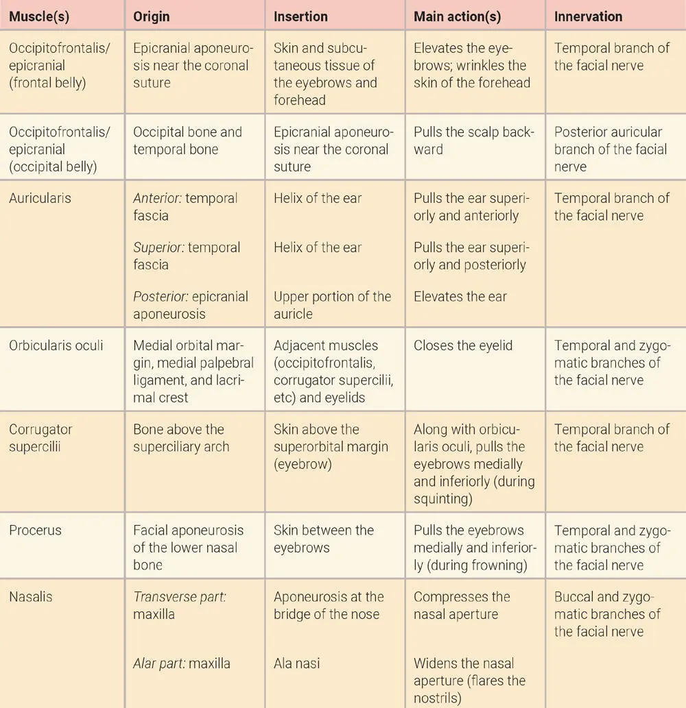
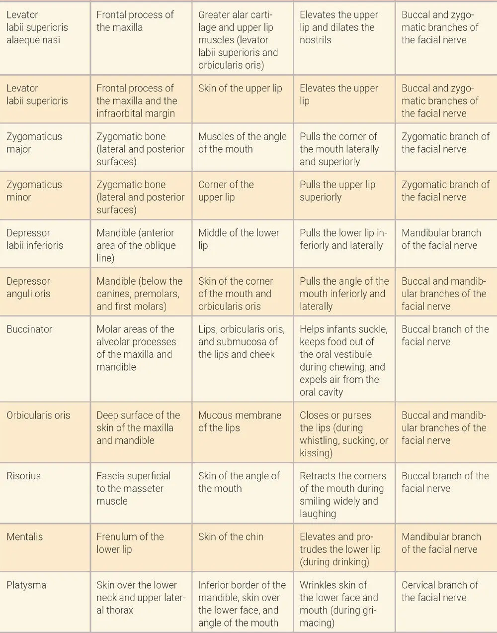
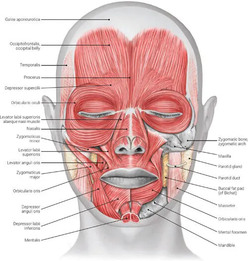
FIG 2-1 Anterior view of the muscles of facial expression.
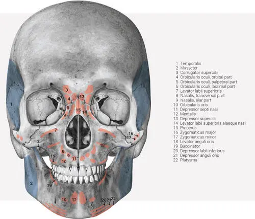
FIG 2-2 Anterior view of insertion of the muscles of facial expression (in red) and muscles of mastication (in blue) .
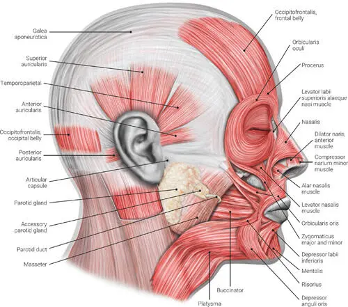
FIG 2-3 Lateral view of the muscles of facial expression.
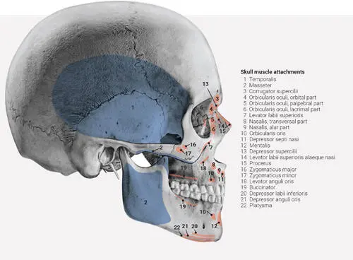
FIG 2-4 Lateral view of insertion of the muscles of facial expression (in red) and muscles of mastication (in blue) .
Muscles of Mastication
The muscles of mastication are located in the parotid and infratemporal regions of the face ( Table 2-2and Figs 2-5 to 2-10; see also Figs 2-2 and 2-4). All of them receive innervation from the mandibular division of the trigeminal nerve.
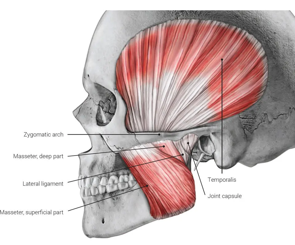
FIG 2-5 Lateral view of insertion of the masseter muscle.
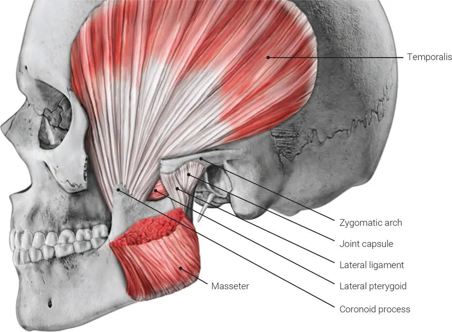
FIG 2-6 Lateral view of insertion of the temporalis muscle.
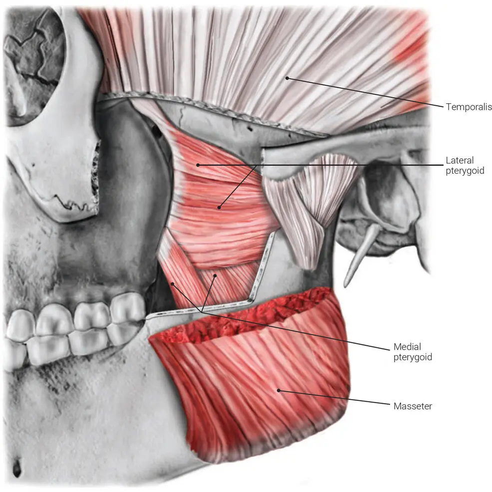
FIG 2-7 Lateral view of insertion of the lateral pterygoid muscle.
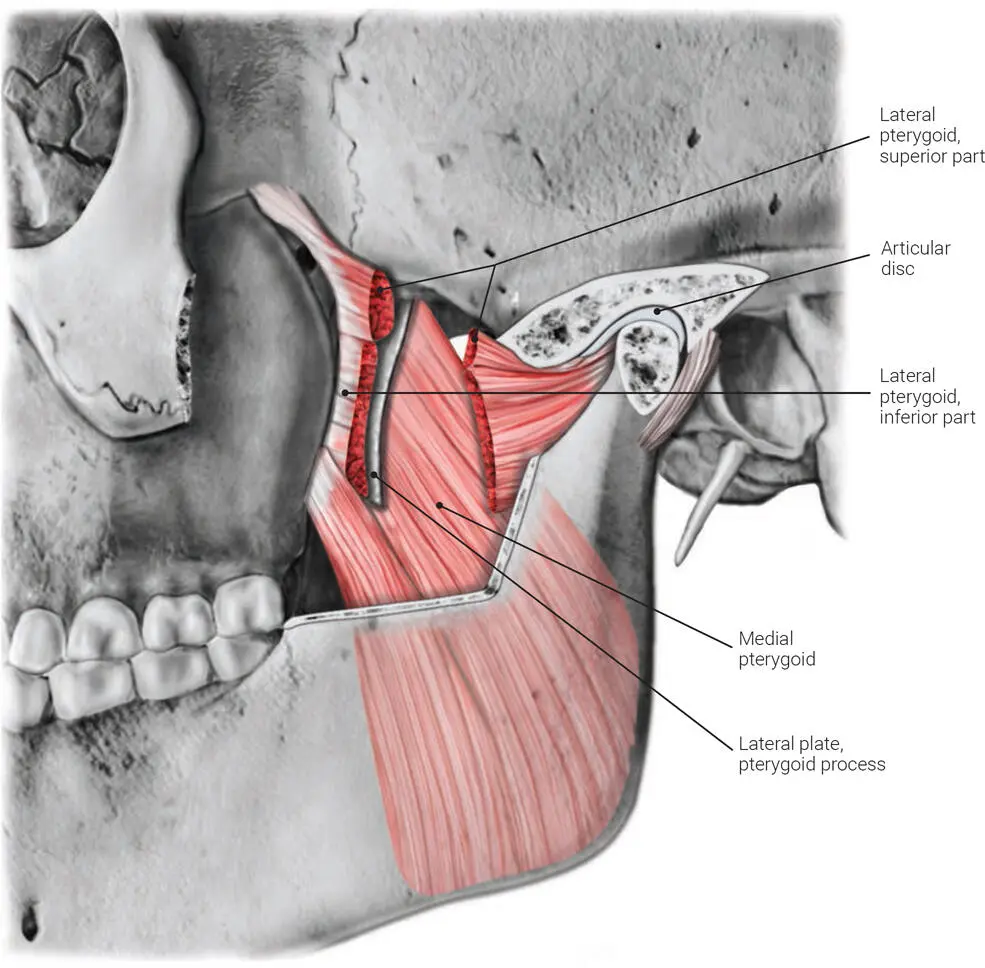
FIG 2-8 Lateral view of insertion of the medial pterygoid muscle.
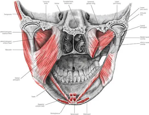
FIG 2-9 Posterior view of insertion of the lateral and medial pterygoid muscles.
Читать дальшеИнтервал:
Закладка:
Похожие книги на «Clinical Anatomy for Oral Implantology»
Представляем Вашему вниманию похожие книги на «Clinical Anatomy for Oral Implantology» списком для выбора. Мы отобрали схожую по названию и смыслу литературу в надежде предоставить читателям больше вариантов отыскать новые, интересные, ещё непрочитанные произведения.


Обсуждение, отзывы о книге «Clinical Anatomy for Oral Implantology» и просто собственные мнения читателей. Оставьте ваши комментарии, напишите, что Вы думаете о произведении, его смысле или главных героях. Укажите что конкретно понравилось, а что нет, и почему Вы так считаете.