Louie Al-Faraje - Clinical Anatomy for Oral Implantology
Здесь есть возможность читать онлайн «Louie Al-Faraje - Clinical Anatomy for Oral Implantology» — ознакомительный отрывок электронной книги совершенно бесплатно, а после прочтения отрывка купить полную версию. В некоторых случаях можно слушать аудио, скачать через торрент в формате fb2 и присутствует краткое содержание. Жанр: unrecognised, на английском языке. Описание произведения, (предисловие) а так же отзывы посетителей доступны на портале библиотеки ЛибКат.
- Название:Clinical Anatomy for Oral Implantology
- Автор:
- Жанр:
- Год:неизвестен
- ISBN:нет данных
- Рейтинг книги:3 / 5. Голосов: 1
-
Избранное:Добавить в избранное
- Отзывы:
-
Ваша оценка:
Clinical Anatomy for Oral Implantology: краткое содержание, описание и аннотация
Предлагаем к чтению аннотацию, описание, краткое содержание или предисловие (зависит от того, что написал сам автор книги «Clinical Anatomy for Oral Implantology»). Если вы не нашли необходимую информацию о книге — напишите в комментариях, мы постараемся отыскать её.
Clinical Anatomy for Oral Implantology — читать онлайн ознакомительный отрывок
Ниже представлен текст книги, разбитый по страницам. Система сохранения места последней прочитанной страницы, позволяет с удобством читать онлайн бесплатно книгу «Clinical Anatomy for Oral Implantology», без необходимости каждый раз заново искать на чём Вы остановились. Поставьте закладку, и сможете в любой момент перейти на страницу, на которой закончили чтение.
Интервал:
Закладка:
Surgical importance of the anatomy of the pterygopalatine fossa
The anatomy of the pterygopalatine fossa is especially important for the following surgeries:
Vidian neurectomy (the surgical sectioning of the pterygoid nerve for the treatment of vasomotor rhinitis, Sluder’s neuralgia of the pterygopalatine ganglion, crocodile tears syndrome, allergic rhinitis [hay fever], and nasal polyposis)
Transmaxillary ligature of the maxillary artery (in cases of severe nasal bleeding that cannot be controlled by anterior and/or posterior tamponades)
Craniofacial surgery
Surgery of the base of the skull or nasopharynx
Lateral approaches to the orbit
Traumatology
Vasomotor rhinitis is a condition that results from a relative imbalance of parasympathetic to sympathetic stimulation of the blood vessels and glands of the nasal mucosa. It is characterized by symptoms of clear rhinorrhea and nasal congestion.
Sluder’s neuralgia of the pterygopalatine ganglion is a rare disorder characterized by unilateral, severe, burning, boring, or nagging headache, starting around the eye and the bridge of the nose and radiating to the maxilla and maxillary teeth, zygoma, mastoidal area and occiput, or even as far down as the shoulder and arm.
Crocodile tears syndrome (gustatory lacrimation; tearing on eating) is a rare complication of a facial nerve lesion proximal to the geniculate ganglion, whereby regenerating preganglionic salivary fibers intended for the chorda tympani nerve are misdirected to the sphenopalatine ganglion, which project to the lacrimal gland.
Veins of the Head
The principal veins of the head and neck are the internal jugular vein, the external jugular vein, and the anterior jugular vein. The internal jugular vein collects blood from the interior of the skull, the anterior and lateral face, and the oral cavity and the neck via the sigmoid sinus, the inferior petrosal sinuses, and the facial, lingual, superior, and middle thyroid and retromandibular (anterior division) veins. The external jugular vein collects blood from the lateral skull and the occiput via the posterior auricular and the retromandibular (posterior division) veins. The anterior jugular vein collects blood from the anterior neck region.
Pterygoid venous plexus
The pterygoid venous plexus is situated on the medial side of the mandibular ramus within the pterygoid muscles. It is linked to the facial vein via the deep facial vein, to the retromandibular vein via the maxillary vein, and to the cavernous sinus via the sphenoidal emissary vein. The pterygoid plexus drains into the jugular veins.
This plexus is of a special importance to dentists because if the needle is overinserted during posterosuperior alveolar block, it may penetrate the pterygoid plexus of the vein and the maxillary artery in the infratemporal fossa (Fig 1-9), thus causing hematoma. This results in extraoral swelling a few minutes after the injection. The hematoma will cause tissue tenderness and discoloration, which will last until the blood is broken down by the body, and possible spread of infection to the cavernous venous sinus if the needle is contaminated. A hematoma can also result during other blocks, such as infraorbital and inferior alveolar blocks. To avoid injection into blood vessels, aspiration should always be attempted for all injections.
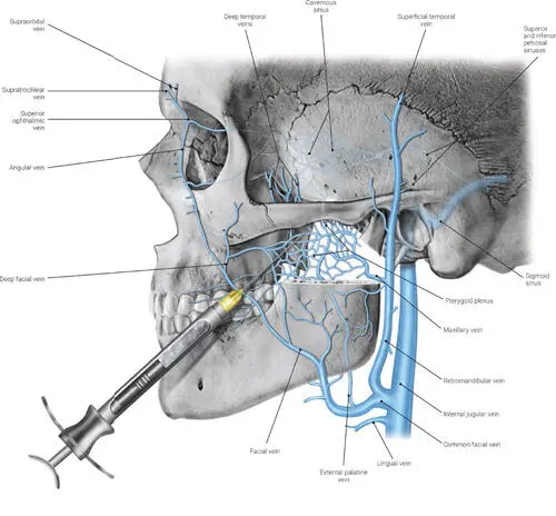
FIG 1-9 Pterygoid venous plexus.
Trigeminal Nerve
The 12 cranial nerves control motor and sensory functions of the head and neck. Figure 1-10 and Table 1-2summarize the skull base foramina from which these nerves exit the skull and their functions.
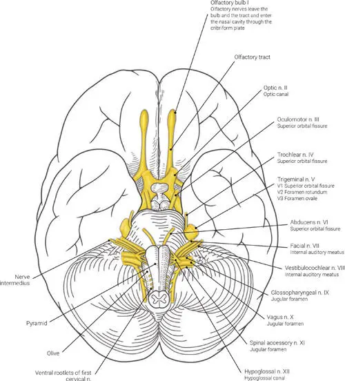
FIG 1-10 The origin of the cranial nerves as seen from an inferior aspect of the brain. n.—nerve.
TABLE 1-2 Exit foramina and functions of the cranial nerves
| Nerve | Name | Skull base foramina | Functions |
| I | Olfactory | Cribriform plate | Sensory for smell |
| II | Optic | Optic canal | Sensory for vision |
| III | Oculomotor | Superior orbital fissure | Motor for six eye muscles |
| IV | Trachlear | Superior orbital fissure | Motor for one eye muscle |
| V1 | Trigeminal/ophthalmic division | Superior orbital fissure | Sensory for lacrimal gland, nearby air sinuses, scalp, forehead, upper eyelid, and nose |
| V2 | Trigeminal/maxillary division | Foramen rotundum | Sensory for parts of the nasal and oral cavities and the skin of the cheek and upper lip |
| V3 | Trigeminal/mandibular division | Foramen ovale | Sensory for the skin over the mandible, lower lip, temporal region, and much of the oral cavityMotor for muscles of mastication as well as the anterior belly of the digastric muscle, mylohyoid muscle, tensor tympani, and tensor veli palatine muscles |
| VI | Abducens | Superior orbital fissure | Motor for one eye muscle |
| VII | Facial | Internal auditory meatus | Motor for muscles of facial expression, stapidius, and posterior belly of the digastric muscle; also motor for the lacrimal glands, oral and nasal mucosa, and submandibular and sublingual glandsSensory for the external auditory meatus; lateral pinna; mastoid; mucosa of the pharynx, nose, and palate; as well as sensory for taste for the anterior two-thirds of the tongue via the chorda tympani |
| VIII | Vestibulocochlear | Internal auditory meatus | Sensory for balance and hearing |
| IX | Glossopharyngeal | Jugular foramen | Motor for the stylopharyngeus muscle and parotid glandSensory for the posterior external ear, tragus, posterior third of the tongue, soft palate, nasopharynx, tympanic membrane, Eustachian tube, and mastoid region and sensory for taste for the posterior third of the tongue |
| X | Vagus | Jugular foramen | Motor for the pharyngeal and laryngeal muscles, including the palatoglossus muscle; also motor to the smooth muscles and glands of the pharynx, larynx, heart, esophagus, and stomachSensory for the ear, external auditory meatus, external surface of the tympanic membrane, dura of posterior cranial fossa, larynx, lungs, heart, esophagus, and stomach |
| XI | Spinal accessory | Jugular foramen | Motor for the sternocleidomastoid and trapezius muscles |
| XII | Hypoglossal | Hypoglossal canal | Motor for all intrinsic tongue muscles and all extrinsic tongue muscles except the palatoglossus muscle (innervated by CN X) |
Maxillary nerve (CN V2)
The maxillary nerve (Fig 1-11a) is the second branch of the fifth cranial nerve (trigeminal nerve). Its function is the transmission of sensory fibers from the maxillary teeth, the nasal cavity, the sinuses, and the skin between the palpebral fissure and the mouth (Figs 1-11b and 1-11c). In the cranium, the maxillary nerve branches off into the middle meningeal nerve, then passes through the foramen rotundum into the pterygopalatine fossa, where it divides into the zygomatic nerve, the ganglionic branches (pterygopalatine branches), and the infraorbital nerve.
Читать дальшеИнтервал:
Закладка:
Похожие книги на «Clinical Anatomy for Oral Implantology»
Представляем Вашему вниманию похожие книги на «Clinical Anatomy for Oral Implantology» списком для выбора. Мы отобрали схожую по названию и смыслу литературу в надежде предоставить читателям больше вариантов отыскать новые, интересные, ещё непрочитанные произведения.


Обсуждение, отзывы о книге «Clinical Anatomy for Oral Implantology» и просто собственные мнения читателей. Оставьте ваши комментарии, напишите, что Вы думаете о произведении, его смысле или главных героях. Укажите что конкретно понравилось, а что нет, и почему Вы так считаете.