Louie Al-Faraje - Clinical Anatomy for Oral Implantology
Здесь есть возможность читать онлайн «Louie Al-Faraje - Clinical Anatomy for Oral Implantology» — ознакомительный отрывок электронной книги совершенно бесплатно, а после прочтения отрывка купить полную версию. В некоторых случаях можно слушать аудио, скачать через торрент в формате fb2 и присутствует краткое содержание. Жанр: unrecognised, на английском языке. Описание произведения, (предисловие) а так же отзывы посетителей доступны на портале библиотеки ЛибКат.
- Название:Clinical Anatomy for Oral Implantology
- Автор:
- Жанр:
- Год:неизвестен
- ISBN:нет данных
- Рейтинг книги:3 / 5. Голосов: 1
-
Избранное:Добавить в избранное
- Отзывы:
-
Ваша оценка:
Clinical Anatomy for Oral Implantology: краткое содержание, описание и аннотация
Предлагаем к чтению аннотацию, описание, краткое содержание или предисловие (зависит от того, что написал сам автор книги «Clinical Anatomy for Oral Implantology»). Если вы не нашли необходимую информацию о книге — напишите в комментариях, мы постараемся отыскать её.
Clinical Anatomy for Oral Implantology — читать онлайн ознакомительный отрывок
Ниже представлен текст книги, разбитый по страницам. Система сохранения места последней прочитанной страницы, позволяет с удобством читать онлайн бесплатно книгу «Clinical Anatomy for Oral Implantology», без необходимости каждый раз заново искать на чём Вы остановились. Поставьте закладку, и сможете в любой момент перейти на страницу, на которой закончили чтение.
Интервал:
Закладка:
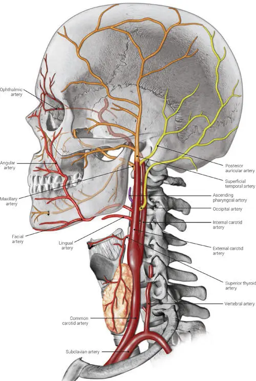
FIG 1-2 The main branches of the external carotid artery.
Three anterior branches: the superior thyroid artery, the lingual artery, and the facial artery
Two terminal branches: the maxillary artery and the superficial temporal artery
Two posterior branches: the occipital auricular artery and the posterior auricular artery
One medial branch: the ascending pharyngeal artery
Maxillary Artery
The maxillary artery (Fig 1-3) arises in the parotid gland as a terminal branch of the external carotid artery. The branches of the maxillary artery can be divided into three parts:
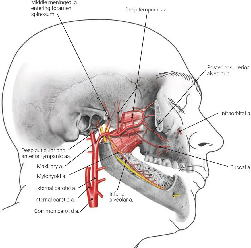
FIG 1-3 The course of the maxillary artery. a.—artery; aa.—arteries.
Part I or the mandibular part (located within the substance of the parotid gland and anterior to the external acoustic meatus): In this part, the maxillary artery gives branches to the ear, the dura, the temporomandibular joint, the mandibular teeth, and the mylohyoid muscle.
Part II or the pterygoid part (located in the infratemporal fossa): The branches here are mainly to the muscles of mastication, the buccal mucosa and skin, and the buccinator muscles through the buccal artery.
Part III or the pterygopalatine part (the branches in the pterygopalatine fossa after entry through the pterygomaxillary fissure): The branches here are mainly to the hard and soft palate through the branches of the descending palatine artery, to the maxillary molars and premolars through the posterior superior alveolar artery, to the upper pharynx and tympanic cavity through the artery of the pterygoid canal, to the nasopharynx and sphenoidal sinus through the pharyngeal artery, and to the maxillary anterior teeth through the infraorbital artery.
The maxillary artery terminates as the sphenopalatine artery on the nasal septum after splitting into nasal branches. Figure 1-4 demonstrates in detail the branches of all three parts of the maxillary artery.
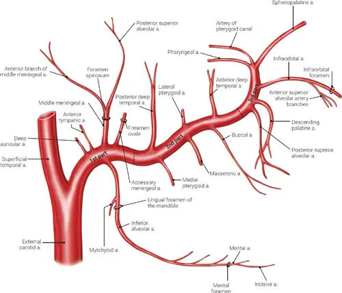
FIG 1-4 The distribution of all three parts of the maxillary artery. a.—artery.
Pterygopalatine Fossa
The pterygopalatine fossa , also called the sphenopalatine fossa , is a narrow, pyramid-shaped fossa on the lateral aspect of the skull. The fossa is a crossroads between the orbit, nasal cavity, oral cavity, nasopharynx, and middle cranial fossa (Figs 1-5 to 1-7). The pterygopalatine ganglion and the terminal branches of the maxillary artery are situated in its superior part. The pterygopalatine fossa along with the infratemporal and pterygoid fossae are referred to as the retromaxillary space .
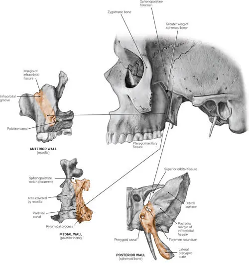
FIG 1-5 The anterior, medial, and posterior bony walls of the left pterygopalatine fossa.
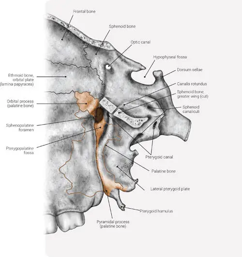
FIG 1-6 The pterygopalatine fossa after removal of the zygomatic bone, greater ala of the sphenoid bone, zygomatic arch, and temporal squama.
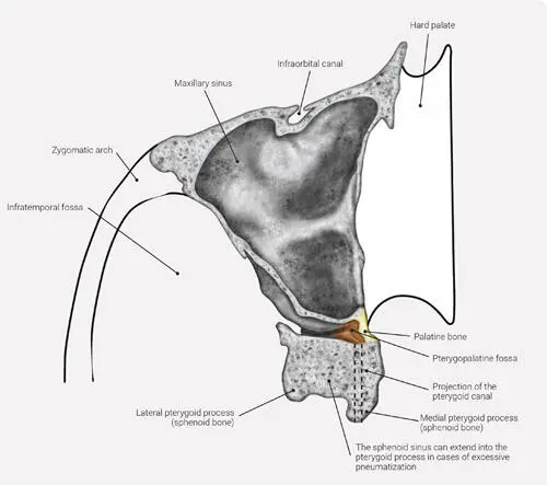
FIG 1-7 Horizontal section of the pterygopalatine fossa at the level of the infraorbital foramen.
Boundaries and communications of the pterygopalatine fossa 1–3
The anterior boundary comprises the superomedial part of the infratemporal surface of the maxilla. The posterior boundary comprises the root of the pterygoid process of the sphenoid bone. Through this posterior wall, the fossa communicates with the middle cranial fossa via the foramen rotundum and the pterygoid canal (also called the vidian canal ). The foramen rotundum lies lateral and superior to the pterygoid canal at the base of the pterygoid process. The vidian canal is located medial and superior to the pterygopalatine ganglion, and thus its nerve lies medial to the major vessels of the pterygopalatine fossa, which allows the surgeon to avoid excessive bleeding during vidian neurectomy (Fig 1-8).
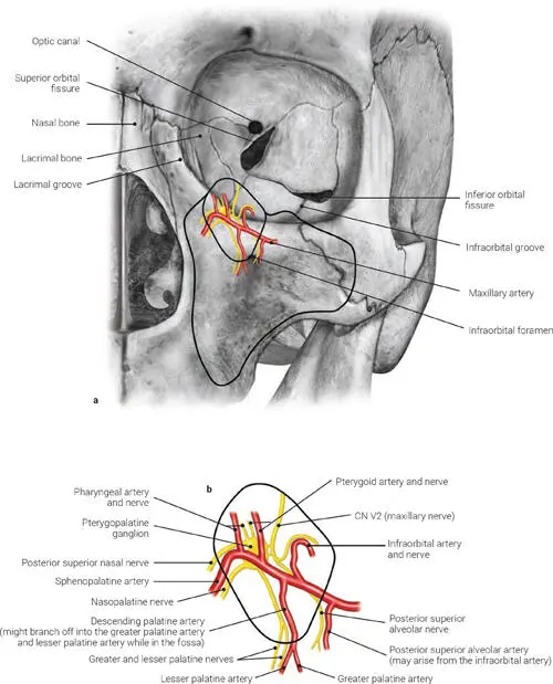
FIG 1-8 (a and b) The branching pattern of the maxillary artery in its relationship to the pterygopalatine ganglion in the pterygopalatine fossa. Some variation in the branching pattern does exist.
Also, at the posterior wall and in an inferoposterior direction, the fossa communicates with the nasopharynx through the palatovaginal (pharyngeal) canal. The palatovaginal canal is located between the vaginal process of the vomer bone and the sphenoid process of the palatine bone, and it passes into the floor of the sphenoid sinus between the pterygoid canal and the vomerine crest of the sphenoid. The opening to the palatovaginal (pharyngeal) canal in the nasal cavity is located near the lateral margin of the ala of the vomer, at the roots of the pterygoid process.
The medial boundary comprises part of the perpendicular plate of the palatine bone and its orbital sphenoidal processes. The pterygopalatine fossa communicates with the nasal cavity at this wall through the sphenopalatine foramen. The sphenopalatine foramen is bounded in front, below, and behind by the palatine bone (and the sphenopalatine incisure) and above by the body of sphenoid bone. Laterally, the pterygopalatine fossa communicates with the infratemporal fossa through the pterygomaxillary fissure.
The superior border of the pterygopalatine fossa comprises a small part of the orbital plate of the palatine bone and part of the maxillary surface of the greater wing of the sphenoid bone and junction with the inferior orbital fissure.
The inferior border of the pterygopalatine fossa is formed by the pyramidal process of the palatine bone; the pterygopalatine (greater palatine) canal is located at this inferior border. The pterygopalatine canal is a continuation of the pterygopalatine fossa and is formed when the maxillary surface of the perpendicular plate of the palatine bone articulates with the maxilla. It leads to the greater and lesser palatine foramina in the roof of the oral cavity.
Table 1-1provides a detailed description of the contents of the pterygopalatine fossa.
TABLE 1-1 Contents of the pterygopalatine fossa
| Opening | Communication | Location | Transmitted structures |
| Foramen rotundum | Middle cranial fossa | Posterior wall | • CN V2 |
| Pterygoid canal | Middle cranial fossa | Posterior wall | • Nerve of the pterygoid canal (vidian nerve) (formed from the greater petrosal and deep petrosal nerves)• Artery of pterygoid canal• Veins of pterygoid canal |
| Palatovaginal (pharyngeal) canal | Nasopharynx | Posterior wall | • Pharyngeal branches of the pterygopalatine ganglion of CN V2 (the ganglion is located in the pterygopalatine fossa)• Pharyngeal artery (maxillary artery)• Pharyngeal vein |
| Sphenopalatine foramen | Nasal cavity | Medial wall | • Nasopalatine nerve and posterior superior nasal nerve (both are pterygopalatine ganglionic branches of CN V2)• Sphenopalatine artery (maxillary artery)• Sphenopalatine vein |
| Pterygomaxillary suture | Infratemporal fossa | Lateral wall | • Posterior superior alveolar nerve• Pterygoid part of the maxillary artery (after branching off into the posterior superior alveolar artery, its only branch outside the fossa)• Posterior superior alveolar vein |
| Inferior orbital fissure | Orbit | Superior wall | • Infraorbital and zygomatic nerves (CN V2)• Infraorbital artery (maxillary artery)• Infraorbital vein |
| Pterygopalatine (greater palatine) canal | Oral cavity | Inferior wall | • Descending palatine nerve (CN V2) (splits into the greater and lesser palatine within the canal)• Descending palatine artery (maxillary artery) (splits into the greater and lesser palatine within the canal)• Descending palatine vein |
Yellow bullet—nerve; red bullet—artery; blue bullet—vein.
Читать дальшеИнтервал:
Закладка:
Похожие книги на «Clinical Anatomy for Oral Implantology»
Представляем Вашему вниманию похожие книги на «Clinical Anatomy for Oral Implantology» списком для выбора. Мы отобрали схожую по названию и смыслу литературу в надежде предоставить читателям больше вариантов отыскать новые, интересные, ещё непрочитанные произведения.


Обсуждение, отзывы о книге «Clinical Anatomy for Oral Implantology» и просто собственные мнения читателей. Оставьте ваши комментарии, напишите, что Вы думаете о произведении, его смысле или главных героях. Укажите что конкретно понравилось, а что нет, и почему Вы так считаете.