Saeid Sanei - EEG Signal Processing and Machine Learning
Здесь есть возможность читать онлайн «Saeid Sanei - EEG Signal Processing and Machine Learning» — ознакомительный отрывок электронной книги совершенно бесплатно, а после прочтения отрывка купить полную версию. В некоторых случаях можно слушать аудио, скачать через торрент в формате fb2 и присутствует краткое содержание. Жанр: unrecognised, на английском языке. Описание произведения, (предисловие) а так же отзывы посетителей доступны на портале библиотеки ЛибКат.
- Название:EEG Signal Processing and Machine Learning
- Автор:
- Жанр:
- Год:неизвестен
- ISBN:нет данных
- Рейтинг книги:3 / 5. Голосов: 1
-
Избранное:Добавить в избранное
- Отзывы:
-
Ваша оценка:
EEG Signal Processing and Machine Learning: краткое содержание, описание и аннотация
Предлагаем к чтению аннотацию, описание, краткое содержание или предисловие (зависит от того, что написал сам автор книги «EEG Signal Processing and Machine Learning»). Если вы не нашли необходимую информацию о книге — напишите в комментариях, мы постараемся отыскать её.
EEG Signal Processing and Machine Learning — читать онлайн ознакомительный отрывок
Ниже представлен текст книги, разбитый по страницам. Система сохранения места последней прочитанной страницы, позволяет с удобством читать онлайн бесплатно книгу «EEG Signal Processing and Machine Learning», без необходимости каждый раз заново искать на чём Вы остановились. Поставьте закладку, и сможете в любой момент перейти на страницу, на которой закончили чтение.
Интервал:
Закладка:
Such disorders are often detected, monitored, or diagnosed through other recording modalities such as heartrate and respiration. Nevertheless, the use of sleep EEG in detection or monitoring of abnormal sleep has been a topic of research recently. Some methods are discussed in the related chapter of this book.
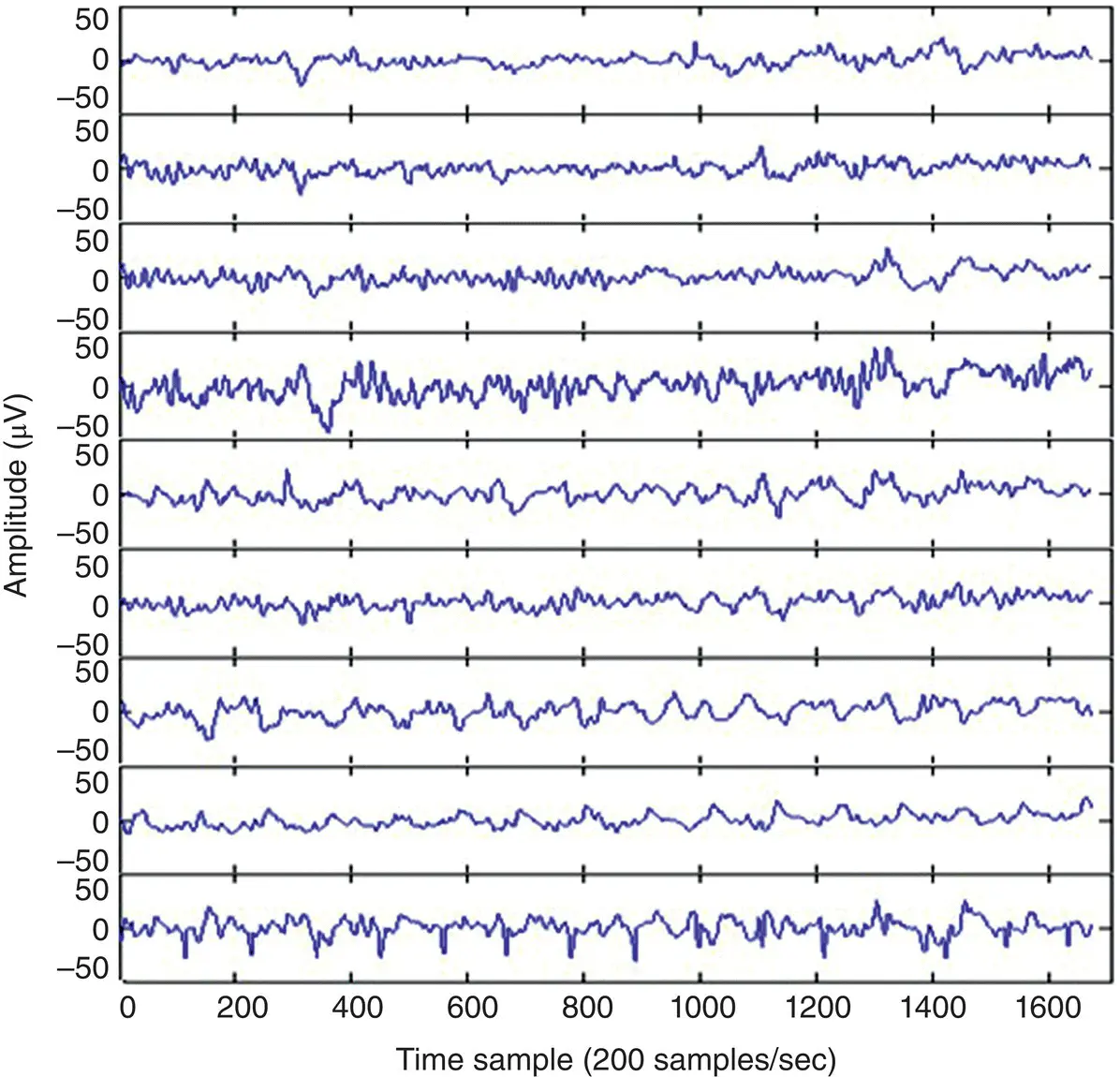
Figure 2.12 A multichannel EEG set with the clear appearance of ECG signals over the electrodes in the occipital region.
2.4 Mental Fatigue
Mental fatigue is often the result of prolonged cognitive load and can negatively affect people over a short or long time and can cause failure of the brain in performing its regular tasks. Mental fatigue mainly affects the function of the brain network rather than changing the normal brain rhythms significantly [21, 22] In these studies it has been shown that there is less synchrony between the left and right brain lobes and less connectivity between various brain lobes when the brain is under mental fatigue.
Mental fatigue also deteriorates the brain response to audio, visual, and other stimulations by attenuating and time shifting the ERPs. It has been shown that the amplitude of ERP (particularly P3b which is a subcomponent of P300,) reduces and the ERP latency, particularly for P3b, increases with the increase in mental fatigue [23–25].
A chapter of this book is devoted to detailed analysis of the EEG for under fatigue brains.
2.5 Emotions
The brain right hemisphere is associated with emotions. Lateralized electrophysiological parameters measured during emotionally charged states found relative activation (as measured by decreased alpha power) using EEG during the recollection of past events associated with anger or relaxation. Similar findings were obtained during self‐reported emotional reactions to visual material [26]. Other experimental studies have verified the relative right‐hemisphere activation when participants were asked to generate emotional imagery [27], and during hypnotically induced depression [28]. More recent quantitative EEG research has demonstrated reliable relationships between the magnitude of cerebral activation and the intensity of emotional arousal [29]. In related research, the ages of emotional memories correlated with the magnitude of activation using quantitative EEG [30]. Most of these studies conclude from the asymmetry in the activities of lateral brain lobes [31].
Perhaps the strongest evidence in support of the valence hypothesis was derived from a plethora of EEG studies that have associated relative increased left‐hemisphere activity with positive emotional states and relative increased right‐hemisphere activity with negative emotional states such as in [32]. Frontal EEG shows relative left‐hemisphere activity during a positive emotional response, whereas the opposite pattern is displayed during a negative emotional response.
Some recent machine learning applications [33] have shown that the ratio between the power of EEG in the beta band and the theta band and their asymmetry is associated with change in emotions.
In [34] the authors have shown that six features namely, power spectral density (PSD), differential entropy (DE), differential asymmetry (DASM), rational asymmetry (RASM), asymmetry (ASM), and differential causality (DCAU) features from EEG, are associated with emotions and can be classified for emotion recognition.
Further unpublished research has focused on brain connectivity for emotion recognition. The patterns of interdependency between different brain regions for emotional and non‐emotional film stimuli from EEGs have been analyzed and the emotion‐related differences evaluated. A simple measure of synchronization index (SI) has then been used to detect interdependencies in EEG signals mainly for happiness and sadness. The SI significantly changes/increases during emotional stimulation and, in particular, during sadness, yielding an enhanced connectivity among frontal channels. Conversely, happiness is associated with a wider synchronization among frontal and occipital sites, although happiness itself was less synchronized [35].
2.6 Neurodevelopmental Disorders
Neurodevelopmental disorders are a group of disorders that affect the development of the nervous system, leading to abnormal brain function which may affect emotion, learning ability, self‐control, and memory [36]. Such disorders, often starting from childhood, though their effects vary over time, usually remain with the subject and affect the person throughout their lifetime.
Examples of neurodevelopmental disorders in children include attention deficit hyperactivity disorder (ADHD), autism, also called autism spectrum disorder (ASD), learning disabilities, intellectual disability (also known as mental retardation), conduct disorders, cerebral palsy, and impairments in vision and hearing. One may add depression to this category of disorders.
ADHD, ASD, and depression have been under EEG studies in recent years and therefore some examples are provided in the later chapters of this book.
2.7 Abnormal EEG Patterns
Variations in the EEG patterns for certain states of the subject indicate abnormality. This may be due to distortion and disappearance of abnormal patterns, appearance and increase of abnormal patterns, or disappearance of all patterns. Sharbrough [37] divided the nonspecific abnormalities in the EEGs into three categories: (i) widespread intermittent slow‐wave abnormalities often in the delta wave range and associated with brain dysfunction, (ii) bilateral persistent EEG usually associated with impaired conscious cerebral reactions, and (iii) focal persistent EEG usually associated with focal cerebral disturbance.
The first category is a burst type signal, which is attenuated by alerting the individual and eye opening, and accentuated with eye closure, hyperventilation, or drowsiness. The peak amplitude in adults is usually localized in the frontal region and influenced by age. In children, however, it appears over the occipital or posterior head region. Early findings showed that this abnormal pattern frequently appears with an increased intracranial pressure with tumour or aqueductal stenosis. Also, it correlates with grey matter disease, both in cortical and subcortical locations. However, it can be seen in association with a wide variety of pathological processes varying from systemic toxic or metabolic disturbances to focal intracranial lesions.
Regarding the second category, i.e. bilateral persistent EEG, the phenomenon in different stages of impaired, conscious, purposeful responsiveness are etiologically nonspecific and the mechanisms responsible for their generation are only partially understood. However, the findings in connection with other information concerning aetiology and chronicity may be helpful in arriving more quickly at an accurate prognosis concerning the patient's chance of recovering his previous conscious life.
As for the third category, i.e. focal persistent EEG, these abnormalities may be in the form of distortion and disappearance of normal patterns, appearance and increase of abnormal patterns, or disappearance of all patterns, but such changes are seldom seen at the cerebral cortex. The focal distortion of normal rhythms may produce an asymmetry of amplitude, frequency, or reactivity of the rhythm. The unilateral loss of reactivity of a physiological rhythm, such as the loss of reactivity of the alpha rhythm to eye opening [38] or to mental alerting [39], may reliably identify the focal side of abnormality. A focal lesion may also distort or eliminate the normal activity of sleep‐inducing spindles and vertex waves.
Читать дальшеИнтервал:
Закладка:
Похожие книги на «EEG Signal Processing and Machine Learning»
Представляем Вашему вниманию похожие книги на «EEG Signal Processing and Machine Learning» списком для выбора. Мы отобрали схожую по названию и смыслу литературу в надежде предоставить читателям больше вариантов отыскать новые, интересные, ещё непрочитанные произведения.


Обсуждение, отзывы о книге «EEG Signal Processing and Machine Learning» и просто собственные мнения читателей. Оставьте ваши комментарии, напишите, что Вы думаете о произведении, его смысле или главных героях. Укажите что конкретно понравилось, а что нет, и почему Вы так считаете.