Saeid Sanei - EEG Signal Processing and Machine Learning
Здесь есть возможность читать онлайн «Saeid Sanei - EEG Signal Processing and Machine Learning» — ознакомительный отрывок электронной книги совершенно бесплатно, а после прочтения отрывка купить полную версию. В некоторых случаях можно слушать аудио, скачать через торрент в формате fb2 и присутствует краткое содержание. Жанр: unrecognised, на английском языке. Описание произведения, (предисловие) а так же отзывы посетителей доступны на портале библиотеки ЛибКат.
- Название:EEG Signal Processing and Machine Learning
- Автор:
- Жанр:
- Год:неизвестен
- ISBN:нет данных
- Рейтинг книги:3 / 5. Голосов: 1
-
Избранное:Добавить в избранное
- Отзывы:
-
Ваша оценка:
EEG Signal Processing and Machine Learning: краткое содержание, описание и аннотация
Предлагаем к чтению аннотацию, описание, краткое содержание или предисловие (зависит от того, что написал сам автор книги «EEG Signal Processing and Machine Learning»). Если вы не нашли необходимую информацию о книге — напишите в комментариях, мы постараемся отыскать её.
EEG Signal Processing and Machine Learning — читать онлайн ознакомительный отрывок
Ниже представлен текст книги, разбитый по страницам. Система сохранения места последней прочитанной страницы, позволяет с удобством читать онлайн бесплатно книгу «EEG Signal Processing and Machine Learning», без необходимости каждый раз заново искать на чём Вы остановились. Поставьте закладку, и сможете в любой момент перейти на страницу, на которой закончили чтение.
Интервал:
Закладка:
In many applications such as brain–computer interfacing (BCI) and study of mental activity, often a small number of electrodes around the movement‐related regions are selected and used from the 10–20 setting system.
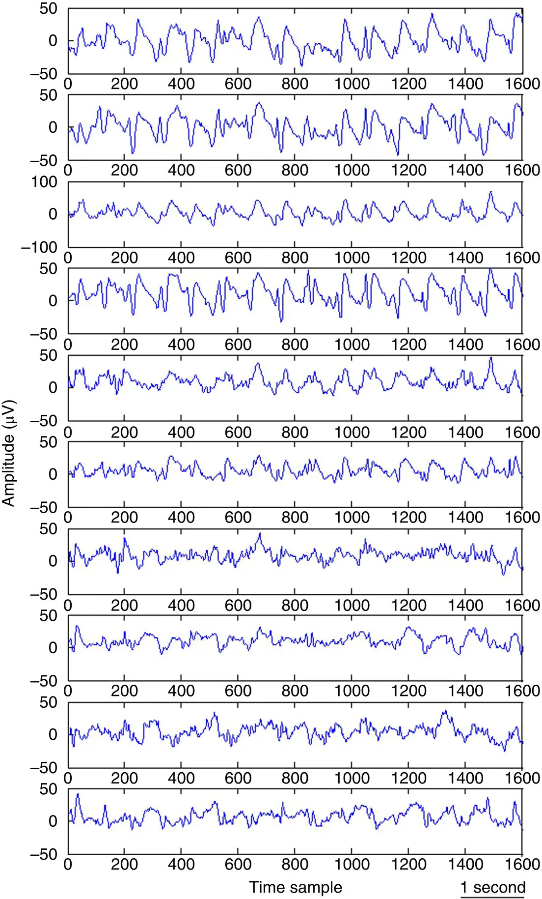
Figure 2.5 A typical set of EEG signals during approximately seven seconds of normal adult brain activity.
Figure 2.5illustrates a typical set of EEG signals during approximately seven seconds of normal adult brain activity.
2.2.2 Unconventional and Special Purpose EEG Recording Systems
Despite many small‐electrode portable EEG systems such as 5‐channel or 14‐channel EEG Emotive systems, advances in wearable technology have led to special purpose and miniaturized designs for EEG recordings. GlassEEG, produced by Mobile Health Technologies for real‐time detection of epileptic seizures, EarEEG, designed mainly for SSVEP‐based brain–computer interface (BCI), and tattooed EEG, very recently developed by Graz University in Austria for BCI are among such systems ( Figure 2.6).
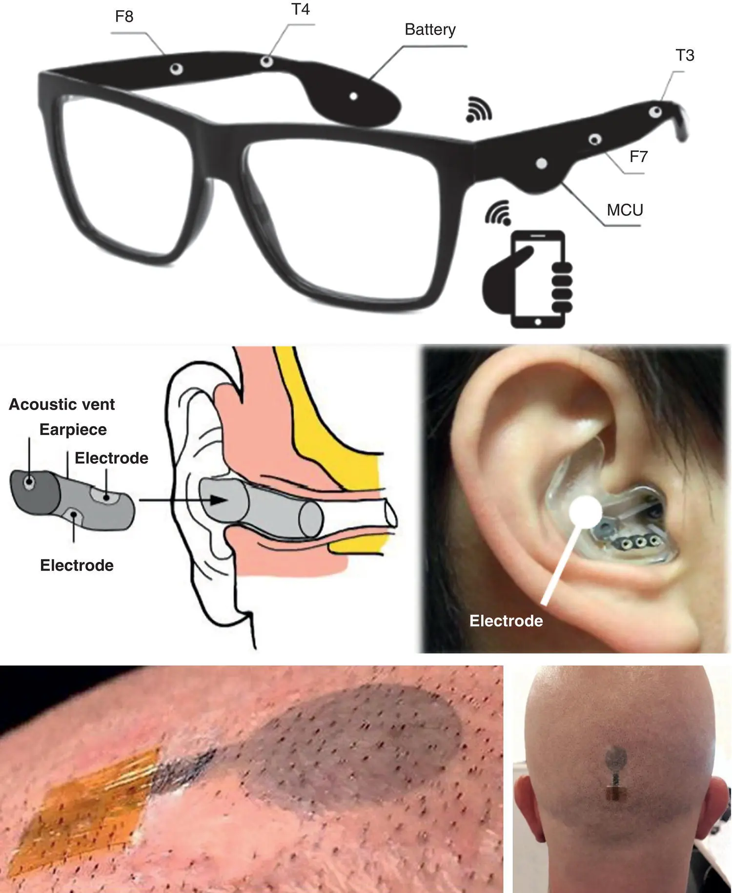
Figure 2.6 Wearable and tattooed EEG systems/electrodes.
2.2.3 Invasive Recording of Brain Potentials
Invasive brain screening is mainly to get closer to the neurons and thereby gain information about timing of neuron firing and the exact locations of defected neurons (such as for seizure) for various purposes such as deep brain stimulation, surgical operation, or rehabilitative implants.
The recording directly from over the cortex, called electrocorticography (ECoG), or intracranial electroencephalography (iEEG), is a type of electrophysiological brain monitoring that uses a grid of electrodes placed directly on the exposed surface of the brain to record electrical activity from the cerebral cortex. Figure 2.7shows an example.
A popular application of ECoG is the localization of the central sulcus by means of phase reversal of somatosensory evoked potentials (EPs) for BCI‐based rehabilitation.
Another technique for iEEG recording is by using strings of electrodes inserted into foramen ovale holes ( Figure 2.8) deep into the hippocampus mainly to record the neuron activities during seizure and localization of infected neurons for surgical purposes. A set of these electrodes can be seen in Figure 2.9.
Later in Chapter 11of this book we will show the application of this recording modality in detection of interictal epileptiform discharges (IEDs) from the hippocampus.
With the advances in microelectronics and microsensor designs, a new type of microsensor array insertable into the brain blood vessels within the motor cortex without any open surgery has been designed. The sensor array, called Stentrode™ is a small metallic mesh tube (stent), with not more than 4 mm diameter and with electrode contacts (small metal discs) within the stent structure.
The Stentrode™ ( Figure 2.10) is expected to accurately record the neuronal activities within motor area and therefore help patients with loss of motor function due to paralysis from spinal cord injury, motor neuron disease, stroke, muscular dystrophy, or loss of limbs.
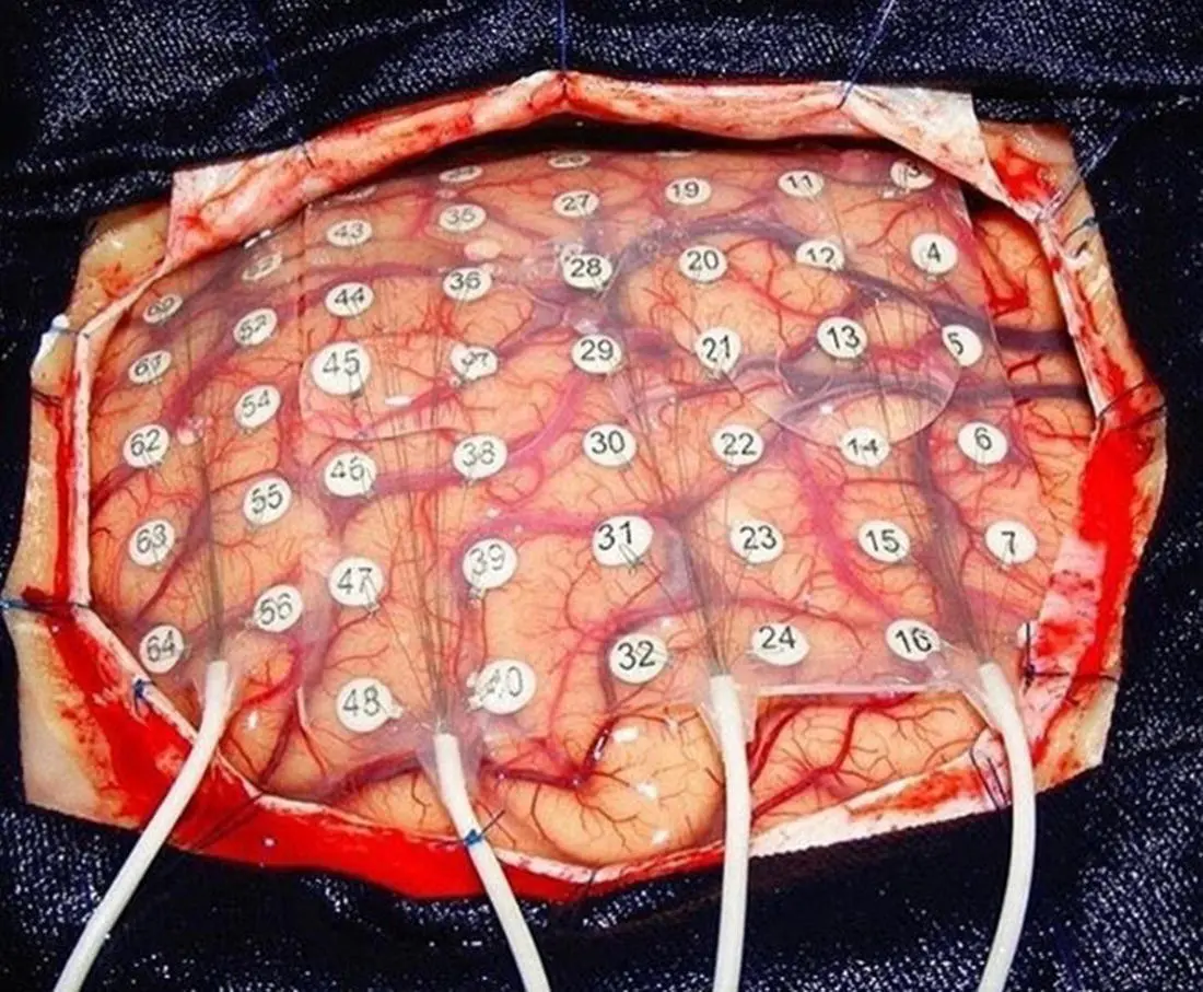
Figure 2.7 Electrocorticography.
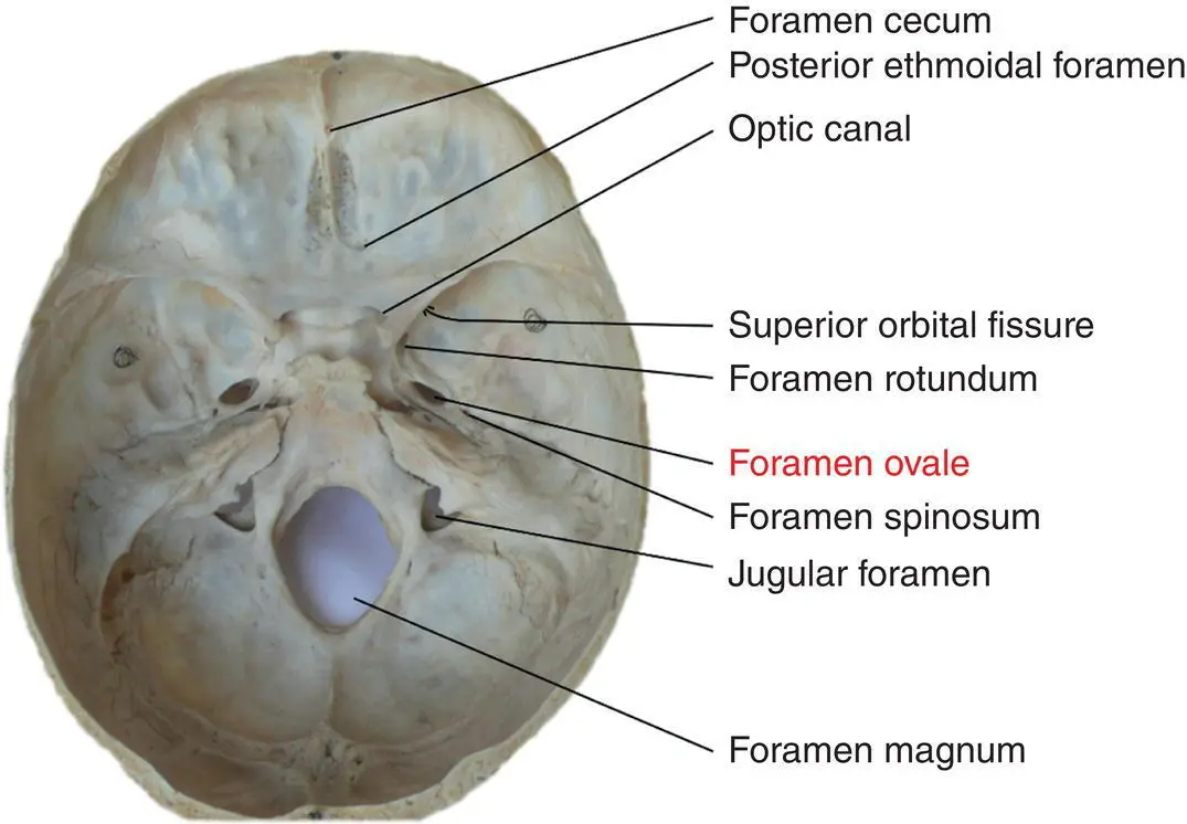
Figure 2.8 Foramen ovale holes within facial skeleton.
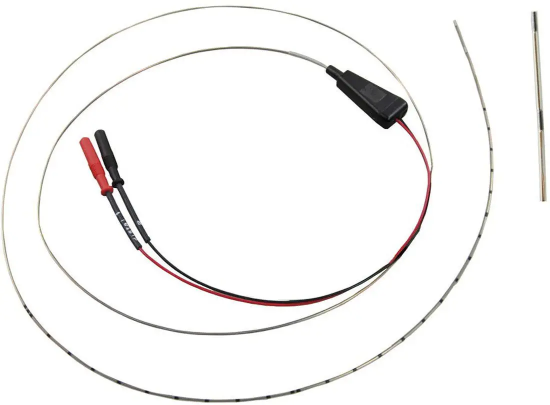
Figure 2.9 Foramen ovale electrodes.
2.2.4 Conditioning the Signals
The raw EEG signals have amplitudes of the order of μV and contain frequency components of up to 300 Hz. To retain the effective information the signals have to be amplified before the ADC and filtered, either before or after the ADC, to reduce the noise and make the signals suitable for processing and visualization. The filters are designed in such a way not to introduce any change or distortion to the signals. Highpass filters with cut‐off frequency of usually less than 0.5 Hz are used to remove the disturbing very low frequency components such as those of breathing. Conversely, high‐frequency noise is mitigated using lowpass filters with cut‐off frequency of approximately 50–70 Hz. Notch filters with the null frequency of 50 Hz are often necessary to ensure perfect rejection of the strong 50 Hz power supply. In this case the sampling frequency can be as low as twice the bandwidth as commonly used by most EEG systems. The commonly used sampling frequencies for EEG recordings are 100, 250, 500, 1000, and 2000 samples per second. The main artefacts can be divided into patient related (physiological) and system artefacts. The patient related or internal artefacts are body movement‐related, EMG, ECG (and pulsation), EOG, ballistocardiogram, and sweating. The system artefacts are 50/60 Hz power supply interference, impedance fluctuation, cable defects, electrical noise from the electronic components, and unbalanced impedances of the electrodes. Often in the preprocessing stage these artefacts are highly mitigated and the informative information is restored. Some methods for removing the EEG artefacts will be discussed in the related chapters of this book. Figure 2.11shows a set of normal EEG signals affected by eye‐blinking artefact. Similarly, Figure 2.12represents a multichannel EEG set with clear appearance of ECG signals over the electrodes in the occipital region.
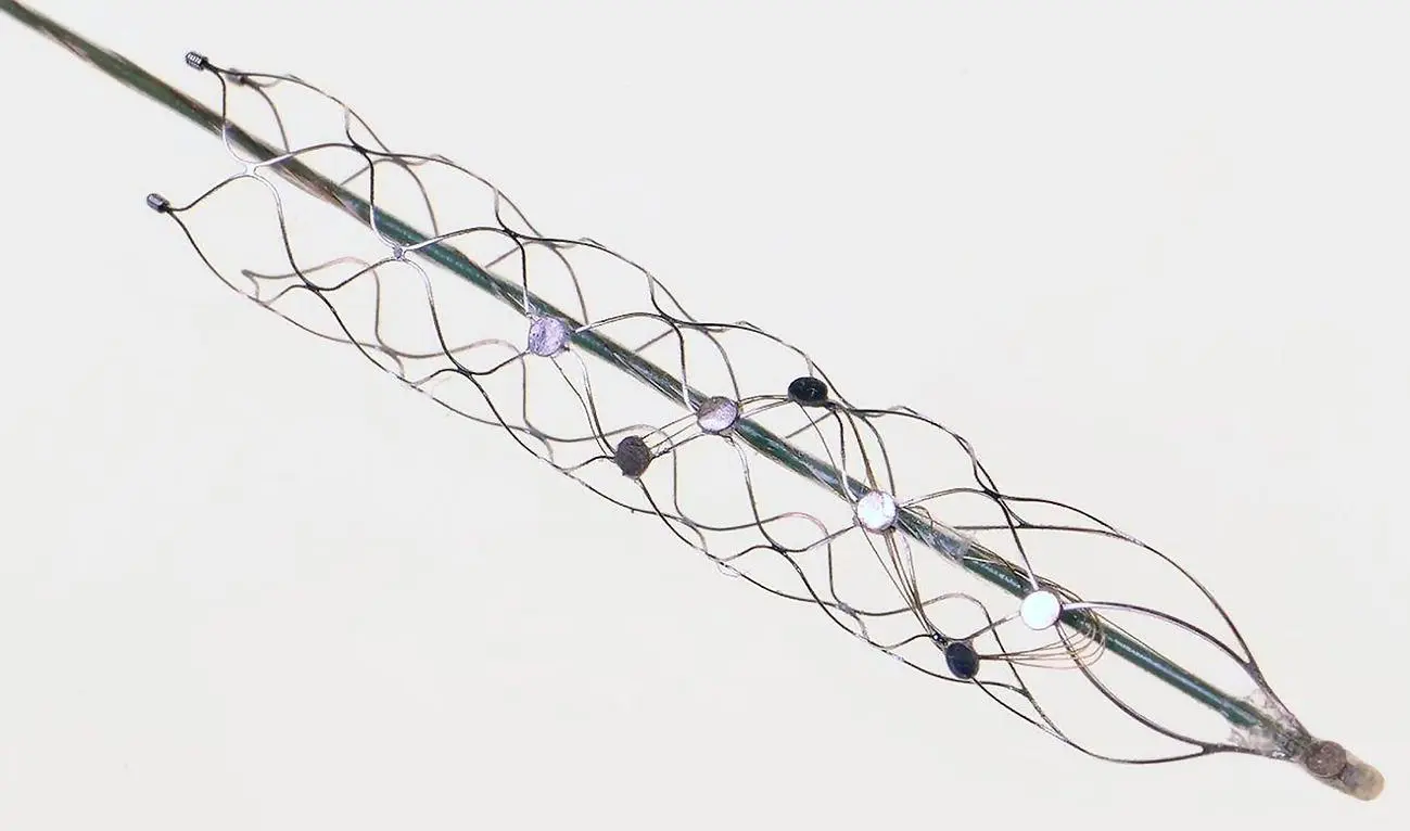
Figure 2.10 A 4‐mm diameter Stentrode with electrode contacts within the stent structure.
In the following sections we highlight the most popular changes in EEG measurements which correlate with physiological and mental abnormalities in the brain.
2.3 Sleep
Brain waves change during the various stages of normal sleep. When sleep starts the high‐frequency waveforms gradually die away and continuous slow waves (in the delta band) followed by spindles and K‐complexes appear from stage two of sleep and extend towards stage four. During the last stages of sleep rapid eye movement (REM) starts and finishes before the person wakes up. All these stages can be observed and accurately detected and scored automatically by analyzing the EEG signals during normal sleep.
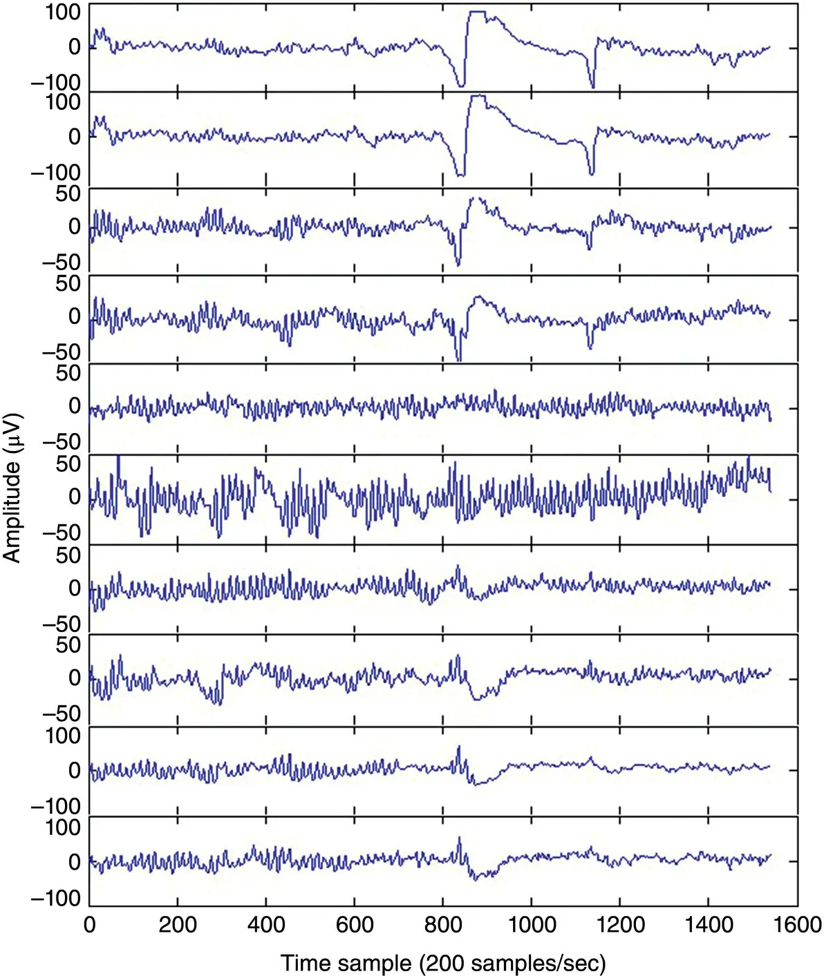
Figure 2.11 A set of normal EEG signals affected by an eye‐blinking artefact.
Conversely, there are cases where the subject has sleep disorder. These include obstructive sleep apnoea (OSA), insomnia (including parainsomnia and hyperinsomnia), REM sleep behaviour disorder, circadian rhythm sleep disorders, non‐24‐hour sleep–wake disorder, periodic limb movement disorder (PLMD), shift work sleep disorder, narcolepsy. Sleep disorders need to be diagnosed and treated.
Читать дальшеИнтервал:
Закладка:
Похожие книги на «EEG Signal Processing and Machine Learning»
Представляем Вашему вниманию похожие книги на «EEG Signal Processing and Machine Learning» списком для выбора. Мы отобрали схожую по названию и смыслу литературу в надежде предоставить читателям больше вариантов отыскать новые, интересные, ещё непрочитанные произведения.


Обсуждение, отзывы о книге «EEG Signal Processing and Machine Learning» и просто собственные мнения читателей. Оставьте ваши комментарии, напишите, что Вы думаете о произведении, его смысле или главных героях. Укажите что конкретно понравилось, а что нет, и почему Вы так считаете.