Saeid Sanei - EEG Signal Processing and Machine Learning
Здесь есть возможность читать онлайн «Saeid Sanei - EEG Signal Processing and Machine Learning» — ознакомительный отрывок электронной книги совершенно бесплатно, а после прочтения отрывка купить полную версию. В некоторых случаях можно слушать аудио, скачать через торрент в формате fb2 и присутствует краткое содержание. Жанр: unrecognised, на английском языке. Описание произведения, (предисловие) а так же отзывы посетителей доступны на портале библиотеки ЛибКат.
- Название:EEG Signal Processing and Machine Learning
- Автор:
- Жанр:
- Год:неизвестен
- ISBN:нет данных
- Рейтинг книги:3 / 5. Голосов: 1
-
Избранное:Добавить в избранное
- Отзывы:
-
Ваша оценка:
EEG Signal Processing and Machine Learning: краткое содержание, описание и аннотация
Предлагаем к чтению аннотацию, описание, краткое содержание или предисловие (зависит от того, что написал сам автор книги «EEG Signal Processing and Machine Learning»). Если вы не нашли необходимую информацию о книге — напишите в комментариях, мы постараемся отыскать её.
EEG Signal Processing and Machine Learning — читать онлайн ознакомительный отрывок
Ниже представлен текст книги, разбитый по страницам. Система сохранения места последней прочитанной страницы, позволяет с удобством читать онлайн бесплатно книгу «EEG Signal Processing and Machine Learning», без необходимости каждый раз заново искать на чём Вы остановились. Поставьте закладку, и сможете в любой момент перейти на страницу, на которой закончили чтение.
Интервал:
Закладка:
More recent EEG systems consist of a number of delicate electrodes, a set of differential amplifiers (one for each channel) followed by filters [9], and needle (pen) type registers. The multichannel EEGs could be plotted on plane paper or paper with a grid. Soon after this system came to the market, researchers started looking for a computerized system, which could digitize and store the signals. Therefore, to analyze EEG signals it was soon understood that the signals must be in digital form. This required sampling, quantization, and encoding of the signals. As the number of electrodes grows the data volume, in terms of the number of bits, increases. The computerized systems allow variable settings, stimulations, and sampling frequency, and some are equipped with simple or advanced signal processing tools for processing the signals.
The conversion from analogue‐to‐digital EEG is performed by means of multichannel analogue‐to‐digital converters (ADCs). Fortunately, the effective bandwidth for EEG signals is limited to approximately 100 Hz. For many applications this bandwidth may be considered even half of this value. Therefore, a minimum frequency of 200 Hz (to satisfy the Nyquist criterion) is often enough for sampling the EEG signals. In some applications where a higher resolution is required for representation of brain activities in the frequency domain, sampling frequencies of up to 2000 samples per second may be used.
In order to maintain the diagnostic information the quantization of EEG signals is normally very fine. Representation of each signal sample with up to 16 bits is very popular for the EEG recording systems. This makes the necessary memory volume for archiving the signals massive, especially for sleep EEG and epileptic seizure monitoring records. However, in general, the memory size for archiving the images is often much larger than that used for archiving the EEG signals.
A simple calculation shows that for a one hour recording from 128‐electrode EEG signals sampled at 500 samples per second a memory size of 128 × 60 × 60 × 500 × 16 ≈ 3.68 Gbits ≈ 0.45 Gbyte is required. Therefore, for longer recordings of a large number of patients there should be enough storage facilities such as in today's technology Zip disks, CDs, large removable hard drives, and optical disks.
Although the format of reading the EEG data may be different for different EEG machines, these formats are easily convertible to spreadsheets readable by most signal processing software packages such as MATLAB.
The EEG recording electrodes and their proper function are crucial for acquiring high quality data. There are different types of electrodes often used in the EEG recording systems as:
disposable (gel‐less, and pre‐gelled types)
reusable disc electrodes (gold, silver, stainless steel, or tin)
headbands and electrode caps
saline‐based electrodes
needle electrodes.
For multichannel recordings with a large number of electrodes, electrode caps are often used. Commonly used scalp electrodes consist of Ag–AgCl discs, less than 3 mm in diameter, with long flexible leads that can be plugged into an amplifier. Needle electrodes are those which have to be implanted under the skull with minimal invasive operations. High impedance between the cortex and the electrodes as well as the electrodes with high impedances can lead to distortion, which can even mask the actual EEG signals. Commercial EEG recording systems are often equipped with impedance monitors. To enable a satisfactory recording the electrode impedances should read less than 5 kΩ and be balanced to within 1 kΩ of each other. For more accurate measurement the impedances are checked after each trial.
Due to the layered and spiral structure of the brain, however, distribution of the potentials over the scalp (or cortex) is not uniform [13]. This may affect some of the results of source localization using the EEG signals.
2.2.1 Conventional Electrode Positioning
The International Federation of Societies for Electroencephalography and Clinical neurophysiology has recommended the conventional electrode setting (also called 10–20) for 21 electrodes (excluding the earlobe electrodes) as depicted in Figure 2.3[14]. Often, the earlobe electrodes called A1 and A2, connected respectively to the left and right earlobes, are used as the reference electrodes. The 10–20 system avoids both eyeball placement and considers some constant distances by using specific anatomic landmarks from which the measurement would be made and then uses 10 or 20% of that specified distance as the electrode interval. The odd electrodes are on the left and the even ones on the right.
For setting a larger number of electrodes using the above conventional system, the rest of the electrodes are placed in between the above electrodes with equidistance between them. For example, C 1is placed between C 3and C z. Figure 2.4represents a larger setting for 75 electrodes including the reference electrodes based on the guidelines by the American EEG Society. Extra electrodes are sometimes used for the measurement of EOC, ECG, and EMG of the eyelid and eye surrounding muscles. In some applications such as ERP analysis and brain–computer interfacing a single channel may be used. In such applications, however, the position of the corresponding electrode has to be well determined. For example, C 3and C 4can be used to record, respectively, the right and left finger movement‐related signals for BCI applications. Also, F 3, F 4, P 3, and P 4can be used for recordings of the ERP P300 signals.
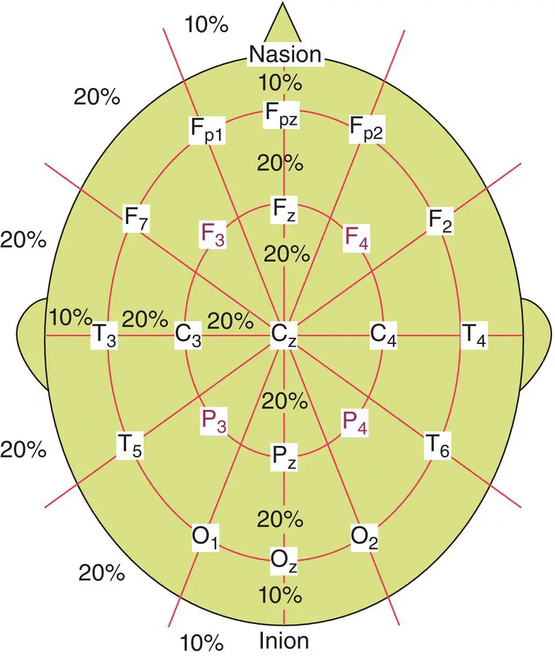
Figure 2.3 Conventional 10–20 EEG electrode positions for the placement of 21 electrodes.
Two different modes of recordings namely differential and referential, are used. In the differential mode the two inputs to each differential amplifier are from two electrodes. In referential mode, conversely, one or two reference electrodes are used. Several different reference electrode placements can be found in the literature. Physical references can be used as vertex (C z), linked ears, linked mastoids, ipsilateral ear, contralateral ear, C 7, bipolar references, and tip of the nose [15]. There are also reference‐free recording techniques which actually use a common average reference. The choice of reference may produce topographic distortion if the reference is not relatively neutral. In modern instrumentation, however, the choice of a reference does not play an important role in the measurement [16]. In such systems other references such as FP z, hand, or leg electrodes may be used [17]. The overall setting includes the active electrodes and the references.
In another similar setting called the Maudsley electrode positioning system the conventional 10–20 system has been modified to better capture the signals from epileptic foci in epileptic seizure recordings. The only difference between this system and the 10–20 conventional system is that the outer electrodes are slightly lowered to enable better capturing of the required signals. The advantage of this system over the conventional one is that it provides a more extensive coverage of the lower part of the cerebral convexity, increasing the sensitivity for the recording from basal sub‐temporal structures [18]. Other deviations from the international 10–20 system as used by researchers are found in [19, 20].
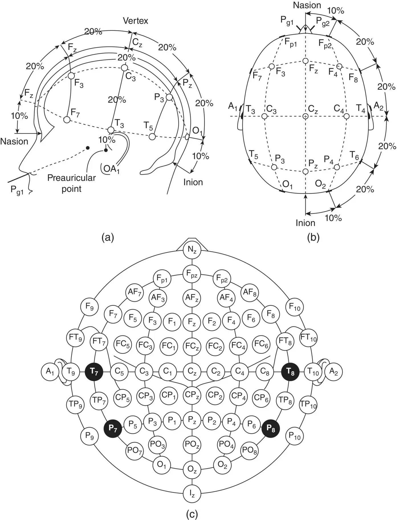
Figure 2.4 A diagrammatic representation of 10–20 electrode settings for 75 electrodes including the reference electrodes: (a and b) represent the three‐dimensional measures and (c) indicates a two‐dimensional view of the electrode setup configuration.
Читать дальшеИнтервал:
Закладка:
Похожие книги на «EEG Signal Processing and Machine Learning»
Представляем Вашему вниманию похожие книги на «EEG Signal Processing and Machine Learning» списком для выбора. Мы отобрали схожую по названию и смыслу литературу в надежде предоставить читателям больше вариантов отыскать новые, интересные, ещё непрочитанные произведения.


Обсуждение, отзывы о книге «EEG Signal Processing and Machine Learning» и просто собственные мнения читателей. Оставьте ваши комментарии, напишите, что Вы думаете о произведении, его смысле или главных героях. Укажите что конкретно понравилось, а что нет, и почему Вы так считаете.