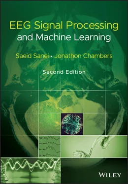
Saeid Sanei - EEG Signal Processing and Machine Learning
Здесь есть возможность читать онлайн «Saeid Sanei - EEG Signal Processing and Machine Learning» — ознакомительный отрывок электронной книги совершенно бесплатно, а после прочтения отрывка купить полную версию. В некоторых случаях можно слушать аудио, скачать через торрент в формате fb2 и присутствует краткое содержание. Жанр: unrecognised, на английском языке. Описание произведения, (предисловие) а так же отзывы посетителей доступны на портале библиотеки ЛибКат.
- Название:EEG Signal Processing and Machine Learning
- Автор:
- Жанр:
- Год:неизвестен
- ISBN:нет данных
- Рейтинг книги:3 / 5. Голосов: 1
-
Избранное:Добавить в избранное
- Отзывы:
-
Ваша оценка:
- 60
- 1
- 2
- 3
- 4
- 5
EEG Signal Processing and Machine Learning: краткое содержание, описание и аннотация
Предлагаем к чтению аннотацию, описание, краткое содержание или предисловие (зависит от того, что написал сам автор книги «EEG Signal Processing and Machine Learning»). Если вы не нашли необходимую информацию о книге — напишите в комментариях, мы постараемся отыскать её.
EEG Signal Processing and Machine Learning — читать онлайн ознакомительный отрывок
Ниже представлен текст книги, разбитый по страницам. Система сохранения места последней прочитанной страницы, позволяет с удобством читать онлайн бесплатно книгу «EEG Signal Processing and Machine Learning», без необходимости каждый раз заново искать на чём Вы остановились. Поставьте закладку, и сможете в любой момент перейти на страницу, на которой закончили чтение.
Интервал:
Закладка:
Despite the above epileptiform signals there are spikes and other paroxysmal discharges in healthy nonepileptic persons. These discharges may be found in healthy individuals without any other symptoms of diseases. However, they are often signs of certain cerebral dysfunctions that may or may not develop into an abnormality. They may appear during periods of particular mental challenge on individuals, such as soldiers in the war front line, pilots, and prisoners.
Generation of epileptiform brain discharges from deeper brain layers such as the hippocampus during pre‐ictal or interictal periods is an indication of upcoming seizure. These discharges which are spike‐type and have particular morphology which can be seen by inserting electrodes such as multichannel foramen ovale electrodes deep into the hippocampus. More than 90% of these discharges cannot be seen over the scalp due to their attenuation and smearing. A comprehensive overview of epileptic seizure disorders and nonepileptic attacks can be found in many books and publications such as [53, 56]. In this book a chapter is dedicated to the methods for analyzing intracranial and scalp EEGs.
2.9.3 Psychiatric Disorders
Not only can functional and certain anatomical brain abnormalities be investigated using EEG signals, pathophysiological brain disorders can also be studied by analyzing such signals. According to the ‘Diagnostic and Statistical Manual (DSM) of Mental Disorders’ of the American Psychiatric Association, changes in psychiatric education have evolved considerably since the 1970s. These changes have mainly resulted from physical and neurological laboratory studies based upon EEG signals [57].
There have been evidences from EEG coherence measures suggesting differential patterns of maturation between normal and learning disabled children [58]. This finding can lead to the establishment of some methodology in monitoring learning disorders.
Several psychiatric disorders are diagnosed by analysis of EPs achieved by simply averaging a number of consecutive trails having the same stimuli.
Some pervasive mental disorders such as: dyslexia which is a developmental reading disorder; autistic disorder which is related to abnormal social interaction, communication, and restricted interests and activities, and starts appearing from the age of three; Rett's disorder, characterized by the development of multiple deficits following a period of normal postnatal functioning; and Asperger's disorder which leads to severe and sustained impairments in social interaction and restricted repetitive patterns of behaviour, interests, and activities; cause significant losses in multiple functioning areas [57].
ADHD and attention‐deficit disorder (ADD), conduct disorder, oppositional defiant disorder, and disruptive behaviour disorder have also been under investigation and considered within the DSM. Most of these abnormalities appear during childhood and often prevent children from learning and socializing well. The associated EEG features have been rarely analytically investigated, but the EEG observations are often reported in the literature [59–63]. However, most of such abnormalities tend to disappear with advancing age.
EEG has also been analyzed recently for the study of delirium [64, 65], dementia [66, 67], and many other cognitive disorders [68]. In EEGs, characteristics of delirium include slowing or dropout of the posterior dominant rhythm, generalized theta or delta slow‐wave activity, poor organization of the background rhythm, and loss of reactivity of the EEG to eye opening and closing. In parallel with that, the quantitative EEG (QEEG) shows increased absolute and relative slow‐wave (theta and delta) power, reduced ratio of fast‐to‐slow band power, reduced mean frequency, and reduced occipital peak frequency [65].
Dementia includes a group of neurodegenerative diseases that cause acquired cognitive and behavioural impairment of sufficient severity to interfere significantly with social and occupational functioning. Alzheimer’s disease is the most common of the diseases that cause dementia. At present, the disorder afflicts approximately 5 million people in the United States and more than 30 million people worldwide. Larger numbers of individuals have lesser levels of cognitive impairment, which frequently evolves into full‐blown dementia. Prevalence of dementia is expected to nearly triple by 2050, since the disorder preferentially affects the elderly, who constitute the fastest‐growing age bracket in many countries, especially in industrialized nations [67].
Among other psychiatric and mental disorders, amnestic disorder (or amnesia), mental disorder due to general medical condition, substance‐related disorder, schizophrenia, mood disorder, anxiety disorder, somatoform disorder, dissociative disorder, sexual and gender identity disorder, eating disorders, sleep disorders, impulse‐controlled disorder, and personality disorders have often been addressed in the literature [57]. However, the corresponding EEGs have been seldom analyzed by means of advanced signal processing tools.
2.9.4 External Effects
EEG signal patterns may significantly change when using drugs for the treatment and suppression of various mental and CNS abnormalities. Variation in the EEG patterns may also rise by just looking at the TV screen or listening to music without any attention. However, among the external effects the most significant ones are the pharmacological and drug effects. Therefore, it is important to know the effects of these drugs on the changes of EEG waveforms due to chronic overdosage, and the patterns of overt intoxication [69].
The effect of administration of drugs for anaesthesia on EEGs is of interest to clinicians. The related studies attempt to find the correlation between the EEG changes and the stages of anaesthesia. It has been shown that in the initial stage of anaesthesia a fast frontal activity appears. In deep anaesthesia this activity become slower with higher amplitude. In the last stage, a burst‐suppression pattern indicates the involvement of brainstem functions, including respiration and finally the EEG activity ceases [69]. In the cases of acute intoxication, the EEG patterns are similar to those of anaesthesia [69].
Barbiturate is commonly used as an anticonvulsant and antiepileptic drug. With small dosage of barbiturate the activities within the 25–35 Hz frequency band around the frontal cortex increases. This changes to 15–25 Hz and spreads to the parietal and occipital regions. Dependence and addiction to barbiturates are common. Therefore, after a long‐term ingestion of barbiturates, its abrupt withdrawal leads to paroxysmal abnormalities. The major complications are myoclonic jerks, generalized tonic–clonic seizures, and delirium [69].
Many other drugs are used in addition to barbiturates as sleeping pills such as melatonin, and bromides. Very pronounced EEG slowing is found in chronic bromide encephalopathies [69]. Antipsychotic drugs also influence the EEG patterns. For example, neuroleptics increase the alpha wave activity but reduce the duration of beta wave bursts and their average frequency. As another example, clozapine increases the delta, theta, and above 21 Hz beta wave activities. As another antipsychotic drug, tricyclic antidepressants such as imipramine, amitriptyline, doxepin, desipramine, nortriptyline, and protriptyline increase the amount of slow and fast activity along with instability of frequency and voltage, and also slow down the alpha wave rhythm. After administration of tricyclic antidepressants the seizure frequency in chronic epileptic patients may increase. With high dosage, this may further lead to single or multiple seizures occurring in nonepileptic patients [69].
During acute intoxication, a widespread, poorly reactive, irregular 8–10 Hz activity and paroxysmal abnormalities including spikes, as well as unspecific coma patterns, are observed in the EEGs [69]. Lithium is often used in the prophylactic treatment of bipolar mood disorder. The related changes in the EEG pattern consist of slowing of the beta rhythm and of paroxysmal generalized slowing, occasionally accompanied by spikes. Focal slowing also occurs, which is not necessarily a sign of a focal brain lesion. Therefore, the changes in the EEG are markedly abnormal with lithium administration [69]. The beta wave activity is highly activated by using benzodiazepines, as an anxiolytic drug. These activities persist in the EEG as long as two weeks after ingestion. Benzodiazepine leads to a decrease in an alpha wave activity and its amplitude, and slightly increases the 4–7 Hz frequency band activity. In acute intoxication the EEG shows prominent fast activity with no response to stimuli [69]. The psychotogenic drugs such as lysergic acid diethylamide and mescaline decrease the amplitude and possibly depress the slow waves [69].
Читать дальшеИнтервал:
Закладка:
Похожие книги на «EEG Signal Processing and Machine Learning»
Представляем Вашему вниманию похожие книги на «EEG Signal Processing and Machine Learning» списком для выбора. Мы отобрали схожую по названию и смыслу литературу в надежде предоставить читателям больше вариантов отыскать новые, интересные, ещё непрочитанные произведения.
Обсуждение, отзывы о книге «EEG Signal Processing and Machine Learning» и просто собственные мнения читателей. Оставьте ваши комментарии, напишите, что Вы думаете о произведении, его смысле или главных героях. Укажите что конкретно понравилось, а что нет, и почему Вы так считаете.
