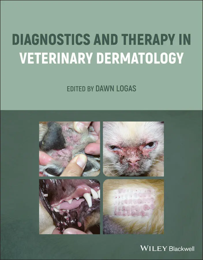
Diagnostics and Therapy in Veterinary Dermatology
Здесь есть возможность читать онлайн «Diagnostics and Therapy in Veterinary Dermatology» — ознакомительный отрывок электронной книги совершенно бесплатно, а после прочтения отрывка купить полную версию. В некоторых случаях можно слушать аудио, скачать через торрент в формате fb2 и присутствует краткое содержание. Жанр: unrecognised, на английском языке. Описание произведения, (предисловие) а так же отзывы посетителей доступны на портале библиотеки ЛибКат.
- Название:Diagnostics and Therapy in Veterinary Dermatology
- Автор:
- Жанр:
- Год:неизвестен
- ISBN:нет данных
- Рейтинг книги:3 / 5. Голосов: 1
-
Избранное:Добавить в избранное
- Отзывы:
-
Ваша оценка:
Diagnostics and Therapy in Veterinary Dermatology: краткое содержание, описание и аннотация
Предлагаем к чтению аннотацию, описание, краткое содержание или предисловие (зависит от того, что написал сам автор книги «Diagnostics and Therapy in Veterinary Dermatology»). Если вы не нашли необходимую информацию о книге — напишите в комментариях, мы постараемся отыскать её.
Focuses on cats and dogs Includes numerous high-quality clinical photographs illustrating all key concepts Covers topics such as how to use your nursing staff to the fullest, the One Health movement, and how changing climate is increasing the spread of certain dermatologic diseases Discusses approaches for building a better working relationship between clients, primary care veterinarians and dermatologists Provides insights on the future of technology in the diagnosis and treatment of dermatologic diseases Covering the very latest developments in the field,
is essential reading for veterinary dermatologists, veterinary students, and any veterinary general practitioner with a dermatology caseload.
Diagnostics and Therapy in Veterinary Dermatology — читать онлайн ознакомительный отрывок
Ниже представлен текст книги, разбитый по страницам. Система сохранения места последней прочитанной страницы, позволяет с удобством читать онлайн бесплатно книгу «Diagnostics and Therapy in Veterinary Dermatology», без необходимости каждый раз заново искать на чём Вы остановились. Поставьте закладку, и сможете в любой момент перейти на страницу, на которой закончили чтение.
Интервал:
Закладка:
10 Chapter 10Figure 10.1 Alopecia secondary to flea allergy: note that the underlying ski...Figure 10.2 Pruritus visual analog scale for a cat.Figure 10.3 Cat with excoriations, alopecia and erosions in the inguinal are...Figure 10.4 Miliary dermatitis in a cat secondary to feline atopic skin synd...Figure 10.5 Indolent ulcer secondary to flea allergy: note the large, erosiv...Figure 10.6 Multiple eosinophilic plaques on the ventrum of a cat secondary ...Figure 10.7 Diagnosis flow chart for feline hypersensitivity.Figure 10.8 Mosquito hypersensitivity: note the multiple small erosions on t...Figure 10.9 Intradermal skin test: note the positive wheal‐and‐flare reactio...Figure 10.10 View of feline intradermal allergy test with Wood’s lamp after ...
11 Chapter 11Figure 11.1 Cytology of pemphigus pustule. White arrows indicate acantholyti...Figure 11.2 Clinical features of canine pemphigus foliaceus. Note the pustul...Figure 11.3 Severe hyperkeratosis secondary to pemphigus foliaceus.Figure 11.4 Subcorneal pustules of pemphigus foliaceus.Figure 11.5 Clinical features of feline pemphigus foliaceus. Note the small ...Figure 11.6 Large pemphigus foliaceus collarettes with crusting and pustules...Figure 11.7 Sebaceous adenitis. Change in coat color and character.Figure 11.8 Sebaceous adenitis.Figure 11.9 Idiopathic sterile nodular panniculitis. (a and c) Nodular lesio...Figure 11.10 Idiopathic sterile nodular panniculitis. Healed lesion with atr...Figure 11.11 Symmetric lupoid onychodystrophy.Figure 11.12 Proliferative thrombovascular necrosis of the pinnae. (a) Scall...Figure 11.13 Proliferative thrombovascular necrosis (PTN) of the pinnae. (a)...Figure 11.14 Generalized vasculitis lesions. Note the paws, which are swolle...Figure 11.15 Vasculitis lesions. (a) Black circles indicate areas of erythem...Figure 11.16 Clinical variations of discoid lupus erythematosus. Note the de...Figure 11.17 Mucocutaneous lupus erythematosus. Note nose erythema and ulcer...Figure 11.18 Erythema multiforme. The edges of the erosions and the collaret...Figure 11.19 German shepherd pyoderma. Note the multiple crust and erosive l...Figure 11.20 Vesicular cutaneous lupus erythematosus. Note the multiple ulce...Figure 11.21 Exfoliative cutaneous lupus erythematosus. Note the fine scale ...Figure 11.22 Active ischemic dermatopathy lesions. Note the alopecia and cru...Figure 11.23 Healed ischemic dermatopathy lesions. Note the scarring with hy...
12 Chapter 12Figure 12.1 Hypothyroidism. Note the thin, brittle hair coat.Figure 12.2 Hypothyroidism with secondary Malassezia and bacterial dermatiti...Figure 12.3 Severe otitis externa due to hypothyroidism in a dog: proliferat...Figure 12.4 Acral lick granuloma secondary to hypothyroidism in a 4‐year‐old...Figure 12.5 Dog with demodicosis secondary to hypothyroidism. Note the alope...Figure 12.6 Hyperadrenocorticism in a dog. Note the pot‐bellied appearance a...Figure 12.7 Hyperadrenocorticism in a dog. Note the generalized alopecia and...Figure 12.8 Calcinosis cutis in a dog. The lesion is made up of multiple fir...Figure 12.10 Dog with Cushing’s disease, calcinosis cutis, and deep pyoderma...Figure 12.11 Thin skin in a cat with hypercortisolism.Figure 12.12 Hepatocutaneous syndrome. Note the diffuse crusting and fissuri...Figure 12.13 Hepatocutaneous syndrome. Diffuse, thick, adherent crusts with ...Figure 12.14 Hepatocutaneous syndrome: erythema and erosion of the genital r...Figure 12.15 Hepatocutaneous syndrome: erosion of the prepuce.Figure 12.16 Zinc‐responsive dermatosis in a husky: crusting and alopecia.Figure 12.18 Zinc‐responsive dermatosis. Closeup view of the gray/white, dry...Figure 12.19 Alopecia X. Note truncal alopecia that spares the head.Figure 12.20 Alopecia X. Note the loss of secondary hairs.
13 Chapter 13Figure 13.1 Examples of types of otitis. Acute otitis: the pinna and vertica...Figure 13.2 Examples of chronic otitis cases that would benefit from the ant...Figure 13.3 Normal tympanic membrane. The malleus is the small C‐shaped bone...Figure 13.4 Examples of otitis media. Ruptured tympanic membrane ) has a gre...Figure 13.5 Where to perform a myringotomy. The blue circle indicates the ca...Figure 13.6 Removal of mucus from the middle ear. The first picture shows a ...Figure 13.7 View of the bulla through a video‐otoscope. In the mildly inflam...
14 Chapter 15Figure 15.1 Keratolytic agents. These agents promote the decrease of adhesio...Figure 15.2 Keratoplastic agents. These agents attempt to normalize keratini...Figure 15.3 Localized Cushing’s disease secondary to topical steroids. This ...Figure 15.4 Clinical conditions that can benefit from antiseborrheic agents....Figure 15.5 Emollients/occlusives. Emollients are oily substances that fill ...Figure 15.6 Humectants. Humectants are hygroscopic substances that attract w...
15 Chapter 16Figure 16.1 Antibiotics’ mechanisms of action (green arrows) and bacterial m...
16 Chapter 17Figure 17.1 Omega‐3 and omega‐6 polyunsaturated fatty acid desaturation and ...Figure 17.2 Alpha‐linolenic acid metabolism. Acetyl Co.‐A, acetyl coenzyme A...Figure 17.3 Representation of the cell membrane phospholipid bilayer. (a) In...
17 Chapter 18Figure 18.1 Genomic effect of glucocorticoids (GC). GC passively diffuse thr...Figure 18.2 Calcinosis cutis plaque on a dog’s abdomen secondary to oral pre...Figure 18.3 Localized iatrogenic hyperadrenocorticism secondary to topical b...Figure 18.4 Multiple full‐thickness skin tears on a cat’s dorsal neck and ba...Figure 18.5 Mechanism of action of lokivetmab and oclacitinib. (a) Oclacitin...Figure 18.6 Cyclosporine’s mechanism of action. Cyclosporine (CsA) binds wit...Figure 18.7 Severe gingival hyperplasia in the mouth of a dog secondary to l...Figure 18.8 Papillomas in the mouth of a dog secondary to long‐term cyclospo...
18 Chapter 20Figure 20.1 Formation of monoclonal antibodies: red, mouse antibody; green, ...Figure 20.2 DNA vaccination. Antigens (proteins) are linked to the virus‐lik...Figure 20.3 Explanation of the Janus kinase/signal transducer and activator ...
19 Chapter 21Figure 21.1 Some of the tips commonly used with the CO 2laser. From top to b...Figure 21.2 Meibomian gland adenoma on the lower eyelid of a dog. Left image...Figure 21.3 Long rigid tip is advanced through the working channel of the Me...Figure 21.4 Pre laser: ceruminous gland cystomatosis occluding the external ...Figure 21.5 Ceruminous gland cystomatosis in the vertical external ear canal...Figure 21.6 Ceruminous gland adenoma that is completely occluding the ear ca...Figure 21.7 Footpad papillomas that were causing significant discomfort. The...Figure 21.8 Oral papillomatosis in a young dog. Unfortunately, the papilloma...Figure 21.9 Interdigital cysts on the palmar aspect of this dog’s paw with a...Figure 21.10 Paintbrush tip for CO 2with the end visible.Figure 21.11 Cross‐section of the excised interdigital cystic tissue. Note t...
20 Chapter 24Figure 24.1 Dermatologic patient history form.Figure 24.2 Pruritus scale.Figure 24.3 Severe cocci on ear cytology (100×).Figure 24.4 Severe rods and neutrophils on ear cytology (100×).Figure 24.5 Demodex mite counting sheet.Figure 24.6 Demodex canis (blue arrows) and a few short fat canine demodex (...Figure 24.7 Microsporum canis colonies on a dermatophyte test medium plate. ...Figure 24.8 Microsporum gypseum macroconidia. Note that the macroconidia hav...Figure 24.9 Direct impression cytology from a crusted area.Figure 24.10 Opening a pustule to apply a slide directly for cytology. Note ...Figure 24.11 Purulent material collected on the needle being spread on a sli...Figure 24.12 Neutrophils and cocci on acetate tape preparation (100×).Figure 24.13 Eosinophils on acetate tape preparation (100×).
21 Chapter 25Figure 25.1 Calgary‐Cambridge guide framework for communication.Figure 25.2 When clients reach the tipping point of frustration, there are n...Figure 25.3 Barriers to referral, as indicated by primary care veterinarians...Figure 25.4 Survival time of dogs treated for congestive heart failure (CHF)...Figure 25.5 Relationships between primary care veterinarians (pcDVMs) and sp...Figure 25.6 Top reasons pet owners accept a referral recommendation.
Читать дальшеИнтервал:
Закладка:
Похожие книги на «Diagnostics and Therapy in Veterinary Dermatology»
Представляем Вашему вниманию похожие книги на «Diagnostics and Therapy in Veterinary Dermatology» списком для выбора. Мы отобрали схожую по названию и смыслу литературу в надежде предоставить читателям больше вариантов отыскать новые, интересные, ещё непрочитанные произведения.


Обсуждение, отзывы о книге «Diagnostics and Therapy in Veterinary Dermatology» и просто собственные мнения читателей. Оставьте ваши комментарии, напишите, что Вы думаете о произведении, его смысле или главных героях. Укажите что конкретно понравилось, а что нет, и почему Вы так считаете.