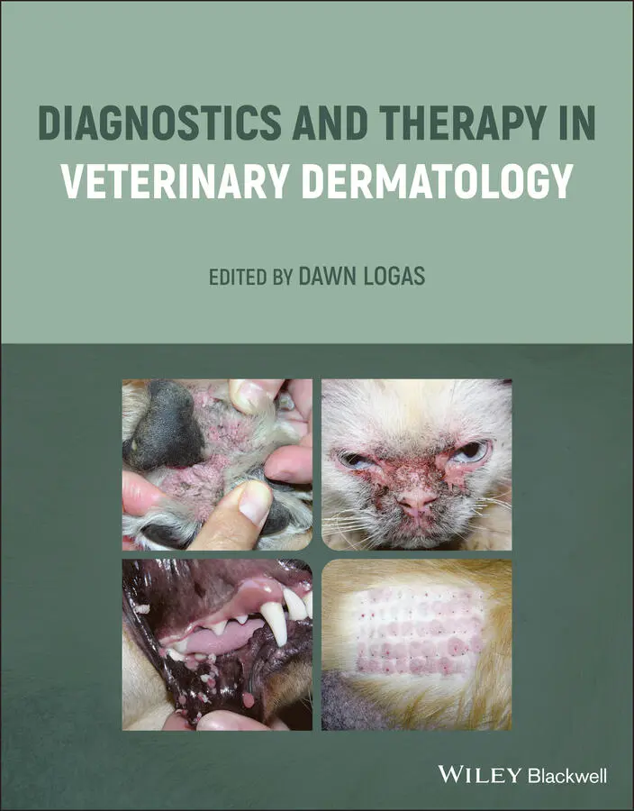
Diagnostics and Therapy in Veterinary Dermatology
Здесь есть возможность читать онлайн «Diagnostics and Therapy in Veterinary Dermatology» — ознакомительный отрывок электронной книги совершенно бесплатно, а после прочтения отрывка купить полную версию. В некоторых случаях можно слушать аудио, скачать через торрент в формате fb2 и присутствует краткое содержание. Жанр: unrecognised, на английском языке. Описание произведения, (предисловие) а так же отзывы посетителей доступны на портале библиотеки ЛибКат.
- Название:Diagnostics and Therapy in Veterinary Dermatology
- Автор:
- Жанр:
- Год:неизвестен
- ISBN:нет данных
- Рейтинг книги:3 / 5. Голосов: 1
-
Избранное:Добавить в избранное
- Отзывы:
-
Ваша оценка:
Diagnostics and Therapy in Veterinary Dermatology: краткое содержание, описание и аннотация
Предлагаем к чтению аннотацию, описание, краткое содержание или предисловие (зависит от того, что написал сам автор книги «Diagnostics and Therapy in Veterinary Dermatology»). Если вы не нашли необходимую информацию о книге — напишите в комментариях, мы постараемся отыскать её.
Focuses on cats and dogs Includes numerous high-quality clinical photographs illustrating all key concepts Covers topics such as how to use your nursing staff to the fullest, the One Health movement, and how changing climate is increasing the spread of certain dermatologic diseases Discusses approaches for building a better working relationship between clients, primary care veterinarians and dermatologists Provides insights on the future of technology in the diagnosis and treatment of dermatologic diseases Covering the very latest developments in the field,
is essential reading for veterinary dermatologists, veterinary students, and any veterinary general practitioner with a dermatology caseload.
Diagnostics and Therapy in Veterinary Dermatology — читать онлайн ознакомительный отрывок
Ниже представлен текст книги, разбитый по страницам. Система сохранения места последней прочитанной страницы, позволяет с удобством читать онлайн бесплатно книгу «Diagnostics and Therapy in Veterinary Dermatology», без необходимости каждый раз заново искать на чём Вы остановились. Поставьте закладку, и сможете в любой момент перейти на страницу, на которой закончили чтение.
Интервал:
Закладка:
7 Chapter 10Table 10.1 Clinical reaction patterns in hypersensitive cats.Table 10.2 Recommended antibiotic doses for cats.Table 10.3 Diagnostic criteria for nonflea feline hypersensitivity.Table 10.4 Recommended antihistamine doses for cats.
8 Chapter 11Table 11.1 Dosing of immunosuppressive medications for autoimmune diseases.Table 11.2 Sample glucocorticoid tapering schedule.Table 11.3 Diagnostic ruleouts for sterile pyogranulomatous panniculitis.
9 Chapter 12Table 12.1 Medications that affect thyroid diagnostic testing.Table 12.2 Interpretations of thyroid diagnostic testing results.
10 Chapter 13Table 13.1 Definition of acute and chronic otitis.Table 13.2 Underlying causes of otitis.
11 Chapter 16Table 16.1 Contributors to antimicrobial resistance.Table 16.2 Enzymatic Comprehensive list of historical and contemporary anti...Table 16.3 Potential impact of antimicrobial drug resistance.Table 16.4 Practices to decrease antibiotic resistance now.
12 Chapter 18Table 18.1 Commonly used oral glucocorticoids in the dog.Table 18.2 Commonly used oral glucocorticoids in the cat.
13 Chapter 19Table 19.1 Drug withdrawal times prior intradermal testing.
14 Chapter 22Table 22.1 Indications for photobiomodulation.
15 Chapter 23Table 23.1 Drugs that can be used for sedating dermatologic patients.Table 23.2 Suitability of sedative drugs for skin testing in small animals.Table 23.3 Induction agents for dermatologic patients.Table 23.4 Recommended maximum dose for the commonly used local anesthetics...Table 23.5 Nonsteroidal anti‐inflammatory drugs (NSAIDs) approved for dogs ...
List of Illustrations
1 Chapter 1 Figure 1.1 Innate skin immune system. 1. The innate immune system is activat... Figure 1.2 Adaptive skin immune system. 1. Langerhans cells (LCs) are activa...
2 Chapter 2 Figure 2.1 Example of a dermatology history form for a client. Figure 2.2 A visual analog scale for owners to report their pet’s level of p... Figure 2.3 Depigmentation, erosion, erythema, and loss of normal cobblestone... Figure 2.4 Hyperkeratosis, crusting, and erythema of the paw pads of a dog.... Figure 2.5 Epidermal collarettes, pustules, erythema, and hyperpigmentation ... Figure 2.6 Alopecia, crusting, and hyperpigmentation along the pinnal margin... Figure 2.7 Ulcerations, draining tracts, and scarring in the inguinal region... Figure 2.8 Perianal erythema in a dog with food allergy. Figure 2.9 Dog with chronic solar dermatitis. Multifocal areas of erythema a...
3 Chapter 3 Figure 3.1 Direct ELISA. In the first stage the antigen (orange triangle) fr... Figure 3.2 Indirect ELISA. The antigen (orange triangle) is adhered to the p... Figure 3.3 Sandwich ELISA. Monoclonal antibody (yellow) for the antigen that...
4 Chapter 4 Figure 4.1 Noninfectious diseases that should be biopsied. Figure 4.2 Nodular lesions that should be biopsied and cultured. Figure 4.3 Primary lesions. Figure 4.4 Secondary lesions. Figure 4.5 Appropriate sampling. (a) Inappropriately taken biopsy punch with... Figure 4.6 Punch biopsy. (a) Instrumentation for punch biopsy. (b) Inject li... Figure 4.7 Double punch. (a) Rapidly growing mycobacteriosis. (b) Lateral vi...
5 Chapter 5 Figure 5.1 Inguinal area of a dog with superficial pyoderma. Note the papule... Figure 5.4 Multiple alopecic annular lesions with mild hyperpigmentation in ... Figure 5.5 Paw of a dog with two large interdigital furuncles (deep pyoderma... Figure 5.6 Deep pyoderma in an acral lick granuloma on the fore limb of a do... Figure 5.7 (a) Large intact pustules and epidermal collarettes on the ventra... Figure 5.8 Collection of a culture sample from beneath a crust in a dog with... Figure 5.9 Collection of a sterile punch biopsy for tissue culture following...
6 Chapter 6Figure 6.1 (a) Example of an epidermal collarette from a dog with bacterial ...Figure 6.2 Generalized dermatophytosis – multifocal to diffuse erythema, alo...Figure 6.3 Young dog with a focal well‐circumscribed patch of erythema, alop...Figure 6.4 Dog with alopecic scaling lesion on the face ( Trichophyton mentag ...Figure 6.5 Dermatophytosis and onychomycosis caused by Microsporum gypseum ....Figure 6.6 Kitten with generalized Microsporum canis ; note erythema, alopeci...Figure 6.7 Focal area of alopecia and crusting just above the nasal planum i...Figure 6.8 Dog with a kerion ( Microsporum gypseum ) on the dorsal muzzle – a ...Figure 6.9 Apple‐green fluorescence of Microsporum canis under Wood’s lamp....Figure 6.10 Ectothrix arthrospores (blue arrow) surround the outside of the ...Figure 6.11 (a) Dermatophyte test medium () plate with red color change asso...Figure 6.12 (a) Microsporum canis macroconidia – spindle shaped with thick, ...Figure 6.13 Dog with Malassezia pododermatitis: lichenification, hyperpigmen...Figure 6.14 Mixed‐breed dog with Malassezia dermatitis: severe lichenificati...Figure 6.15 13‐year‐old cat with paraneoplastic alopecia. Both hind paws and...Figure 6.16 German shepherd with pythiosis: multiple nodules and draining tr...Figure 6.17 Large granulomatous lesion on the tail along with a smaller lesi...Figure 6.18 Large granulomatous lesion on the paw of a dog with lagenidiosis...Figure 6.19 Diff‐Quik–stained touch prep from a dog with a draining tract on...Figure 6.20 10‐year‐old feline influenza virus–positive cat with subcutaneou...Figure 6.21 Outdoor cat with firm swelling and multiple draining tracts on a...Figure 6.22 Dog with a large, firm mass on the side of the muzzle, draining ...
7 Chapter 7Figure 7.1 Sarcoptes scabiei . Round to oval mite with four short legs, termi...Figure 7.2 Chronic case of scabies. Entire pinnae affected. The pinnae are t...Figure 7.3 Dog with scabies. Generalized papular eruption with excoriations....Figure 7.4 Dorsum of dog in Figure 7.3: more profound excoriations with papu...Figure 7.5 Scabies scabiei on skin scraping.Figure 7.6 Notoedres cati . A round mite with four short forelimb legs with m...Figure 7.7 Cat with Notoedres cati . Severe crusting and excoriation of the h...Figure 7.8 Canine demodex. Current consensus is that Demodex cornei is not a...Figure 7.9 Varies clinical presentations of canine demodicosis.Figure 7.10 Papules, pustules, and comedones associated with demodicosis.Figure 7.11 Erythema with severe follicular plugging.Figure 7.12 Dog with furunculosis secondary to demodicosis.Figure 7.13 Hair pluck with demodex.Figure 7.14 Feline leukemia‐positive cat with Demodex cati . Severe crusting ...Figure 7.15 Diabetic cat with Demodex cati . Generalized alopecia, fine crust...Figure 7.16 Demodex gatoi infections. Notice the alopecia on the ventrum and...Figure 7.17 Felicola subrostratus .Figure 7.18 Cat with nits. White arrows designate nits.
8 Chapter 8Figure 8.1 Cutaneous manifestations of leishmaniasis in dogs. (a). Facial ex...Figure 8.2 (a) Perioral ulcerative lesions in a cat with sporotrichosis. (b)...
9 Chapter 9Figure 9.1 Chronic flea‐allergic dog. Pruritus has focused on the sides and ...Figure 9.2 Acute‐onset contact allergy in the inguinal area. Note the erythe...Figure 9.3 Hives secondary to food allergy. Note the multiple hives coalesci...Figure 9.4 Fore paw of an atopic dog. Note the mild erythema and lichenifica...Figure 9.5 Typical atopic dermatitis presentation. Note the erythema periocu...Figure 9.6 Chronic food‐allergic dog. Note the severe papular eruption on th...Figure 9.7 Acute food‐allergic dog. Note the hair loss from the chest back w...Figure 9.8 Milder case of contact allergy with secondary pyoderma. Note the ...Figure 9.9 Papules on the pinnae of a patient allergic to plants.Figure 9.10 Contact allergy in the patient in Figure 9.9. Note the erythema ...Figure 9.11 Example of Commelina plant growing on a sidewalk. This is a grou...Figure 9.12 Example of a positive patch test. Note the erythema and papules ...
Читать дальшеИнтервал:
Закладка:
Похожие книги на «Diagnostics and Therapy in Veterinary Dermatology»
Представляем Вашему вниманию похожие книги на «Diagnostics and Therapy in Veterinary Dermatology» списком для выбора. Мы отобрали схожую по названию и смыслу литературу в надежде предоставить читателям больше вариантов отыскать новые, интересные, ещё непрочитанные произведения.


Обсуждение, отзывы о книге «Diagnostics and Therapy in Veterinary Dermatology» и просто собственные мнения читателей. Оставьте ваши комментарии, напишите, что Вы думаете о произведении, его смысле или главных героях. Укажите что конкретно понравилось, а что нет, и почему Вы так считаете.