Diagnostics and Therapy in Veterinary Dermatology
Здесь есть возможность читать онлайн «Diagnostics and Therapy in Veterinary Dermatology» — ознакомительный отрывок электронной книги совершенно бесплатно, а после прочтения отрывка купить полную версию. В некоторых случаях можно слушать аудио, скачать через торрент в формате fb2 и присутствует краткое содержание. Жанр: unrecognised, на английском языке. Описание произведения, (предисловие) а так же отзывы посетителей доступны на портале библиотеки ЛибКат.
- Название:Diagnostics and Therapy in Veterinary Dermatology
- Автор:
- Жанр:
- Год:неизвестен
- ISBN:нет данных
- Рейтинг книги:3 / 5. Голосов: 1
-
Избранное:Добавить в избранное
- Отзывы:
-
Ваша оценка:
Diagnostics and Therapy in Veterinary Dermatology: краткое содержание, описание и аннотация
Предлагаем к чтению аннотацию, описание, краткое содержание или предисловие (зависит от того, что написал сам автор книги «Diagnostics and Therapy in Veterinary Dermatology»). Если вы не нашли необходимую информацию о книге — напишите в комментариях, мы постараемся отыскать её.
Focuses on cats and dogs Includes numerous high-quality clinical photographs illustrating all key concepts Covers topics such as how to use your nursing staff to the fullest, the One Health movement, and how changing climate is increasing the spread of certain dermatologic diseases Discusses approaches for building a better working relationship between clients, primary care veterinarians and dermatologists Provides insights on the future of technology in the diagnosis and treatment of dermatologic diseases Covering the very latest developments in the field,
is essential reading for veterinary dermatologists, veterinary students, and any veterinary general practitioner with a dermatology caseload.
Diagnostics and Therapy in Veterinary Dermatology — читать онлайн ознакомительный отрывок
Ниже представлен текст книги, разбитый по страницам. Система сохранения места последней прочитанной страницы, позволяет с удобством читать онлайн бесплатно книгу «Diagnostics and Therapy in Veterinary Dermatology», без необходимости каждый раз заново искать на чём Вы остановились. Поставьте закладку, и сможете в любой момент перейти на страницу, на которой закончили чтение.
Интервал:
Закладка:
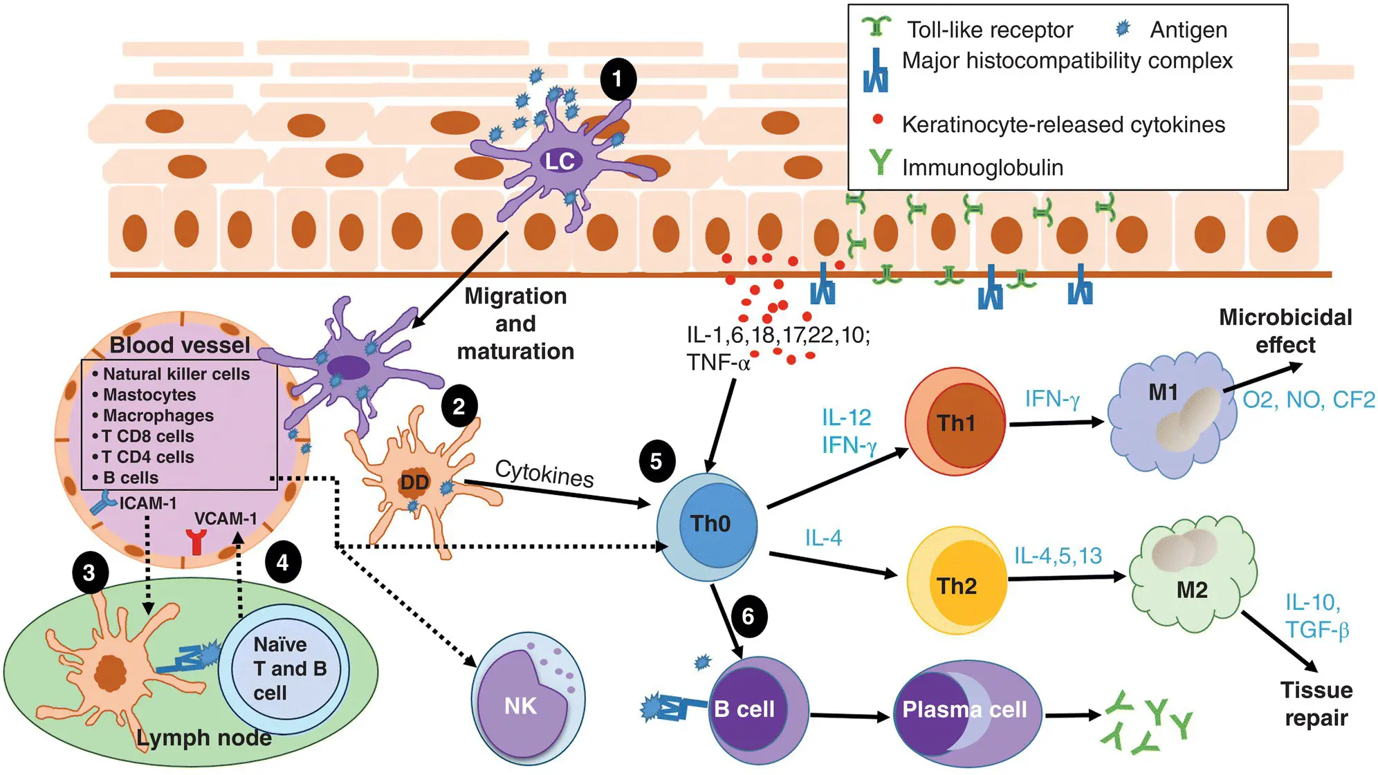
Figure 1.2 Adaptive skin immune system. 1. Langerhans cells (LCs) are activated once they uptake and process the antigen. 2. Some will mature and migrate along with some activated dermal dendritic (DD) cells to the local lymph node. 3. The antigen in association with major histocompatibility complex (MHC) I or II will prime naïve T and B cells. 4. The primed T‐helper (CD4+) and cytotoxic cells (CD8+) enter the blood system and return to the area of inflammation along with other inflammatory cells via adhesion molecules like intercellular adhesion molecule (ICAM)‐1 and vascular cell adhesion molecule (VCAM)‐1. 5. The primed T lymphocytes mature into various types of T‐helper (Th) cells, depending on which cytokines are produced by keratinocytes and dendritic cells. These mature T‐helper cells are responsible for amplifying the inflammatory response. 6. Primed T‐helper cells are also able to prime B cells in the skin that are displaying the same antigen in association with MHC on the B cell’s surface. The B cell will then mature into plasma cell and produce immunoglobulins against that antigen. CF, chorion factor; IFN, interferon; IL, interleukin; M, macrophage; NK, natural killer cell; NO, nitric oxide; TGF, transforming growth factor; TNF, tumor necrosis factor.
Melanocytes and Langerhans cells are the other major immunologically active cell types in the epidermis. Melanocytes’ major function is the production of melanin to protect keratinocytes from the harmful effects of ultraviolet radiation (UVR). However, they also play an important regulatory role in both the innate and adaptive immune systems by promoting phagocytosis and producing a series of pro‐inflammatory cytokines. Like keratinocytes, they can also express various pattern‐recognition receptors such as Toll‐like receptors.
Langerhans cells and dermal dendritic cells are the most important antigen‐presenting cells in the skin, as they are the only cells able to activate naïve T lymphocytes. Depending on the microenvironment and the immunologic triggers present, Langerhans cells can mediate a tolerogenic response through the production of interleukin 10 (IL‐10), or they can promote an inflammatory response. Once in contact with an invader, Langerhans cells phagocytose the antigen, process it, and represent it on their surface in association with MHC‐II. This causes the maturation of the Langerhans cells, which then migrate to the regional lymph node to prime naïve T lymphocytes.
The epidermis also hosts resident memory T lymphocytes, part of the adaptive immune system. These cutaneous memory T lymphocytes may possess either α/β or γ/δ T‐cell receptors. Those with γ/δ receptors are considered a hybrid between an innate and an adaptive immune cell. They bind to common PAMP molecules or respond to the class Ib MHC molecules produced by stressed, cancer‐, or virus‐infected cells.
Dermis
The dermis is mainly composed of extracellular matrix proteins that provide structure and elasticity to the skin. It is separated from the epidermis by the basal membrane, a thin layer of extracellular matrix proteins. This membrane regulates the movement of cells and substances from the dermis to the epidermis and anchors the basal epidermal keratinocytes to the dermis via hemidesmosomes. The dermis contains hair follicles, glandular structures, specialized neural receptors, as well as blood and lymphatic vessels essential for the support and maintenance of dermal and epidermal cells.
Fibroblasts are the most abundant immunologically active cells in the dermis. Their primary function is production of the extracellular matrix that provides structural support for the dermis and a scaffolding that allows cells of the immune system to move easily through the dermis. Fibroblasts secrete a variety of cytokines, including chemoattractants for T lymphocytes, and direct the inflammatory response that is part of wound healing. Fibroblasts produce matrix metalloproteinases, a group of enzymes responsible for the degradation of the extracellular matrix, but they also produce tissue inhibitors of matrix metalloproteinases. Therefore, fibroblasts facilitate both activation and termination of inflammation.
Endothelial cells and neurons are also essential for the dermal immune response. Endothelial cells regulate the influx of inflammatory cells from the blood into the dermis through the activation of adhesion molecules and cytokines. During the initial phase of inflammation, endothelial cells activate multiple Toll‐like receptors and various adhesion molecules, including intercellular adhesion molecule‐1 (ICAM‐1), vascular cell adhesion molecule‐1 (VCAM‐1), and selectins E and P. As the inflammatory response progresses, endothelial cells secrete more pro‐inflammatory cytokines such as IL‐1, tumor necrosis factor alpha (TNF‐α), and interferon gamma (IFN‐γ), which in turn increase the expression of adhesion molecules to further facilitate leukocyte migration and extravasation.
Nerves are important for sensation and they also have an intimate connection with memory T cells. Neurons modulate signals between the innate and adaptive branches of the immune system by producing substance‐P, calcitonin gene‐related peptide, and alpha melanocyte‐stimulating hormone. These substances support a healthy level of inflammation and help to resolve the inflammatory response when needed.
The dermis is home to other cells of the innate immune system (granulocytes, NK cells, dendritic cells, macrophages) and lymphocytes (adaptive immune system cells). Neutrophils and macrophages are part of the first line of defense against microorganisms. As soon as an invader is detected, these cells are called to the site of infection by potent chemoattractants. Once there, they express receptors that recognize, bind, capture, and phagocytose microorganisms. Neutrophils are also able to externally project their DNA and DNA‐associated proteins to form “sticky” neutrophil extracellular traps (NETs) that trap pathogens like flies on a spider’s web.
Macrophages are a bridge between the innate and adaptive arms of the immune system. In addition to phagocytosing microorganisms, macrophages remove debris, dead cells, and exogenous or endogenous inflammatory stimuli (e.g. free melanin or keratin). They also act as antigen‐presenting cells. Macrophages can recognize, bind, capture, and destroy microorganisms directly or via their recognition of antibody molecules, complement proteins, or lectins. Macrophages secrete various cytokines that direct the immune response. Different types of macrophages have recently been identified: M0, M1, M2, M4, M17, and Mreg. M1 macrophages secrete TNF‐α, IL‐6, and IL‐12, cytokines that promote a T‐helper type 1 pro‐inflammatory immune response. M2 macrophages promote an anti‐inflammatory immune response through the secretion of IL‐10 and IL‐4. M4 macrophages secrete TNF‐α, IL‐6, matrix metalloproteinases 7 and 12, and macrosialin, all of which reduce phagocytosis and induce a pro‐inflammatory immune response. Finally, M17 and Mreg cells have been proposed, but their characterization and involvement in the inflammatory process need further clarification.
Eosinophils are more abundant in the later stages of inflammation. They are important in the pathogenesis of allergic and parasitic diseases, both driven by T‐helper type 2 cytokines. Once eosinophils reach the site of inflammation, they release granular proteins highly toxic to parasites. Eosinophils are also essential in secreting cytokines characteristic of an allergic response. Finally, eosinophils release prostaglandins and leukotrienes that cause vasodilation and chemotaxis of neutrophils and prolong an allergic response. Eosinophils have a very strong connection with mast cells, mononuclear granulocytes essential for the allergic response and wound healing.
Читать дальшеИнтервал:
Закладка:
Похожие книги на «Diagnostics and Therapy in Veterinary Dermatology»
Представляем Вашему вниманию похожие книги на «Diagnostics and Therapy in Veterinary Dermatology» списком для выбора. Мы отобрали схожую по названию и смыслу литературу в надежде предоставить читателям больше вариантов отыскать новые, интересные, ещё непрочитанные произведения.


Обсуждение, отзывы о книге «Diagnostics and Therapy in Veterinary Dermatology» и просто собственные мнения читателей. Оставьте ваши комментарии, напишите, что Вы думаете о произведении, его смысле или главных героях. Укажите что конкретно понравилось, а что нет, и почему Вы так считаете.