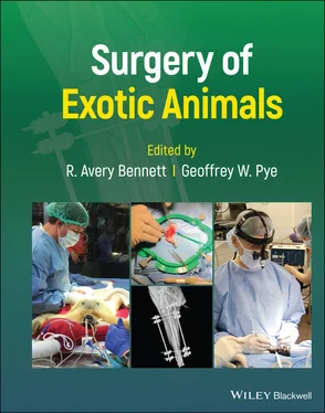
Surgery of Exotic Animals
Здесь есть возможность читать онлайн «Surgery of Exotic Animals» — ознакомительный отрывок электронной книги совершенно бесплатно, а после прочтения отрывка купить полную версию. В некоторых случаях можно слушать аудио, скачать через торрент в формате fb2 и присутствует краткое содержание. Жанр: unrecognised, на английском языке. Описание произведения, (предисловие) а так же отзывы посетителей доступны на портале библиотеки ЛибКат.
- Название:Surgery of Exotic Animals
- Автор:
- Жанр:
- Год:неизвестен
- ISBN:нет данных
- Рейтинг книги:5 / 5. Голосов: 1
-
Избранное:Добавить в избранное
- Отзывы:
-
Ваша оценка:
- 100
- 1
- 2
- 3
- 4
- 5
Surgery of Exotic Animals: краткое содержание, описание и аннотация
Предлагаем к чтению аннотацию, описание, краткое содержание или предисловие (зависит от того, что написал сам автор книги «Surgery of Exotic Animals»). Если вы не нашли необходимую информацию о книге — напишите в комментариях, мы постараемся отыскать её.
The first book to provide veterinarians with in-depth guidance on exotic animal surgical principles and techniques Surgery of Exotic Animals
Surgery of Exotic Animals
Surgery of Exotic Animals — читать онлайн ознакомительный отрывок
Ниже представлен текст книги, разбитый по страницам. Система сохранения места последней прочитанной страницы, позволяет с удобством читать онлайн бесплатно книгу «Surgery of Exotic Animals», без необходимости каждый раз заново искать на чём Вы остановились. Поставьте закладку, и сможете в любой момент перейти на страницу, на которой закончили чтение.
Интервал:
Закладка:
10 Chapter 10Figure 10.1 Figure‐of‐eight bandage application. Coaptation is started by wr...Figure 10.2 Radiographs of an adult bald eagle ( Haliaeetus leucocephalus) , ...Figure 10.3 Curved edge splint application (Ponder and Redig 2016). Select a...Figure 10.4 Tape splint application. Apply two layers of tape on the medial ...Figure 10.5 Lateral radiograph showing a short oblique mid‐tibiotarsal fract...Figure 10.6 Radiographs of a simple transverse distal radius and ulna fractu...Figure 10.7 Radiograph of a long oblique mid‐humeral fracture stabilized wit...Figure 10.8 Various plate types. From top left to right: 6‐hole 2.0 mm dynam...Figure 10.9 Locking and nonlocking screws (a) placed in a 5‐hole LCP that ac...Figure 10.10 Radiographs of a blue‐fronted amazon ( Amazona aestiva ) show a c...Figure 10.11 A red‐tailed hawk ( Buteo jamaicensis ) presented with right‐side...Figure 10.12 Craniocaudal (a) and lateral (b) radiographs of a mid‐diaphysea...Figure 10.13 Picture of the components of the IMEX SK ESF system. From top l...Figure 10.14 Lateral radiograph showing a segmental fracture of the mid‐to‐d...Figure 10.15 Picture of a type I (unilateral, uniplanar) ESF (a), type Ib (u...Figure 10.16 Picture of the supplies needed for making an acrylic connecting...Figure 10.17 Acrylx 2‐part resin comes with an applicator, multiuse cartridg...Figure 10.18 Radiographs of adult red‐tailed hawk found down on the road wit...Figure 10.19 Radiographs of an adult red‐shouldered hawk with a complete, se...Figure 10.20 Schematic of various configurations of IM‐ESF. In terms of biom...Figure 10.21 Radiographs of a three‐year‐old Harris hawk ( Parabuteo unicinct ...Figure 10.22 Photograph of proper IM pin (a) being bent with either a Frazie...Figure 10.23 Bilateral application of external ring fixators to a bald eagle...Figure 10.24 Photographs of corticocancellous bone harvest. Aseptically prep...Figure 10.25 Avulsion fracture of the ventral tubercle (arrow) of the proxim...Figure 10.26 Radiographs of mild caudodorsal elbow luxation (a) and normal c...Figure 10.27 Lateral (a) and cranial/caudal (b) radiographs of a dorsal luxa...Figure 10.28 Radiographs of a craniodorsal right‐sided coxofemoral luxation ...Figure 10.29 Ventrodorsal (a) and lateral (b) radiograph of a toggle pin app...Figure 10.30 CT DICOM images were acquired to make a digital 3‐D rendering (...Figure 10.31 Photographs of a juvenile bald eagle ( Haliaeetus leucocephalus )...Figure 10.32 Pictures of a blue and gold macaw ( Ara ararauna ) with mild scis...Figure 10.33 Subadult Scarlet macaw ( Ara macao ) with mandibular prognathism ...Figure 10.34 Left‐sided traumatic beak injury in a bald eagle ( Haliaeetus le ...Figure 10.35 Photographs of a right‐sided, minimally contaminated mandibular...Figure 10.36 Photograph of a scarlet macaw ( Ara macao ) with the upper and lo...
11 Chapter 11Figure 11.1 A chicken prepared for aseptic surgery. Huck towels have been pl...Figure 11.2 The Harrison tip bipolar forceps (a) has a 45° °bend on one...Figure 11.3 To incise avian skin with bipolar forceps, tent the skin with fo...Figure 11.4 A left lateral celiotomy provides exposure to much of the viscer...Figure 11.5 Position for left lateral celiotomy (drawing) (a) and an umbrell...Figure 11.6 When positioned for a lateral celiotomy a fold of skin is create...Figure 11.7 After making the skin incision for a left lateral celiotomy iden...Figure 11.8 Superficial branches of the femoral artery and vein need to be l...Figure 11.9 Make the incision in the body wall beginning at the last rib. Th...Figure 11.10 After incising the body wall with bipolar electrosurgery a self...Figure 11.11 Close with a simple continuous pattern in the intercostal and a...Figure 11.12 A ventral midline celiotomy enters the intestinal peritoneal ca...Figure 11.13 Immediately inside the ventral coelomic wall is the duodenal lo...
12 Chapter 12Figure 12.1 Male chicken left reproductive anatomy. Testis (T), epididymis (...Figure 12.2 Intraoperative view of castration in a rooster. The left testis ...Figure 12.3 Cadaver dissection demonstrating how to perform orchidectomy in ...Figure 12.4 Intraoperative image of castration of an umbrella cockatoo. The ...Figure 12.5 To perform a vasectomy using the left lateral approach (a), gras...Figure 12.6 The mesovarium is difficult to identify because it is short and ...Figure 12.7 Anatomy of the female reproductive tract in a juvenile cockatiel...Figure 12.8 Intraoperative anatomy of a juvenile chicken during ovariectomy....Figure 12.9 Large follicles in a mature bird often obstruct visualization of...Figure 12.10 Intraoperative view of ovariectomy in an umbrella cockatoo ( Cac ...Figure 12.11 In a juvenile bird, the vessels are not well developed and the ...Figure 12.12 LigaSure™ has been evaluated for use for avian ovariectomy. Fol...Figure 12.13 Anatomy of the oviduct showing the infundibulum, magnum, isthmu...Figure 12.14 Intraoperative image of a juvenile lovebird ( Agapornis sp.). A ...Figure 12.15 Intraoperative image of a juvenile cockatiel ( Nymphicus holland ...Figure 12.16 A salpingohysterotomy was performed in this love bird ( Agaporni ...
13 Chapter 13Figure 13.1 Overview of the avian gastrointestinal tract. (a) Crop, (b) prov...Figure 13.2 The proventriculus (P), isthmus (arrow), and the large and muscu...Figure 13.3 A duodenal loop (a) showing pancreas (b) within the loop. The bl...Figure 13.4 (a,b) Orthogonal radiographs of a double‐crested cormorant revea...Figure 13.5 For a proventriculotomy, (a) start the incision in a hypovascula...Figure 13.6 Use thumbs forceps for counter pressure to pass sutures to avoid...Figure 13.7 Orthogonal radiographs of an ostrich coelom revealing various fo...Figure 13.8 The stomach of an ostrich is exteriorized and sutured to the ski...Figure 13.9 Drawing of a cloacotomy visualizing the coprourodeal fold (cu), ...Figure 13.10 (a) Circumferential cloacal papillomatosis affecting the procto...Figure 13.11 Cloacal prolapse in a cockatoo.Figure 13.12 (a) Ventral midline and bilateral parasternal flap approach to ...Figure 13.13 (a) Dilated vent in a cockatoo with chronic cloacal prolapse. (...Figure 13.14 For placement of a duodenostomy feeding tube (a) the duodenum i...Figure 13.15 Pancreatic biopsy of the caudal edge of the ventral lobe. This ...
14 Chapter 14Figure 14.1 Correction of choanal atresia in an African grey parrot ( Psittac ...Figure 14.2 Knowledge of the anatomy and relationship of the infraorbital si...Figure 14.3 (a, b) Magnetic resonance imaging in a scarlet macaw ( Ara macao )...Figure 14.4 To access the mass in the rostral diverticulum (or one in the ma...Figure 14.5 The defect in the beak was covered with a dental mesh that was t...Figure 14.6 To access the preorbital diverticulum of the avian infraorbital ...Figure 14.7 Leg cranial positioning of a pigeon ( Columba livia ) for air sac ...Figure 14.8 Leg caudal positioning of a pigeon ( Columba livia ) for air sac c...Figure 14.9 Hand position on hemostats to prevent overpenetration when creat...Figure 14.10 Positioning of a bird for tracheotomy to allow the best visuali...Figure 14.11 For an avian tracheostomy, (a) incise the skin on ventral midli...Figure 14.12 For an avian tracheotomy, (a) place stay sutures around the tra...Figure 14.13 (a) To remove a tracheal foreign body, gently remove the foreig...Figure 14.14 This crane had been intubated for anesthesia for a quarantine p...Figure 14.15 Multiple cartilage rings may need to be removed where a trachea...Figure 14.16 For an approach to the cranial coelom, (a) incise the skin over...Figure 14.17 To close the cranial coelom, reattach the wedge, by (a) passing...Figure 14.18 Bifid sternum in an African grey parrot ( Psittacus erithacus ). ...Figure 14.19 To surgically correct bifid sternum in a bird, (a) carefully in...
Читать дальшеИнтервал:
Закладка:
Похожие книги на «Surgery of Exotic Animals»
Представляем Вашему вниманию похожие книги на «Surgery of Exotic Animals» списком для выбора. Мы отобрали схожую по названию и смыслу литературу в надежде предоставить читателям больше вариантов отыскать новые, интересные, ещё непрочитанные произведения.
Обсуждение, отзывы о книге «Surgery of Exotic Animals» и просто собственные мнения читателей. Оставьте ваши комментарии, напишите, что Вы думаете о произведении, его смысле или главных героях. Укажите что конкретно понравилось, а что нет, и почему Вы так считаете.


