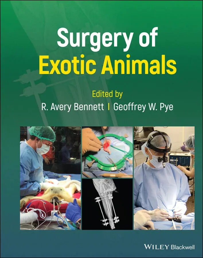
Surgery of Exotic Animals
Здесь есть возможность читать онлайн «Surgery of Exotic Animals» — ознакомительный отрывок электронной книги совершенно бесплатно, а после прочтения отрывка купить полную версию. В некоторых случаях можно слушать аудио, скачать через торрент в формате fb2 и присутствует краткое содержание. Жанр: unrecognised, на английском языке. Описание произведения, (предисловие) а так же отзывы посетителей доступны на портале библиотеки ЛибКат.
- Название:Surgery of Exotic Animals
- Автор:
- Жанр:
- Год:неизвестен
- ISBN:нет данных
- Рейтинг книги:5 / 5. Голосов: 1
-
Избранное:Добавить в избранное
- Отзывы:
-
Ваша оценка:
Surgery of Exotic Animals: краткое содержание, описание и аннотация
Предлагаем к чтению аннотацию, описание, краткое содержание или предисловие (зависит от того, что написал сам автор книги «Surgery of Exotic Animals»). Если вы не нашли необходимую информацию о книге — напишите в комментариях, мы постараемся отыскать её.
The first book to provide veterinarians with in-depth guidance on exotic animal surgical principles and techniques Surgery of Exotic Animals
Surgery of Exotic Animals
Surgery of Exotic Animals — читать онлайн ознакомительный отрывок
Ниже представлен текст книги, разбитый по страницам. Система сохранения места последней прочитанной страницы, позволяет с удобством читать онлайн бесплатно книгу «Surgery of Exotic Animals», без необходимости каждый раз заново искать на чём Вы остановились. Поставьте закладку, и сможете в любой момент перейти на страницу, на которой закончили чтение.
Интервал:
Закладка:
2 Chapter 2 Figure 2.1 Inflammatory reactions after implantation of different suture mat...
3 Chapter 3 Figure 3.1 Optics of magnification aids that use a two‐lens system. Note the... Figure 3.2 Binocular head–body of the operating microscope. Contains the eye... Figure 3.3 Eyepieces contribute to the total magnification of an operating m... Figure 3.4 Eyepieces of an operating microscope are adjustable to accommodat... Figure 3.5 Field of view is the extent of the operating field seen in focus ... Figure 3.6 Operating microscope foot pedal displaying the joystick for adjus... Figure 3.7 The surgeon should sit at the operating table with hips slightly ... Figure 3.8 Resting the elbows and antebrachii on the surgical table at 90° t... Figure 3.9 Adjust the diopter settings for each eye, one eye at a time, to e... Figure 3.10 The author's through‐the‐lens (TTL) mounted loupes. Note the ste... Figure 3.11 Small collection of microsurgical instruments. From right to lef... Figure 3.12 A stable hand position with fingers stacked on one another holdi... Figure 3.13 Double Acland microvascular clamps on an approximating frame. Th... Figure 3.14 Alternate stable hand position with fingers splayed instead of s... Figure 3.15 The surgeon's thumb and first two fingers should surround the in...
4 Chapter 4 Figure 4.1 This schematic shows the cellular structure of a sponge ( Halisarc ... Figure 4.2 This schematic shows three stages of regeneration in the sponge, Figure 4.3 This aquarium is filled with coral “frags” representing a number ... Figure 4.4 This image illustrates the major external anatomical features of ... Figure 4.5 This apple snail (Ampullariidae) suffered a fractured shell after... Figure 4.6 The same snail in Figure 4.5 after the application of an epoxy br... Figure 4.7 This series of images shows how earthworms can be a model for vas... Figure 4.8 This image shows the careful removal of a retained exoskeleton fr... Figure 4.9 These images illustrate fracture repair of a horseshoe crab ( Limu ... Figure 4.10 This cockroach ( Blaberus discoidalis ) has been outfitted with a ...
5 Chapter 5 Figure 5.1 (a) Positioning for a CT‐scan in an anesthetized koi ( Cyprinus ca ... Figure 5.2 Anesthetic equipment used for a large 8 kg koi ( Cyprinus carpio ) ... Figure 5.3 Intervention in the oropharyngeal cavity of a Ranchu goldfish ( Ca ... Figure 5.4 Use of adjunctive cryotherapy for excision of an odontoma in an a... Figure 5.5 Use of a hand‐held electrocautery during a lateral celiotomy in a... Figure 5.6 Large ulceration on the ventrum of a female koi ( Cyprinus carpio )... Figure 5.7 Excision of a neoplastic mass of the vent of a koi ( Cyprinus carp ... Figure 5.8 Intralesional bleomycin injection into a myxoma on the head of an... Figure 5.9 Opercular plasty in an anesthetized Asian arowana ( Scleropages fo ... Figure 5.10 Fibrous tissue obstructing the oral cavity of a koi ( Cyprinus ca ... Figure 5.11 Green spotted pufferfish ( Tetraodon nigroviridis ) before (a) and... Figure 5.12 Fluorescein staining of a large corneal ulceration in a lookdown... Figure 5.13 Enucleation of a rockfish ( Sebastes caurinus ) with a retinal tum... Figure 5.14 Suture of the periorbital tissue after an enucleation in a saith... Figure 5.15 Enucleation of a sea horse ( Hippocampus erectus ) with a retro‐or... Figure 5.16 Incision of the coelom between the pelvic fins and the digestive... Figure 5.17 Right lateral radiograph of a positively buoyant goldfish ( Caras ... Figure 5.18 Ovariectomy in an Oranda goldfish ( Carassius auratus ): the head ... Figure 5.19 Whole body right lateral radiograph (a) and ultrasound image (b)... Figure 5.20 A goldfish ( Carassius auratus ) showing its impacted intestine ex...
6 Chapter 6 Figure 6.1 A wound on the lateral aspect of the tarsus in a laboratory Afric... Figure 6.2 Possible intravenous injection sites in amphibians. Figure 6.3 Cutaneous everting suture pattern with monofilament absorbable su... Figure 6.4 Amputation of the tip of the tail of a California newt ( Taricha t ... Figure 6.5 Surgical toe amputation in an African bullfrog ( Pyxicephalus adsp ... Figure 6.6 An albino axolotl ( Ambystoma mexicanum ) presented with a traumati... Figure 6.7 Enucleation of the right eye in an Oriental fire‐bellied toad (Bo... Figure 6.8 Exploratory celiotomy in an Argentine horned frog ( Ceratophrys or ... Figure 6.9 Location of the ovaries (white arrow) in a reproductively active ... Figure 6.10 Gastrotomy in an axolotl ( Ambystoma mexicanum ) anesthetized with... Figure 6.11 Gastrotomy in an axolotl ( Ambystoma mexicanum ): (a) suture of th...
7 Chapter 7 Figure 7.1 A dorso‐ventral (DV) projection of the entire skeleton of a... Figure 7.2 A CT reconstruction of a juvenile African spurred tortoise ( Centr ... Figure 7.3 A DV projection of a green iguana ( Iguana iguana ) showing chronic... Figure 7.4 Full body DV projection of a female African spurred thighed torto... Figure 7.5 Typical motor vehicle trauma to the plastron of a red‐eared slide...Figure 7.6 Examples of commonly available small bone plates. (a) A 2.0/2.7 m...Figure 7.7 Severe fracture of the carapace in a painted turtle ( Chrysemys pi ...Figure 7.8 Fractured carapace in a red‐eared slider ( Trachemys scripta ) show...Figure 7.9 A full body DV view of a chelonian (species unknown) showing luxa...Figure 7.10 A lateral radiograph of a red‐tailed Boa ( Boa constrictor ) showi...Figure 7.11 Intraoperative photograph showing approach to the spine to obtai...
8 Chapter 8Figure 8.1 (a) Skin incision for a prefemoral celiotomy in a chelonian. (b) ...Figure 8.2 (a–d) Computed tomography of a chelonian used for surgical planni...Figure 8.3 A diamond‐edged cutting wheel is used to create the plastron oste...Figure 8.4 (a) An oscillating saw is used to create the plastron osteotomy i...Figure 8.5 A ring retractor can be used to improve exposure of the coelomic ...Figure 8.6 If the plastron osteotomy flap is cut with an inward beveled edge...Figure 8.7 (a, b) Fiberglass and epoxy resin can be used to stabilize the pl...Figure 8.8 Placing tape directly over the edges of the plastron osteotomy si...Figure 8.9 (a–g) A three‐year‐old female leopard tortoise ( Stigmochelys pard ...Figure 8.10 A combination of a prefemoral approach with an adjacent plastron...Figure 8.11 Closure of a prefemoral approach in combination with adjacent pl...Figure 8.12 (a–c) For a snake celiotomy, (a) elevate the second scale from t...Figure 8.13 Closure of snake skin following a celiotomy using a horizontal m...Figure 8.14 The ventral abdominal vein in a lizard runs directly dorsal to t...Figure 8.15 A paramedian celiotomy incision closure following castration in ...Figure 8.16 (a, b) For closure of a celiotomy in a snake or lizard, (a) roll...Figure 8.17 To approach the kidney for biopsy in a lizard, make an incision ...
9 Chapter 9Figure 9.1 A postmortem image of a boa constrictor demonstrating the three c...Figure 9.2 Dystocia in this Brazilian rainbow boa ( Epicrates cenchria ) was t...Figure 9.3 This python suffered dystocia and an oviduct volvulus around a si...Figure 9.4 This albino Burmese python developed dystocia requiring multiple ...Figure 9.5 Cadaveric image of a sexually immature green iguana undergoing bi...Figure 9.6 Intraoperative images from a sexually mature female green iguana ...Figure 9.7 Removed oviducts full of eggs following salpingectomy in a green ...Figure 9.8 In a young green iguana, it only requires one or two hemostatic c...Figure 9.9 Intraoperative picture of a sexually active male green iguana und...Figure 9.10 An eastern diamondback rattlesnake presenting for unilateral hem...Figure 9.11 Prolapsed oviduct in a California desert tortoise ( Gopherus agas ...Figure 9.12 This tortoise presented for paraparesis. This radiograph showed ...Figure 9.13 A ventrodorsal (a) and lateral (b) radiograph of a green iguana ...Figure 9.14 Cystic calculus in a California desert tortoise ( Gopherus agassi ...Figure 9.15 Intraoperative images of a ventral cystotomy in a green iguana w...Figure 9.16 Image of an egg in the urinary bladder of a tortoise (a). Note t...Figure 9.17 A cystic calculus in a green iguana ( Iguana iguana ) that was mis...Figure 9.18 The ileocecocolic junction in a green iguana ( Iguana iguana ) dem...Figure 9.19 Intraoperative image of a boa constrictor undergoing gastrotomy ...Figure 9.20 Ventrodorsal radiograph of a green iguana ( Iguana iguana ) with c...Figure 9.21 Intraoperative image of leopard gecko diagnosed with a small int...Figure 9.22 Radiographs of a green iguana ( Iguana iguana ) that was housed on...Figure 9.23 This tortoise was diagnosed with colonic impaction. Medical mana...Figure 9.24 Intraoperative image of a green sea turtle with a small intestin...
Читать дальшеИнтервал:
Закладка:
Похожие книги на «Surgery of Exotic Animals»
Представляем Вашему вниманию похожие книги на «Surgery of Exotic Animals» списком для выбора. Мы отобрали схожую по названию и смыслу литературу в надежде предоставить читателям больше вариантов отыскать новые, интересные, ещё непрочитанные произведения.


Обсуждение, отзывы о книге «Surgery of Exotic Animals» и просто собственные мнения читателей. Оставьте ваши комментарии, напишите, что Вы думаете о произведении, его смысле или главных героях. Укажите что конкретно понравилось, а что нет, и почему Вы так считаете.