Pathology of Genetically Engineered and Other Mutant Mice
Здесь есть возможность читать онлайн «Pathology of Genetically Engineered and Other Mutant Mice» — ознакомительный отрывок электронной книги совершенно бесплатно, а после прочтения отрывка купить полную версию. В некоторых случаях можно слушать аудио, скачать через торрент в формате fb2 и присутствует краткое содержание. Жанр: unrecognised, на английском языке. Описание произведения, (предисловие) а так же отзывы посетителей доступны на портале библиотеки ЛибКат.
- Название:Pathology of Genetically Engineered and Other Mutant Mice
- Автор:
- Жанр:
- Год:неизвестен
- ISBN:нет данных
- Рейтинг книги:3 / 5. Голосов: 1
-
Избранное:Добавить в избранное
- Отзывы:
-
Ваша оценка:
Pathology of Genetically Engineered and Other Mutant Mice: краткое содержание, описание и аннотация
Предлагаем к чтению аннотацию, описание, краткое содержание или предисловие (зависит от того, что написал сам автор книги «Pathology of Genetically Engineered and Other Mutant Mice»). Если вы не нашли необходимую информацию о книге — напишите в комментариях, мы постараемся отыскать её.
An updated and comprehensive reference to pathology in every organ system in genetically modified mice Pathology of Genetically Engineered and Other Mutant Mice
Pathology of Genetically Engineered and Other Mutant Mice
Pathology of Genetically Engineered and Other Mutant Mice — читать онлайн ознакомительный отрывок
Ниже представлен текст книги, разбитый по страницам. Система сохранения места последней прочитанной страницы, позволяет с удобством читать онлайн бесплатно книгу «Pathology of Genetically Engineered and Other Mutant Mice», без необходимости каждый раз заново искать на чём Вы остановились. Поставьте закладку, и сможете в любой момент перейти на страницу, на которой закончили чтение.
Интервал:
Закладка:
Anatomic Pathology Evaluation ofDeveloping Mice
For prenatal phenotypes, the first task is to measure maternal total body weight and then count the numbers of total and viable conceptuses (i.e. embryo/placental units) as well as abnormal and resorbed implantation sites. For isolated embryos and neonates, relevant individual animal characteristics are recorded including sex (based on anogenital distance; Figure 5.8), body weight, and/or crown/rump length as well as the kind and severity of external gross defects. The embryos and neonates then are relegated to different subgroups for conventional necropsy to harvest tissues ( Figure 5.9), free‐hand sectioning of the torso ( Figure 5.10) and head ( Figure 5.11) in multiple planes to view organs in situ to find gross internal defects, double staining to highlight skeletal (bone and cartilage) differentiation ( Figure 5.12), or fixation to permit histopathological evaluation [70]. Placentas should be fixed together with their embryos.
Histological processing is a key step in preparing the tissues of developing mice for microscopic evaluation. The choice or processing techniques depend on the age of the animal and the types of tissues that need to be preserved. Commonly, fixation is done by immersion, which is simple and rapid, but perfusion fixation may be needed to preserve deep, dense tissues that degenerate quickly, such as bone marrow and brain [58, 71]. For early embryos, tissues generally are fixed by immersion in neutral buffered 10% formalin (NBF), Bouin's solution, or modified Davidson's solution. All three of these solutions penetrate well, and both Bouin's and modified Davidson's solutions harden soft embryonic tissues so that they may be handled for macroscopic evaluation. For late‐stage embryos (GD14.0 and later) and neonates, the same three fixatives still may be used, but for NBF the thick skin will needed to be opened (usually by a long slit in the ventral thoracic and abdominal regions) to permit penetration. Tissue dehydration and clearing, embedding media, and routine staining protocols for use with embryos and neonates generally are equivalent to those used for adult tissues [72]. The two biggest differences between embryos and neonates vs. adults are that gentle decalcification is required for all samples of developing mice beginning at GD15.5 and later due to progressive skeletal mineralization, and that all embryos and neonates should be either serial‐ or step‐sectioned to ensure that all major organs and tissues are available for microscopic evaluation. A useful strategy is to stain every fifth or tenth serial section with hematoxylin and eosin (H&E) while allocating the other sections for molecular biomarkers. When step‐sectioning, the interval between steps needed to ensure that all major organs are sampled effectively varies with the developmental stage of the animal. In our experience, suitable intervals between steps are between 25 to 50 μm at GD5.5–6.5, 75 to 100 μm at GD7.0–8.0, and 150 to 250 μm at GD9.5–10.5. For older embryos and neonates, the presence of major viscera can be verified on the block face when sectioning. Greater cellular detail may be visible, particularly in very young embryos (GD8.5 or earlier), if samples are post‐fixed by immersion in 1% osmium tetroxide for one to two hours followed by embedding in hard plastic resin. Smaller embryos, especially preimplantation embryos (GD0.5–GD4.0) are pre‐embedded in 1% agar (in distilled water) following fixation to minimize cell trauma during processing [72].
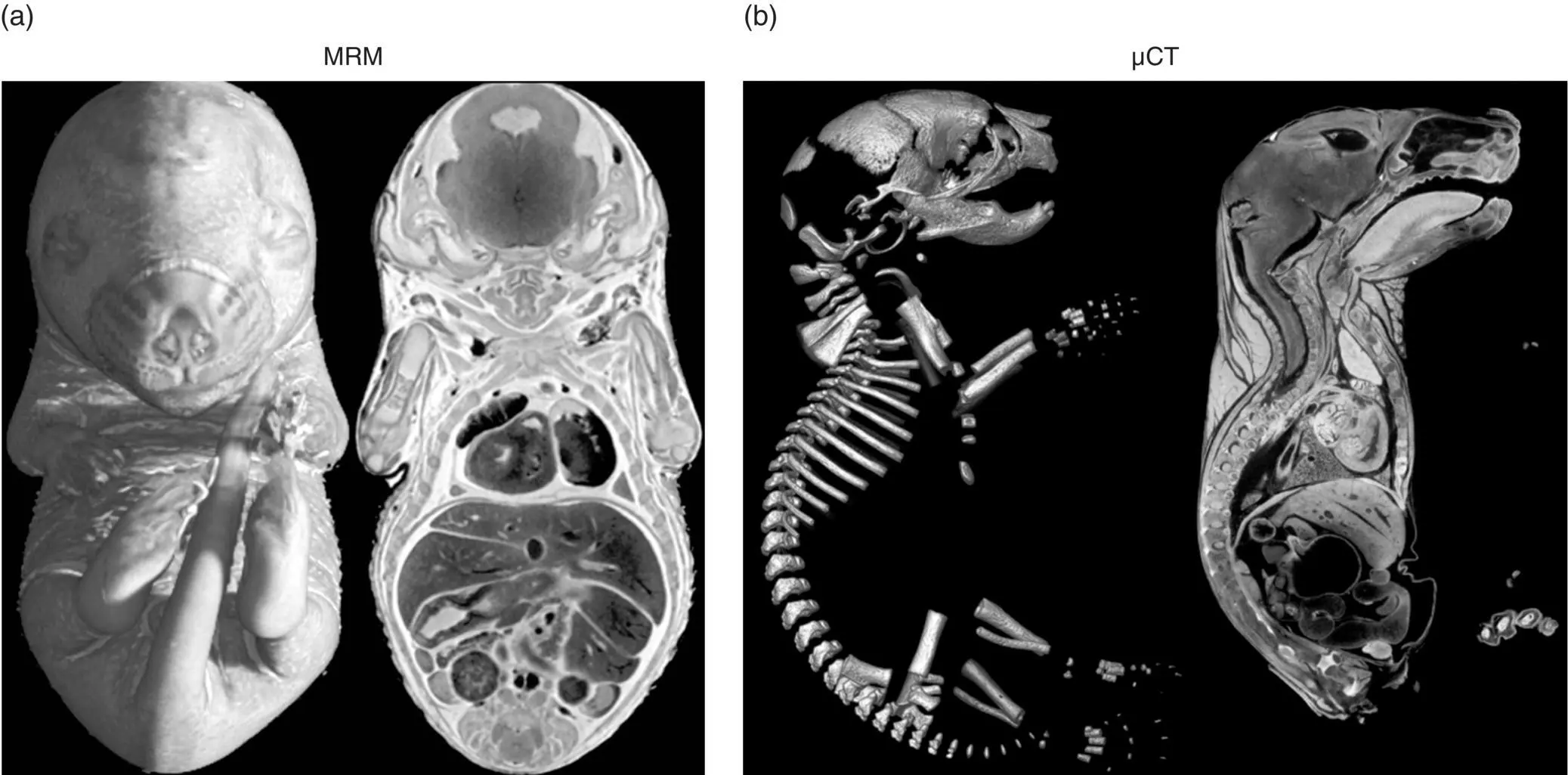
Figure 5.7 Noninvasive imaging modalities are useful means for characterizing developmental phenotypes in embryonic mice. Panel (a): Magnetic resonance microscopy (MRM) of a GD17.5 mouse embryo showing volume‐rendered composite (left) and “dissected” images acquired at 20‐μm resolution using a magnetic field strength of 9.4 T [18]. Panel (b): Microscopic computer‐assisted tomography (μCT) of a neonatal (PND0) mouse demonstrating differential highlighting of bone (left, unstained specimen) or soft tissues (right, sample processed with the proprietary Virtual Histology TMstaining protocol [Numira Biosciences, Inc., Salt Lake City, UT, USA]).
Sources: MRM image from Dr. G. A. Johnson, Duke University and µCT image from Numira Biosciences, Inc.; both images reproduced from Bolon et al. [67] by permission of CRC Press.
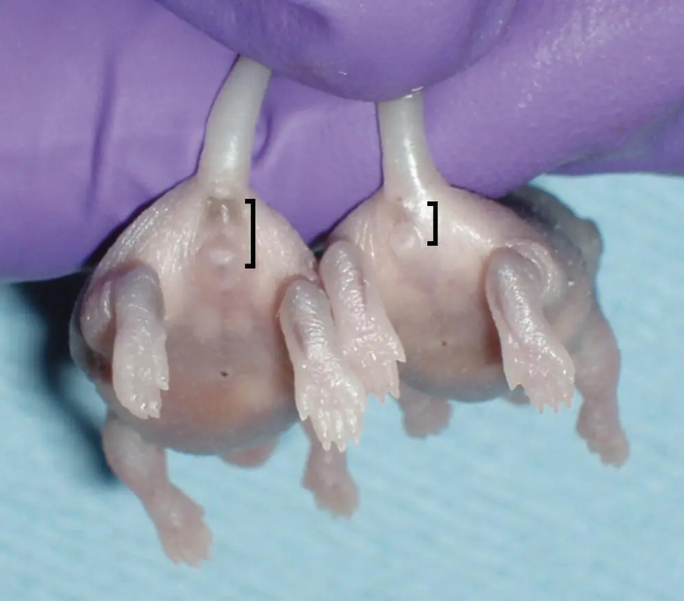
Figure 5.8 Sex differentiation of neonatal (PND1) mice using anogenital distances (brackets), which are longer in males (left) relative to females (right).
Source: Dr. Cynthia Besch‐Williford, IDEXX and Newbigging et al. [70] with permission of CRC Press.
Histopathologic evaluation is a critical element in phenotyping embryos and neonates since microscopic findings often represent the only evidence of a new phenotype [73]. The first step of the examination is to assess sections at low magnification (i.e. using objectives ranging from 1× to 5×) to observe the general appearance of differentiated organs ( Figure 5.13). This portion of the analysis will detect obvious developmental defects in tissues and organs. Special care must be taken to recognize the absence of any missing structures, which may be obvious but sometimes is not ( Figure 5.14) [74, 75]. The second step is to assess tissue features at intermediate magnification (4× to 20×) to further define potential target cell populations in affected organs. This task requires detailed understanding of the unique cytoarchitectural features of each region and its major cell types across many stages of prenatal and postnatal development – knowledge that can only be gained by experience. The third step of a well‐designed histopathologic analysis is to fully describe the nature of the structural defects at the organ, cellular, and (if necessary) subcellular levels, using both routinely stained (H&E) and specially processed sections (e.g. to demonstrate molecular biomarkers, especially utilizing immunohistochemistry [IHC]). A successful phenotypic evaluation for prenatal and neonatal mice often will require comparison of possible lesions to normal structures shown in a well‐illustrated and highly annotated rodent developmental anatomy atlases [11–15].
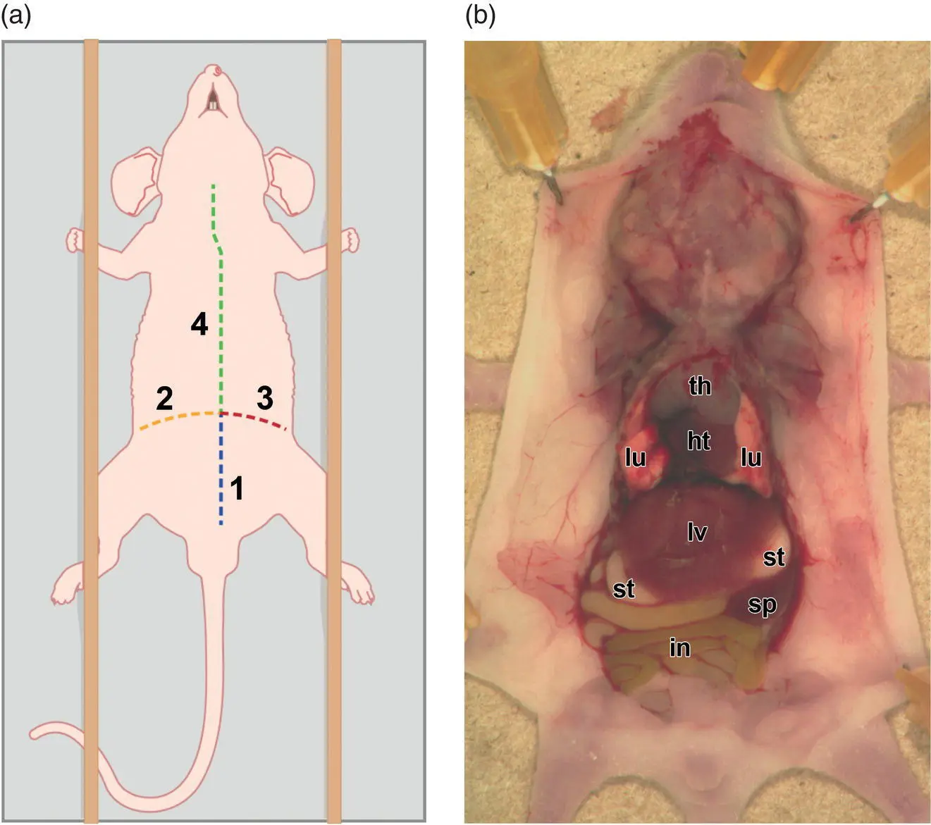
Figure 5.9 Schematic diagram showing necropsy approach for neonatal and juvenile mice. The animal is placed in dorsal recumbency, and the limbs are secured to maintain the “splayed” position. Panel (a): Organs are exposed with four cuts (made in numerical order) through the body wall. The curved arc represented by cuts “2” and “3” follows the contour of the diaphragm, which separates the thoracic cavity (located under “4”) from the abdominal cavity (found under “1”). Panel (b): Photograph demonstrating the location of principal viscera in the thoracic and abdominal cavities of a juvenile (10‐day‐old) mouse as viewed from the ventral aspect. Abbreviations: ht = heart, in = intestine, lv = liver, lu = lung, sp = spleen, st = stomach, th = thymus.
Sources: Diagram, prepared by Tim Vojt, The Ohio State University, is from Newbigging et al. [70] with permission of CRC Press; photograph from Bolon et al. [58] with permission of John Wiley & Sons.
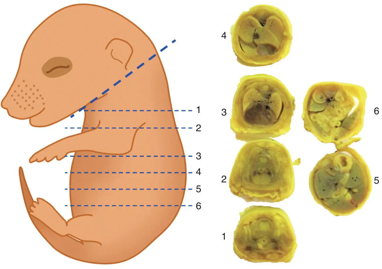
Figure 5.10 Sectioning (Wilson's) technique to allow macroscopic evaluation of defects in large internal organs of near‐term (GD17 and older) mouse embryos and neonates. The schematic diagram (left panel) demonstrates the placement of freehand incisions to expose internal organs. Numbers of the dashed lines in the diagram correspond to the numbered planes in the labeled torso blocks (right panel) of a near‐term embryo that had been fixed in Bouin's solution. Tissue blocks may be processed subsequently for histopathological assessment.
Читать дальшеИнтервал:
Закладка:
Похожие книги на «Pathology of Genetically Engineered and Other Mutant Mice»
Представляем Вашему вниманию похожие книги на «Pathology of Genetically Engineered and Other Mutant Mice» списком для выбора. Мы отобрали схожую по названию и смыслу литературу в надежде предоставить читателям больше вариантов отыскать новые, интересные, ещё непрочитанные произведения.


Обсуждение, отзывы о книге «Pathology of Genetically Engineered and Other Mutant Mice» и просто собственные мнения читателей. Оставьте ваши комментарии, напишите, что Вы думаете о произведении, его смысле или главных героях. Укажите что конкретно понравилось, а что нет, и почему Вы так считаете.