Pathology of Genetically Engineered and Other Mutant Mice
Здесь есть возможность читать онлайн «Pathology of Genetically Engineered and Other Mutant Mice» — ознакомительный отрывок электронной книги совершенно бесплатно, а после прочтения отрывка купить полную версию. В некоторых случаях можно слушать аудио, скачать через торрент в формате fb2 и присутствует краткое содержание. Жанр: unrecognised, на английском языке. Описание произведения, (предисловие) а так же отзывы посетителей доступны на портале библиотеки ЛибКат.

- Название:Pathology of Genetically Engineered and Other Mutant Mice
- Автор:
- Жанр:
- Год:неизвестен
- ISBN:нет данных
- Рейтинг книги:3 / 5. Голосов: 1
-
Избранное:Добавить в избранное
- Отзывы:
-
Ваша оценка:
Pathology of Genetically Engineered and Other Mutant Mice: краткое содержание, описание и аннотация
Предлагаем к чтению аннотацию, описание, краткое содержание или предисловие (зависит от того, что написал сам автор книги «Pathology of Genetically Engineered and Other Mutant Mice»). Если вы не нашли необходимую информацию о книге — напишите в комментариях, мы постараемся отыскать её.
An updated and comprehensive reference to pathology in every organ system in genetically modified mice Pathology of Genetically Engineered and Other Mutant Mice
Pathology of Genetically Engineered and Other Mutant Mice
Pathology of Genetically Engineered and Other Mutant Mice — читать онлайн ознакомительный отрывок
Ниже представлен текст книги, разбитый по страницам. Система сохранения места последней прочитанной страницы, позволяет с удобством читать онлайн бесплатно книгу «Pathology of Genetically Engineered and Other Mutant Mice», без необходимости каждый раз заново искать на чём Вы остановились. Поставьте закладку, и сможете в любой момент перейти на страницу, на которой закончили чтение.
Интервал:
Закладка:
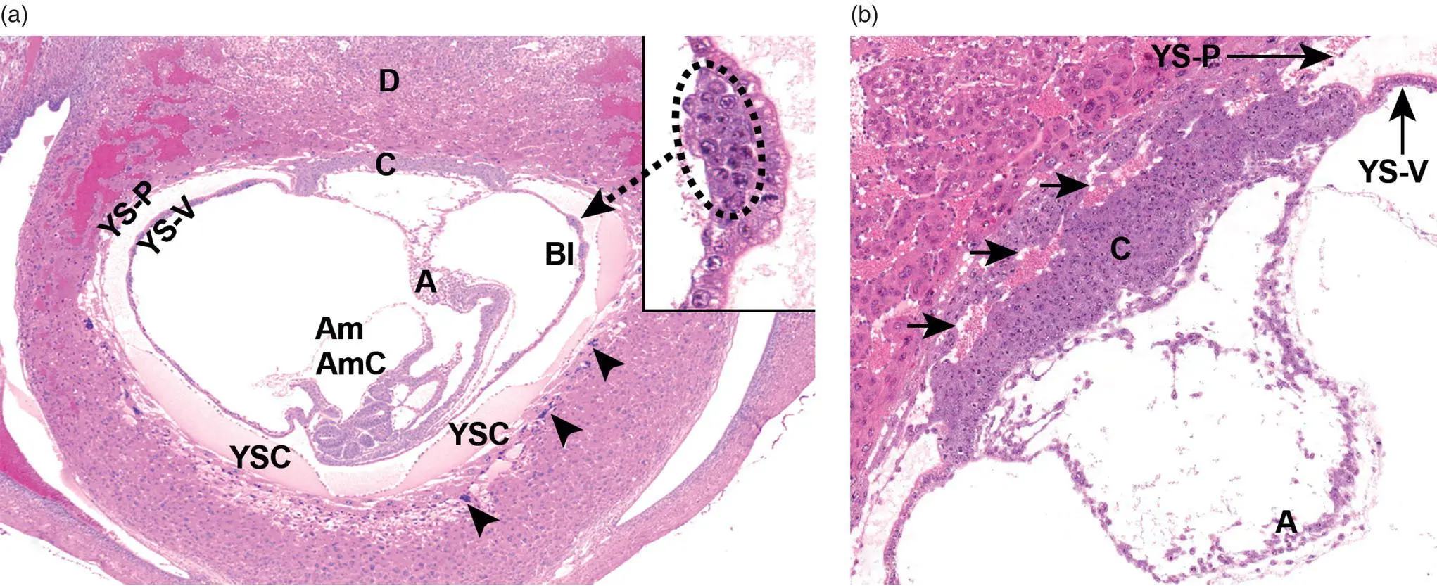
Figure 5.3 The early placenta (shown here at GD8.5) consists chiefly of maternal decidua (D) and the embryonic yolk sac (YS), separated at their interface by trophoblast giant cells (arrowheads). The YS consists of visceral (inner, YS‐V) and parietal (outer, YS‐P) layers. Blood islands (BI, circled in inset of Panel (a)) form in the YS‐V as the source of primitive hematopoietic cell precursors. The YS‐P rests atop Reichert's membrane, a rodent‐specific tough basement membrane that is not visible here. The allantois (A) is a mesodermal projection (i.e. the nascent umbilical cord) connecting the caudal end of the embryo proper to the flat, broad epithelial plate known as the chorion (C), which forms centrally at the base of the embryonic placenta. The amnion (Am) is a thin membrane that eventually will envelop the entire embryo. Short arrows (in (b)) indicate large embryonic blood vessels in the chorion that connect to umbilical cord blood vessels. Abbreviations: AmC = amniotic cavity, YSC = yolk sac cavity. Stain: H&E.
Sources: Bolon [96] with permission of Elsevier, and Bolon and Ward [39] with permission of CRC Press.
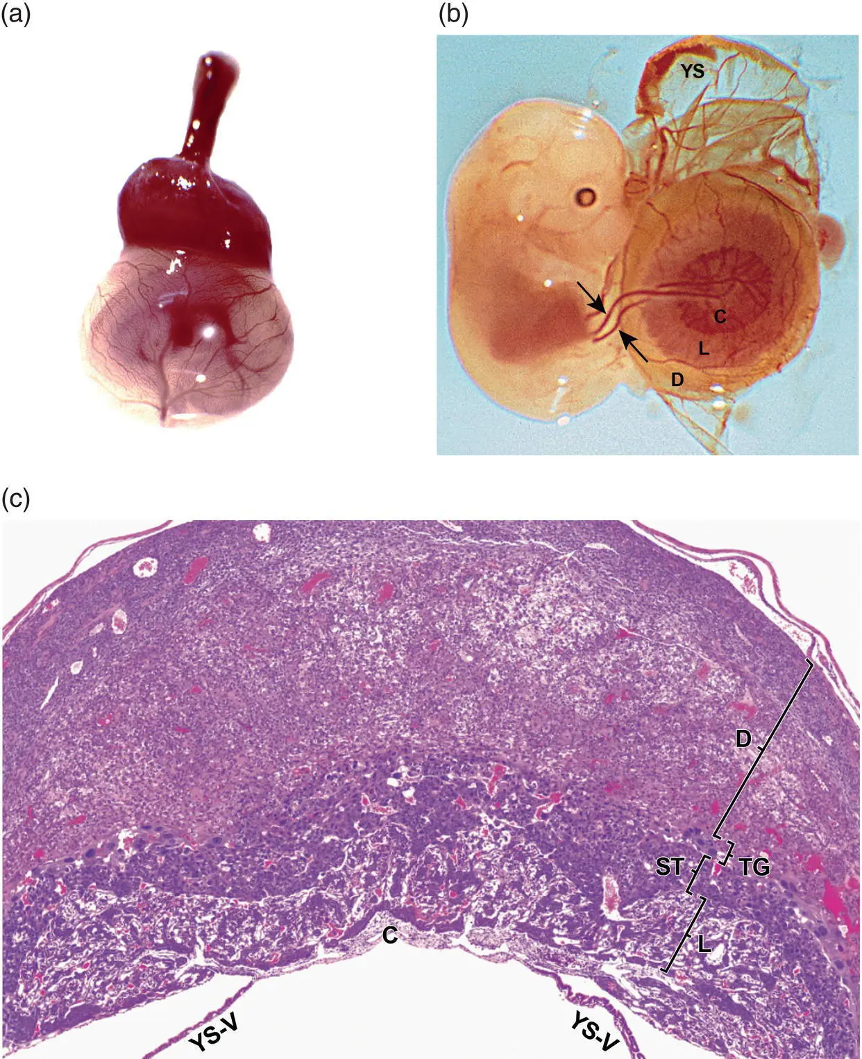
Figure 5.4 The definitive placenta forms fully by GD12.5 (Panel (a)), appearing while intact as a discoid, highly vascularized mass capping the mesometrial pole of the thin, highly vascularized, translucent yolk sac (YS). After dissection (Panel (b)), the discoid cap at GD12.5 may be seen to consist of three concentric zones: the chorion (C) centrally, to which the umbilical blood vessels (arrows) are anchored; labyrinth (L); and decidua (D). Microscopically when viewed in mid‐axial cross‐section (Panel (c)), the definitive placenta at GD12.5 displays many cytoarchitecturally distinct structures, the chief of which are the labyrinth, junctional zone (comprised of the spongiotrophoblast [ST] and trophoblast giant cell [TG] layers), and decidua. Other abbreviation: YS‐V = visceral portion of the yolk sac. Stain (Panel (c)): H&E.
Sources: Ward and Devor‐Henneman [44] with permission of Iowa State University Press, and Bolon and Ward [39] with permission of CRC Press.
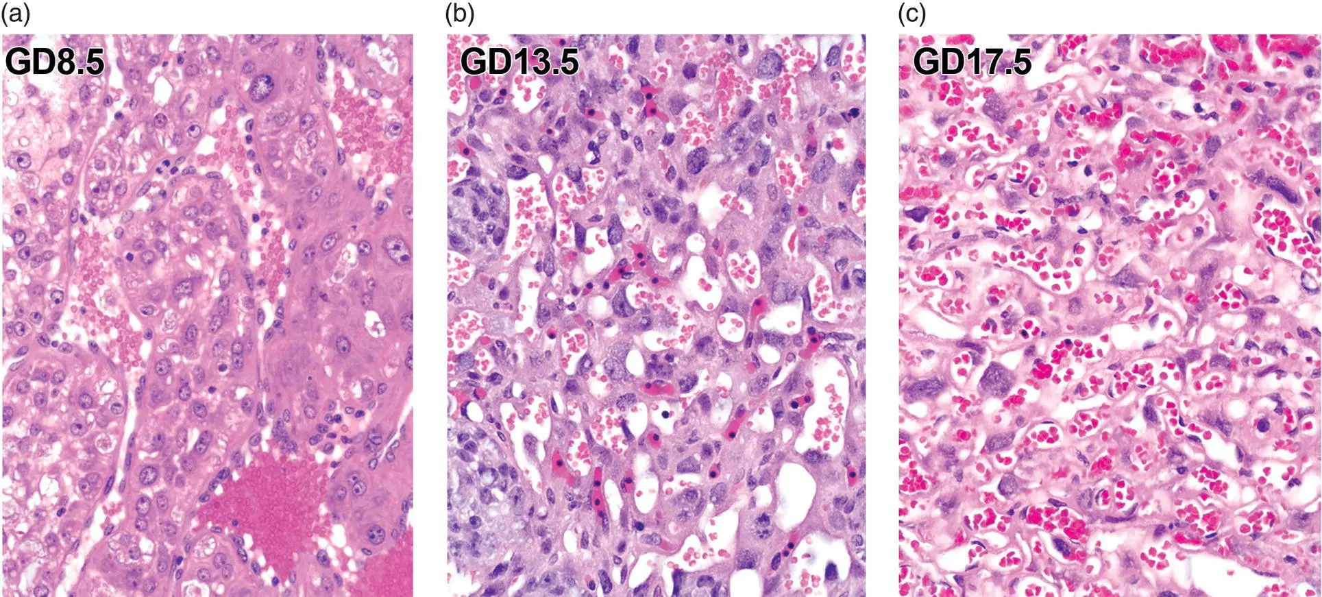
Figure 5.5 The labyrinth of the placenta (shown here for wild‐type CD‐1 mice) evolves over time. Early in development (GD8.5, in (a)), this zone consists of dense trophoblast cords separated by large, trophoblast‐lined maternal blood spaces (MBS or “sinusoids”) that are packed with non‐nucleated red blood cells (RBCs). In mid‐gestation (GD13.5, in (b)), the definitive placenta exhibits many tortuous, thin‐walled, closely apposed vascular channels, of two kinds: embryonic blood spaces (EBS) lined by endothelial cells and containing a mixture of primitive (large‐diameter, nucleated) and definitive (small‐diameter, non‐nucleated) RBCs, and MBS lined by three embryo‐derived trophoblast layers but not endothelium and containing only definitive RBCs. Near term (GD17.5, in (c)), both EBS and MBS contain large numbers of definitive RBCs. Stain: H&E.
Source: Bolon and Ward [39] with permission of CRC Press.
The maternal immune response against the embryo is restrained by the presence of a transient immune‐modulating structure in the decidua of mice and rats designated variously as the decidualized mesometrial triangle, mesometrial lymphoid aggregates of pregnancy (MLAp), or metrial gland ( Figure 5.6) [49–52]. This metrial “gland” isn't really a gland but an area of the placenta composed of arteriolar‐like blood vessels and abundant uterine natural killer (uNK) cells (also called granulated metrial gland [GMG] cells). These uNK cells are maternal bone marrow‐derived, perforin‐positive leukocytes that express NK cell biomarkers [52, 53]. The uNK cell population and the entire metrial gland begin to regress by mid‐gestation.
Placental size differs among mouse strains. Outbred ICR (Institute of Cancer Research) mice have placentas that are about 25% larger than those of age‐matched inbred C57BL/6J (B6) mice. This discrepancy is correlated with embryonic growth as ICR embryos are approximately 25% bigger than age‐matched B6 embryos [54]. A similar placental size increase of 20% has been reported in hybrid mice, where embryos are larger relative to those generated by the inbred C57BL/Mcl or C3H/BiMcl parental strains [55], and even larger increases have been described in cloned mice [56]. Strain differences in placental biology are not limited to structural effects, as metabolic capacities and pathways in placental tissue also vary among mouse strains [57].
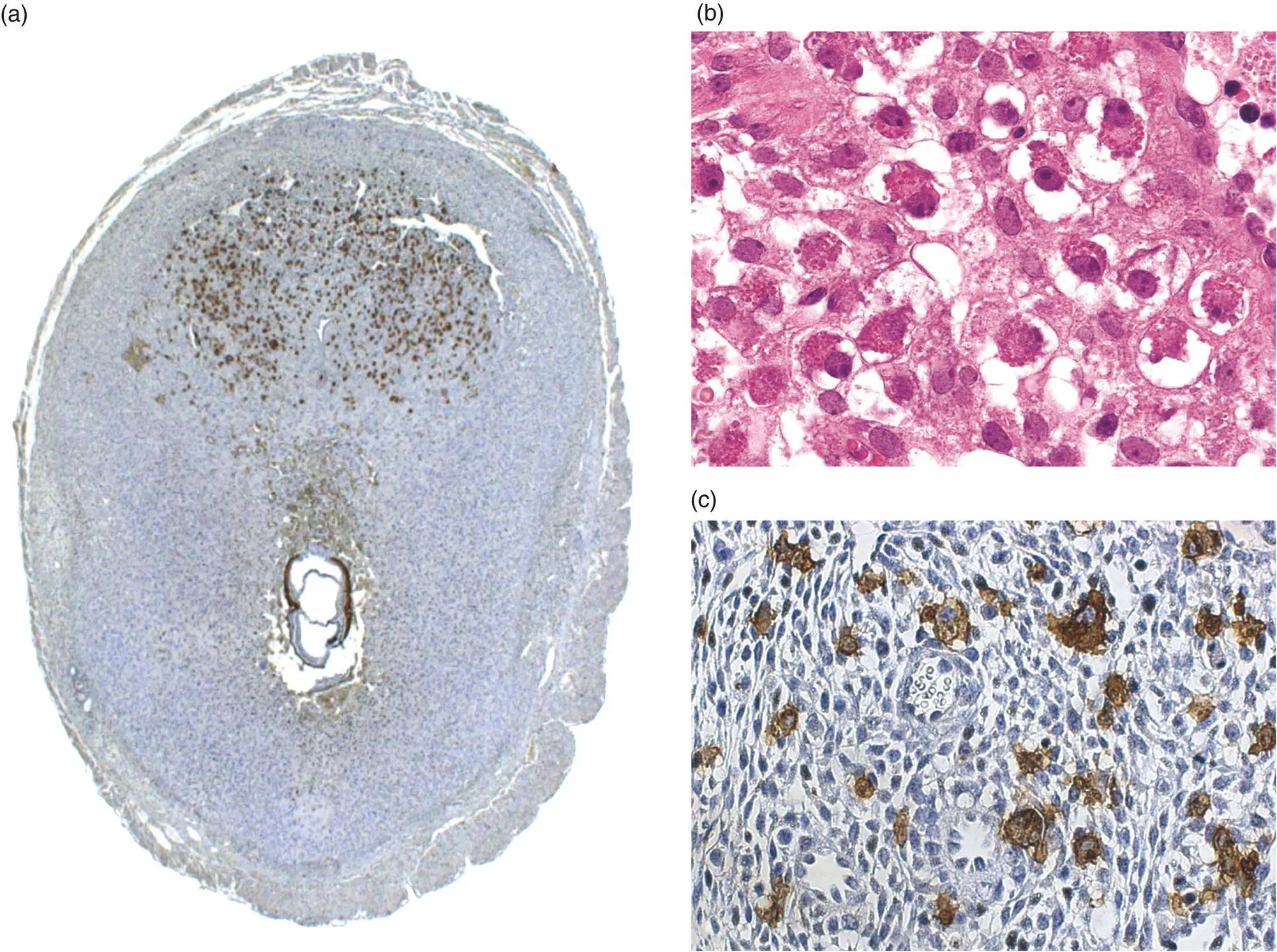
Figure 5.6 The nascent metrial gland (also designated the mesometrial lymphoid aggregate of pregnancy), shown here at GD8.5 (Panel (a)), appears as a dense collection of granulated metrial gland (GMG) cells (Panel (b); also called uterine natural killer [uNK] cells, Panel (c)) that surrounds large maternal blood vessels in the mesometrial decidua. Lectin histochemistry for the uNK cell marker Dolichos biflorus agglutinin (DBA) lectin (Panels (a) and (c)). Stain (panel (b)): H&E.
Sources: Dr. Bruno Zavan, University of Alfenas, Brazil, and from Bolon [96] with permission of Elsevier.
Technical Considerations for Pathologic Evaluation of Developing Mice
The plan for pathologic evaluation for any phenotyping project in developing mice will utilize techniques comparable to those applied when examining adult mice. However, the choice of methods will depend on several factors unique to prenatal and early postnatal animals, including the developmental age of the subjects, the presumed timing of the lethal event(s), and in many instances the expertise and interest of the investigators and cost. In general, the initial pathologic evaluation of a novel phenotype will center on known anatomic, clinical, and molecular pathology procedures [5, 58]. For prenatal phenotypes, both the embryo and placenta must be examined since developmental and lethal events may occur in either or both of these specimens.
An important consideration for success in mouse developmental pathology is a sufficient degree of prior knowledge and experience by the morphologist. Developmental pathology is an endeavor where DIY (“do it yourself”) assessments [59] are rarely efficient or effective, and often are incorrect or misleading. Substantial money, time, and animals will likely be wasted if individuals with limited or no foundation in mouse developmental biology and pathology attempt to lead a developmental pathology project.
Noninvasive Imaging of Developing Mice
The traditional basis for characterizing structural changes in embryonic lethal and neonatal lethal phenotypes in mice has concentrated on qualitative macroscopic and light microscopic analysis, supplemented as needed by quantitative measurements of organ dimensions [60, 61], volumes [62], or cell numbers [48]. Historically, these strategies have been undertaken at one or more specific time points and have been complicated by some degree of sample destruction (e.g. via continuous or interval [“step”] sectioning to make multiple μm‐thick, two‐dimensional [2D] tissue slices) and subsequent three‐dimensional [3D] tissue reconstruction (e.g. by mental or physical realignment [“stacking”] many 2D tissue slices). In recent decades, the advent of noninvasive imaging methods for rodents has been adapted to allow one‐time or serial evaluation of adult [63] and developing [22, 64, 65] mice. These technologies facilitate the digitized, relatively high‐resolution, 2D and 3D macroscopic and microscopic analysis of anatomic features and/or functional attributes without destroying or greatly distorting the specimen ( Figure 5.7). Key modalities include computer‐assisted tomography (CAT or μCT), confocal microscopy, echocardiography, magnetic resonance microscopy (MRM), optical coherence tomography (OCT), and ultrasound biomicroscopy (UBM), among others [66–69]. These noninvasive imaging techniques will not replace standard microscopy since both light and electron microscopy afford higher resolution of cell and tissue architecture. Instead, the use of imaging techniques provides the best data when employed in tandem with conventional pathology techniques.
Читать дальшеИнтервал:
Закладка:
Похожие книги на «Pathology of Genetically Engineered and Other Mutant Mice»
Представляем Вашему вниманию похожие книги на «Pathology of Genetically Engineered and Other Mutant Mice» списком для выбора. Мы отобрали схожую по названию и смыслу литературу в надежде предоставить читателям больше вариантов отыскать новые, интересные, ещё непрочитанные произведения.


Обсуждение, отзывы о книге «Pathology of Genetically Engineered and Other Mutant Mice» и просто собственные мнения читателей. Оставьте ваши комментарии, напишите, что Вы думаете о произведении, его смысле или главных героях. Укажите что конкретно понравилось, а что нет, и почему Вы так считаете.