Lori Renda-Francis - Textbook for the Veterinary Assistant
Здесь есть возможность читать онлайн «Lori Renda-Francis - Textbook for the Veterinary Assistant» — ознакомительный отрывок электронной книги совершенно бесплатно, а после прочтения отрывка купить полную версию. В некоторых случаях можно слушать аудио, скачать через торрент в формате fb2 и присутствует краткое содержание. Жанр: unrecognised, на английском языке. Описание произведения, (предисловие) а так же отзывы посетителей доступны на портале библиотеки ЛибКат.
- Название:Textbook for the Veterinary Assistant
- Автор:
- Жанр:
- Год:неизвестен
- ISBN:нет данных
- Рейтинг книги:4 / 5. Голосов: 1
-
Избранное:Добавить в избранное
- Отзывы:
-
Ваша оценка:
Textbook for the Veterinary Assistant: краткое содержание, описание и аннотация
Предлагаем к чтению аннотацию, описание, краткое содержание или предисловие (зависит от того, что написал сам автор книги «Textbook for the Veterinary Assistant»). Если вы не нашли необходимую информацию о книге — напишите в комментариях, мы постараемся отыскать её.
constitutes an exceptionally broad-ranging and in-depth guide to one of America’s greatest poets. The volume makes the best and most up-to-date scholarly thinking on Walt Whitman available to students. It encourages them to be more aware of the contexts of Whitman’s work, and helps them to understand the experimental nature of his writings. The
starts with a section which communicates a strong sense of the poet’s time, place and history. The contributions in this section range over subjects such as national identity, imperialism, slavery, race, gender, sexuality, and popular culture. A further selection of essays situates Whitman’s work in its literary context. Finally, there are essays devoted to specific works, covering both Whitman’s poetry and prose writings. The
also includes a compact biography of the poet and a bibliography of his works.
Textbook for the Veterinary Assistant — читать онлайн ознакомительный отрывок
Ниже представлен текст книги, разбитый по страницам. Система сохранения места последней прочитанной страницы, позволяет с удобством читать онлайн бесплатно книгу «Textbook for the Veterinary Assistant», без необходимости каждый раз заново искать на чём Вы остановились. Поставьте закладку, и сможете в любой момент перейти на страницу, на которой закончили чтение.
Интервал:
Закладка:
Vertebral formulas are written in a specific format and vary from species to species ( Figure 3.8). For example, the dog or cat vertebral formula would look like this:
C‐7, T‐13, L‐7, S‐3, Cy 0‐23
Appendicular skeleton
As mentioned above, the appendicular skeleton is composed of the bones of the limbs, clavicle, scapula, humerus, radius, ulna, carpus, metacarpals, pelvis, femur, patella, tibia, fibula, tarsus, metatarsals, and phalanges.
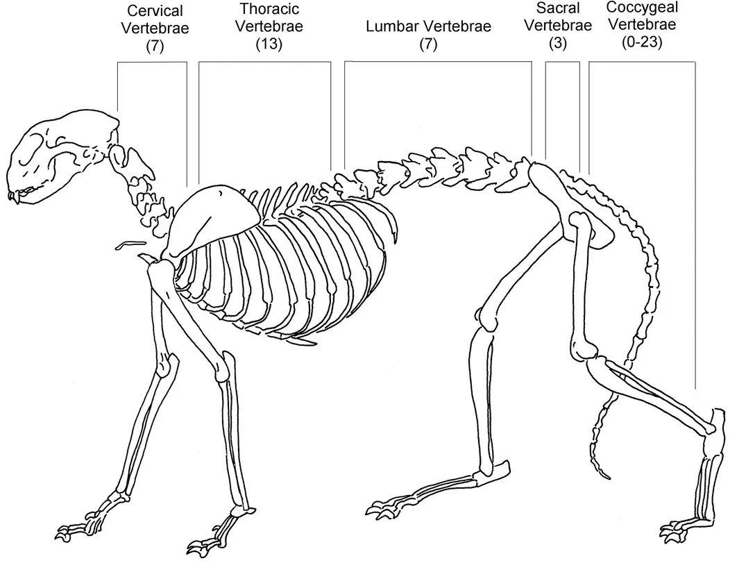
Figure 3.8 Vertebral formula.
Source: Courtesy of Jennifer Smith, LVT.
Bones of the forelimb
Bones of the forelimb can vary depending on the species. The clavicleis also known as the collarbone and is a skinny bone that connects the sternum to the scapula. Not all species have a clavicle. The scapula is also known as the shoulder blade and is a triangular‐shaped bone located on the side of the thorax. The humerusis the long bone of the forelimb that extends from the shoulder to the elbow. The ulna and radius are located distal to the humerus. The radiusis the cranial long bone of the forelimb that runs from the elbow to the carpus, and the ulnais the caudal long bone of the forelimb that runs from the elbow to the carpus.
Just distal to the radius and ulna is the carpuswhich consists of the joint and several carpal bones. The carpal bones are two rows of irregularly shaped bones.
The metcarpalsare long bones found just distal to the carpus. The number of metacarpals varies among species. Dogs have five metacarpal bones.
The most distal part of the forelimb are the phalanges. Phalangesare the bones of the digit. Digitsrelate to human fingers. The number varies in animals ( Figure 3.9).
Bones of the hindlimb
The types and number of bones of the hindlimb can vary depending on the species. The information below refers to domestic animals such as dogs and cats.
The bones of the hindlimb consist of the pelvis, femur, patella, tibia, fibula, tarsus, metatarsals, and phalanges.
The pelvisand the coccygeal vertebrae articulate with the sacrum. The acetabulumis the hip socket in which the head of the femur sits. The femuris the longest bone in the body and is located just distal to the pelvis. The stifleis the joint located between the femur and the tibia. The patellais a large flat bone that is located over the stifle joint. In humans, this is referred to as the knee cap. Just distal to the patella are the tibia and fibula. The tibiais the larger of the two bones and is considered the more weight‐bearing bone, and the fibulais a long, thinner bone.
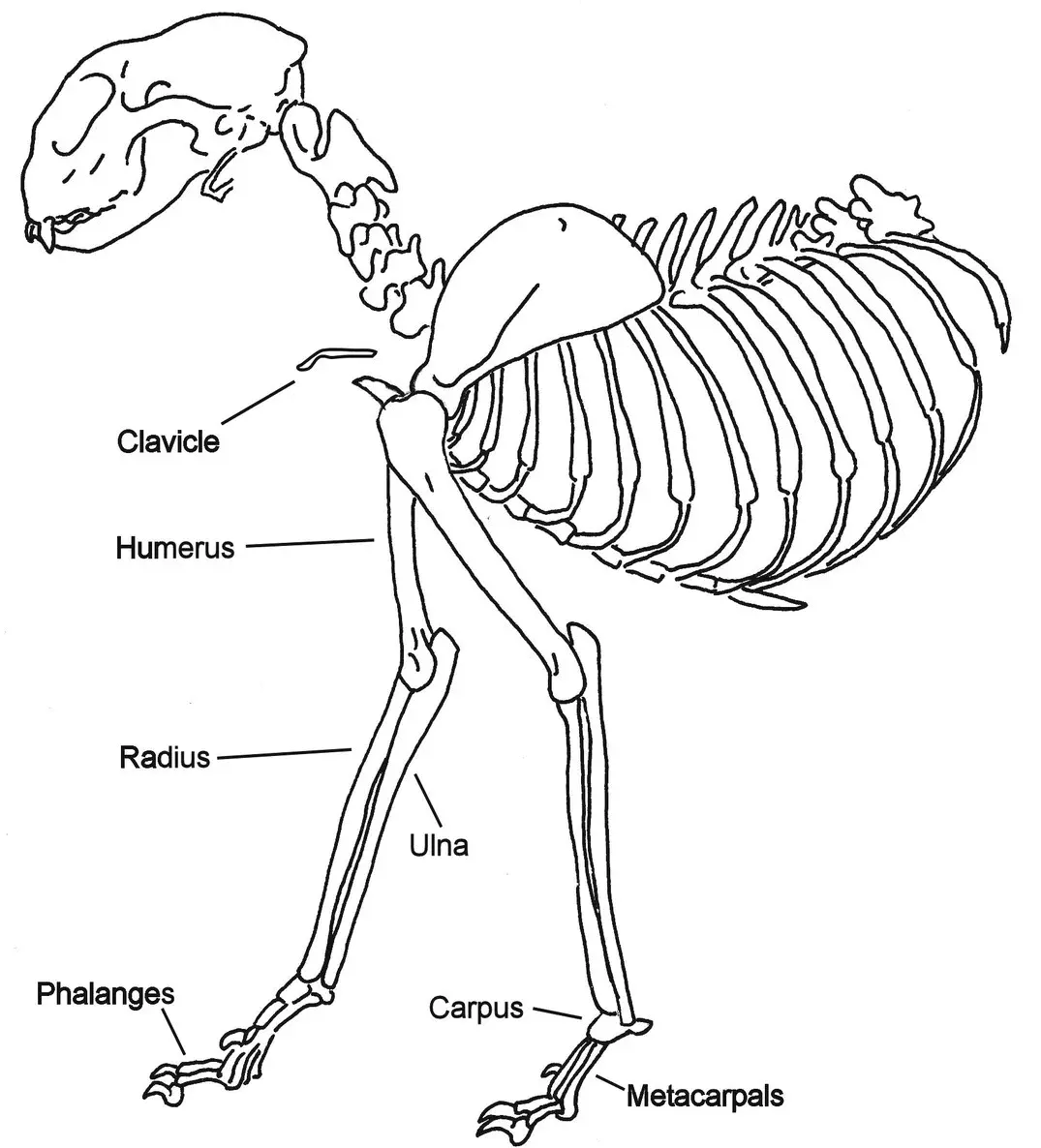
Figure 3.9 Bones of the forelimb.
Source: Courtesy of Jennifer Smith, LVT.
The tarsusis located distal to the tibia and fibula and consists of numerous, irregularly shaped bones that are arranged in several rows. In humans, the tarsus is known as the ankle.
The metatarsalsare the long bones found just distal to the tarsus. Similar to the metacarpals in the front leg, the metatarsals will vary among species. The most distal part of the rear limb are the phalanges. Phalangesare the bones of the digits ( Figure 3.10).
Now that you are familiar with all the basic bones in the dog and cat and the basic directional terms, let’s put them into use. For practice, use the information you learned above to complete the activity below.
1 The skull is ‐‐‐‐‐‐‐‐‐ to the pelvis. (cranial/caudal)
2 The femur is ‐‐‐‐‐‐‐‐‐ to the tarsus. (distal/proximal)
3 The ‐‐‐‐‐‐‐‐‐ is also known as the breastbone.
4 The ‐‐‐‐‐‐‐‐‐vertebrae are the vertebrae of the neck.
5 There are ‐‐‐‐‐‐‐‐‐ lumbar vertebrae.
6 The ‐‐‐‐‐‐ is the long bone of the forelimb that extends from the shoulder to the elbow.
7 The ‐‐‐‐‐‐‐ is just distal to the radius and ulna.
8 The ‐‐‐‐‐‐‐ are the long bones found just distal to the tarsus.
Branches of science
Studying the animal body is a complex activity. There are six branches of science that deal with the study of the body: anatomy, physiology, pathology, embryology, histology, and biology. We will define each of these branches even though this course focus mainly on anatomy and physiology.
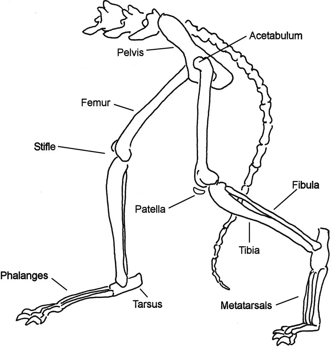
Figure 3.10 Bones of the hindlimb.
Source: Courtesy of Jennifer Smith, LVT.
Anatomy – the study of the structure of the body and the relationship of its parts. Examination of the structures of various animal species is referred to as comparative anatomy.
Physiology – the study of the normal functions and activities of organisms.
Pathology – the study of the causes and effects of diseases or injury.
Embryology – the study of the origin and development of an individual organism. It begins after conception, or fertilization of the egg, and continues through parturition, or birth.
Histology – the microscopic study of the minute structure, composition, and function of cells and tissues.
Biology – the study of all forms of life.
Body systems
The animal’s body is composed of different systems. A system is a combination of organs that performs a particular function. The systems of the body and their functions are listed below.
Musculoskeletal system – includes all the muscles, bones, and joints. It permits motion and movement of the body.
Integumentary system – includes skin, hair, nails, sweat, and sebaceous glands. The skin is considered the largest organ in the body and has many functions. It covers and protects the body, aids in temperature regulation, and has functions in sensation and excretion.
Cardiovascular system – includes the heart and blood vessels. The main function is to transport the blood to the body.
Respiratory system – includes the mouth, nose, trachea, and lungs. It is responsible for absorbing oxygen and discharging carbon dioxide. It also aids in regulating body temperature.
Digestive system – includes the mouth, teeth, salivary glands, esophagus, stomach, intestines, pancreas, colon, liver, and gallbladder. Its function is to digest and absorb food and excrete wastes.
Urogenital system – includes the kidneys, urinary bladder, genitals, ureters, and urethra. It provides for reproduction and urine excretion.
Endocrine system – includes the thyroid glands, adrenal glands, and parathyroid glands. These glands are responsible for manufacturing hormones.
Nervous system – includes the brain, spinal cord, and nerves. With the special senses, it processes stimuli and enables the body to act and respond.
External anatomy
The head consists of several different anatomical parts. The foreheadarea is just above the eyes and just before the stop. The indentation in a dog’s forehead just above level of the eyes is the stop. The depth of the stop will vary depending on the breed of dog. The muzzleis the area between the stop and the nose. The length of the muzzle will vary depending on the breed of dog. The noseis located on the front of the face. It is black in most breeds. The mouth is located below the nose and, again, depending on the breed of dog, the shape and size will vary. Finally, the flewconsists of the upper lips. The flew may be larger and longer in specific breeds such as the boxer or St Bernard. The earsare located on or near the top of the head and can vary in size, placement, and shape depending on the breed ( Figure 3.11).
Читать дальшеИнтервал:
Закладка:
Похожие книги на «Textbook for the Veterinary Assistant»
Представляем Вашему вниманию похожие книги на «Textbook for the Veterinary Assistant» списком для выбора. Мы отобрали схожую по названию и смыслу литературу в надежде предоставить читателям больше вариантов отыскать новые, интересные, ещё непрочитанные произведения.


Обсуждение, отзывы о книге «Textbook for the Veterinary Assistant» и просто собственные мнения читателей. Оставьте ваши комментарии, напишите, что Вы думаете о произведении, его смысле или главных героях. Укажите что конкретно понравилось, а что нет, и почему Вы так считаете.