Lori Renda-Francis - Textbook for the Veterinary Assistant
Здесь есть возможность читать онлайн «Lori Renda-Francis - Textbook for the Veterinary Assistant» — ознакомительный отрывок электронной книги совершенно бесплатно, а после прочтения отрывка купить полную версию. В некоторых случаях можно слушать аудио, скачать через торрент в формате fb2 и присутствует краткое содержание. Жанр: unrecognised, на английском языке. Описание произведения, (предисловие) а так же отзывы посетителей доступны на портале библиотеки ЛибКат.
- Название:Textbook for the Veterinary Assistant
- Автор:
- Жанр:
- Год:неизвестен
- ISBN:нет данных
- Рейтинг книги:4 / 5. Голосов: 1
-
Избранное:Добавить в избранное
- Отзывы:
-
Ваша оценка:
Textbook for the Veterinary Assistant: краткое содержание, описание и аннотация
Предлагаем к чтению аннотацию, описание, краткое содержание или предисловие (зависит от того, что написал сам автор книги «Textbook for the Veterinary Assistant»). Если вы не нашли необходимую информацию о книге — напишите в комментариях, мы постараемся отыскать её.
constitutes an exceptionally broad-ranging and in-depth guide to one of America’s greatest poets. The volume makes the best and most up-to-date scholarly thinking on Walt Whitman available to students. It encourages them to be more aware of the contexts of Whitman’s work, and helps them to understand the experimental nature of his writings. The
starts with a section which communicates a strong sense of the poet’s time, place and history. The contributions in this section range over subjects such as national identity, imperialism, slavery, race, gender, sexuality, and popular culture. A further selection of essays situates Whitman’s work in its literary context. Finally, there are essays devoted to specific works, covering both Whitman’s poetry and prose writings. The
also includes a compact biography of the poet and a bibliography of his works.
Textbook for the Veterinary Assistant — читать онлайн ознакомительный отрывок
Ниже представлен текст книги, разбитый по страницам. Система сохранения места последней прочитанной страницы, позволяет с удобством читать онлайн бесплатно книгу «Textbook for the Veterinary Assistant», без необходимости каждый раз заново искать на чём Вы остановились. Поставьте закладку, и сможете в любой момент перейти на страницу, на которой закончили чтение.
Интервал:
Закладка:
The dorsalplane is an imaginary line that divides the body into dorsal and ventral portions. The medianplane is an imaginary line that extends directly down the middle of the body, dividing it into equal right and left portions ( Figure 3.1). The sagittalplane is an imaginary line that divides the body into right and left unequal parts, and the transverseplane is an imaginary cross‐section that divides the body into cranial and caudal parts.
The terms below are used to describe direction or the position of a body part. We will begin with the basic directional terms.
The term cranialrefers to the head portion of the animal’s body, while caudalrefers to the tail portion of the body. For example, we might say that the tail is caudal to the head or the head is cranial to the tail.
The terms “anterior” and “posterior” are used to describe the front and rear ends of the body. Anteriorrefers to the front of the body and posteriorrefers to the rear end of the body ( Figure 3.2). For example, if a lacerationwas near the tail of an animal, we might say that the laceration is located on the posterior portion of the body.
We stated that the median plane divides the body into equal right and left portions. The terms “medial” and “lateral” are used to describe the location as it relates to the midline. Medialis defined as nearer or toward the midline or median plane, and lateralis defined as farther from the midline or median plane or toward the side of the body ( Figure 3.3).
The dorsal plane divides the body into dorsal and ventral portions. The terms “ventral” and “dorsal” are utilized to describe the location as it relates to the dorsal plane. Ventralis defined as pertaining to the belly or underside of the body, and dorsalis pertaining to the back of the animal or toward the spine. For example, the abdomen is ventral to the spinal column.
“Proximal” and “distal” are commonly used when referring to extremities, with the point of origin being the main body mass. Proximalis used to describe a structure nearer to the point of origin or closer to the body, and distalis used to describe a structure that is farther from the point of origin or away from the body ( Figure 3.4). For example, the femur is proximal to the fibula.
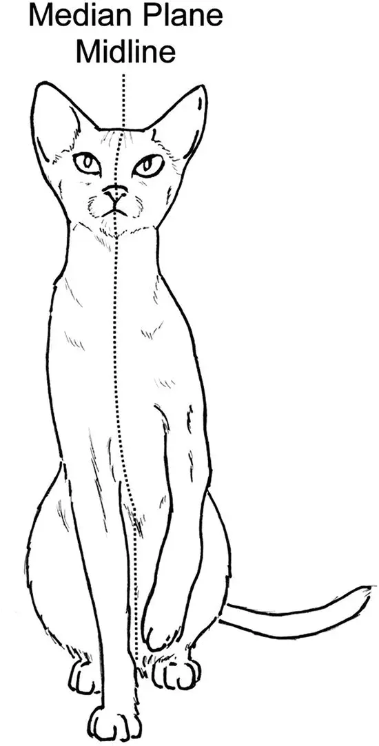
Figure 3.1 Median plane.
Source: Courtesy of Dr Lori Renda‐Francis, LVT.
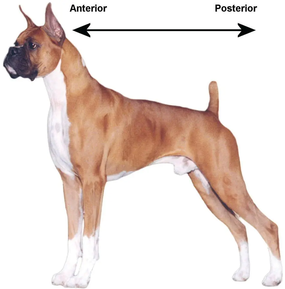
Figure 3.2 Anterior/posterior.
Source: Courtesy of Dr Lori Renda‐Francis, LVT.
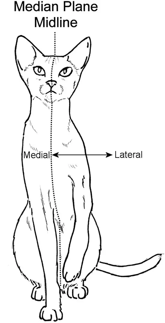
Figure 3.3 Medial/lateral.
Source: Courtesy of Dr Lori Renda‐Francis, LVT.
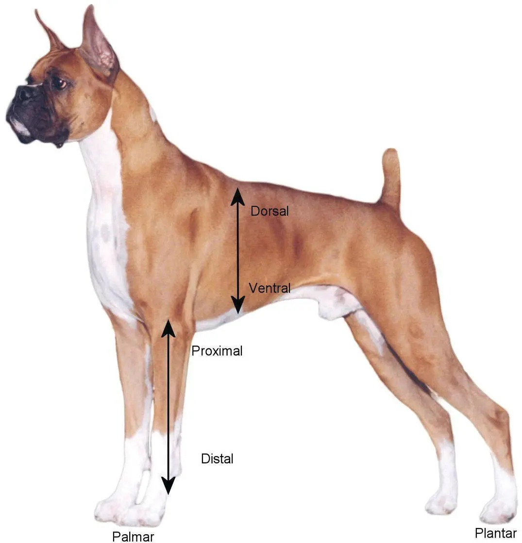
Figure 3.4 Proximal/distal.
Source: Courtesy of Dr Lori Renda‐Francis, LVT.
The terms “palmar” and “plantar” are used when referring to the undersurface of the feet. Palmaris defined as the caudal surface of the forelimb below the carpus,and plantaris the caudal surface of the hindlimb below the tarsus( Figure 3.5).
“Internal” and “external” are used to describe a structure as it relates to the surface of the body. Internalrefers to deep inside the body, and externalrefers to the outer surface of the body, which is more superficial.
Finally, the term rostralis defined as pertaining to the nose end of the head ( Figure 3.6). For example, the nose is rostral to the eyes.
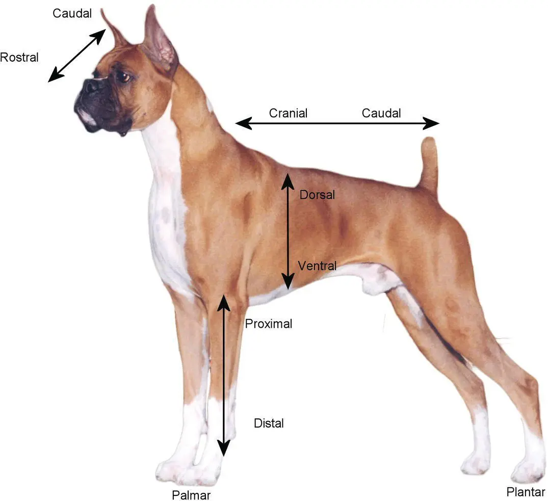
Figure 3.5 Anatomical terms.
Source: Courtesy of Dr Lori Renda‐Francis, LVT.
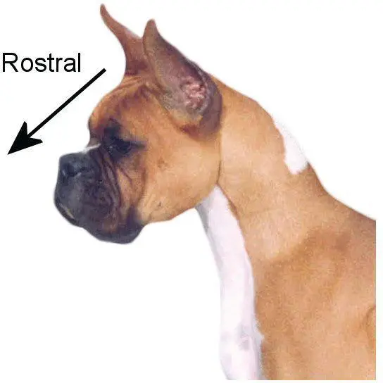
Figure 3.6 Rostral.
Source: Courtesy of Dr Lori Renda‐Francis, LVT.
Skeletal system
Skeletonis defined as the jointed framework of the bones. Osteologyis the study of bones. Veterinary assistants will be expected to describe the bones of the body, which make up the skeleton, and have an understanding of the ways in which they connect and are moved.
The skeleton is divided into two main parts—the axialand the appendicular. The axial skeleton comprises the bones of the skull, the hyoid bones, the ribs, the sternum, and the vertebral column. The appendicular skeleton comprises the bones of the limbs: clavicle, scapula, humerus, radius, ulna, carpus, metacarpals, pelvis, femur, patella, tibia, fibula, tarsus, metatarsals, and phalanges.
Axial skeleton
The craniumis the portion of the skull that encases the brain. The mandible and maxilla are two significant facial bones. The mandibleforms the lower jaw, and the maxillaforms the upper jaw. The eye sockets in the skull are known as the orbits( Figure 3.7).
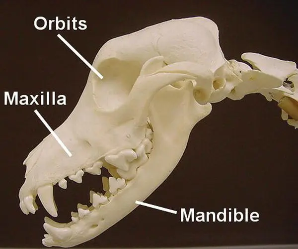
Figure 3.7 Cranium.
Source: Courtesy of Jennifer Smith, LVT.
The hyoid bone(also known as the hyoid apparatus) is made up of several parts that may be cartilage or bone. It is a U‐shaped structure that is located above the larynx and below the mandible and is suspended by ligaments.
Ribsare pairs of flat, curved bones that attach dorsally to the thoracic vertebrae. The cartilage at the end where the rib attaches to the sternum is called the costal cartilage. The sternumis also known as the breastbone, and it forms the ventral midline of the rib cage. The ribs that are not attached to cartilage are known as floating ribs.Cats and dogs have 13 pairs of ribs.
The vertebral columnis also known as the backbone and is made up of numerous vertebrae. There are five different types of vertebrae, and there are variations among species in the number of each different type. Here, we focus on the number of vertebrae in canine and feline patients.
The main functions of the vertebral column, also known as the spinal column, are to support the head and body and to protect the spinal cord. The vertebral column is made up of individual bones called vertebrae.
The cervical vertebraeare the vertebrae of the neck. There are a total of seven cervical vertebrae in all domestic mammals. The first one is called the atlas and the second one is called the axis. The thoracic vertebraeare the vertebrae of the chest. The ribs are attached to these vertebrae. There are 13 thoracic vertebrae in the cat or dog. The lumbar vertebraeare the vertebrae of the lower back. There are a total of seven in cats and dogs. The sacral vertebrae(also known as the sacrum) consist of three fused vertebrae to which the pelvis is attached. Finally, the coccygeal vertebraeare the vertebrae of the tail, and the number of coccygeal vertebrae can vary between six and 23 depending on the species and whether the tail has been docked.
Читать дальшеИнтервал:
Закладка:
Похожие книги на «Textbook for the Veterinary Assistant»
Представляем Вашему вниманию похожие книги на «Textbook for the Veterinary Assistant» списком для выбора. Мы отобрали схожую по названию и смыслу литературу в надежде предоставить читателям больше вариантов отыскать новые, интересные, ещё непрочитанные произведения.


Обсуждение, отзывы о книге «Textbook for the Veterinary Assistant» и просто собственные мнения читателей. Оставьте ваши комментарии, напишите, что Вы думаете о произведении, его смысле или главных героях. Укажите что конкретно понравилось, а что нет, и почему Вы так считаете.