Lori Renda-Francis - Textbook for the Veterinary Assistant
Здесь есть возможность читать онлайн «Lori Renda-Francis - Textbook for the Veterinary Assistant» — ознакомительный отрывок электронной книги совершенно бесплатно, а после прочтения отрывка купить полную версию. В некоторых случаях можно слушать аудио, скачать через торрент в формате fb2 и присутствует краткое содержание. Жанр: unrecognised, на английском языке. Описание произведения, (предисловие) а так же отзывы посетителей доступны на портале библиотеки ЛибКат.
- Название:Textbook for the Veterinary Assistant
- Автор:
- Жанр:
- Год:неизвестен
- ISBN:нет данных
- Рейтинг книги:4 / 5. Голосов: 1
-
Избранное:Добавить в избранное
- Отзывы:
-
Ваша оценка:
Textbook for the Veterinary Assistant: краткое содержание, описание и аннотация
Предлагаем к чтению аннотацию, описание, краткое содержание или предисловие (зависит от того, что написал сам автор книги «Textbook for the Veterinary Assistant»). Если вы не нашли необходимую информацию о книге — напишите в комментариях, мы постараемся отыскать её.
constitutes an exceptionally broad-ranging and in-depth guide to one of America’s greatest poets. The volume makes the best and most up-to-date scholarly thinking on Walt Whitman available to students. It encourages them to be more aware of the contexts of Whitman’s work, and helps them to understand the experimental nature of his writings. The
starts with a section which communicates a strong sense of the poet’s time, place and history. The contributions in this section range over subjects such as national identity, imperialism, slavery, race, gender, sexuality, and popular culture. A further selection of essays situates Whitman’s work in its literary context. Finally, there are essays devoted to specific works, covering both Whitman’s poetry and prose writings. The
also includes a compact biography of the poet and a bibliography of his works.
Textbook for the Veterinary Assistant — читать онлайн ознакомительный отрывок
Ниже представлен текст книги, разбитый по страницам. Система сохранения места последней прочитанной страницы, позволяет с удобством читать онлайн бесплатно книгу «Textbook for the Veterinary Assistant», без необходимости каждый раз заново искать на чём Вы остановились. Поставьте закладку, и сможете в любой момент перейти на страницу, на которой закончили чтение.
Интервал:
Закладка:
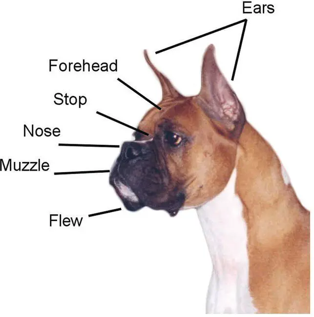
Figure 3.11 External anatomy of the head.
Source: Courtesy of Dr Lori Renda‐Francis, LVT.
Anterior portion
The shoulderis the muscular upper section of the upper arm, and the point of the shoulderconsists of the front of the joint where the upper arm and shoulder blade come together. The brisketis the front portion of the body located between the forelegs and below the chest. The forearmis the top area of the upper front leg. The wristis located lower on the front paw and is also known as the carpus. The pawsinclude the toes, feet, and paw pads of all four of the dog’s legs. The elbowis the joint that connects the upper arm and the forearm ( Figure 3.12).
Posterior portion
The stifleis the joint that connects the lower thigh to the upper thigh. The thighis the area of the hindquarters located between the hip and the stifle. The tarsusis the joint that connects the crus (the lower leg) to the metatarsals, which are the beginning of the foot. The back of the upper thigh is referred to as the hamstrings. The tailis just below the croup and comes in a variety of shapes and lengths depending on the breed. The croupis the area of the back from the root of the tail to the front of the pelvis. The loinis the area on both sides of the vertebrae between the last few ribs and the hindquarters ( Figure 3.13).
Middle portion
The underside of the abdomen is the belly. The area from the withers to the root of the tail is the back. The withersare the top of the shoulder blade and the highest point of the body and are located just behind the neck. The ruffis the thick, dense hair located around the top of the neck. The crestis the upper arched area of the neck ( Figure 3.14).
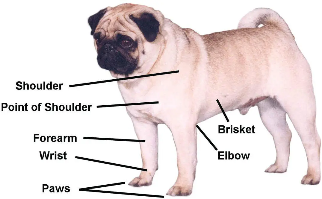
Figure 3.12 External anatomy, anterior portion.
Source: Courtesy of Dr Lori Renda‐Francis, LVT.
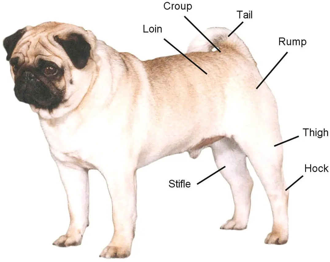
Figure 3.13 External anatomy, posterior portion.
Source: Courtesy of Dr Lori Renda‐Francis, LVT.
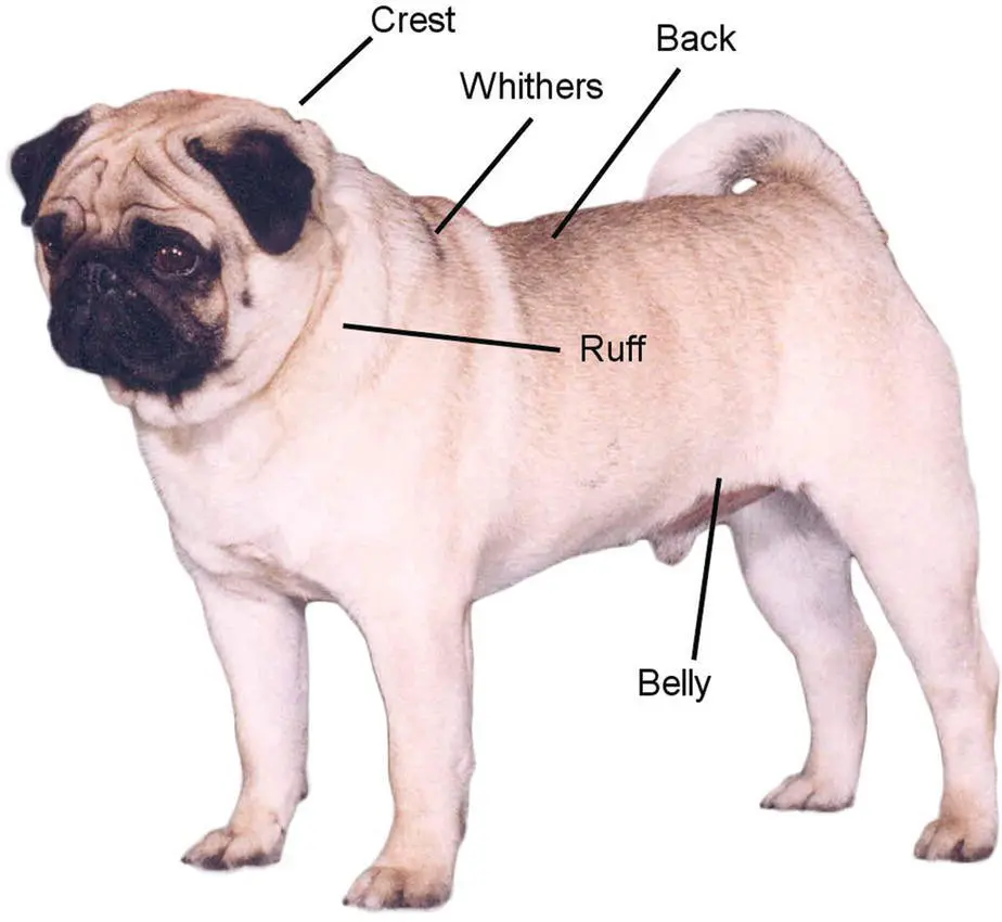
Figure 3.14 External anatomy, middle portion.
source: Courtesy of Dr Lori Renda‐Francis, LVT.
Common veins
While veterinary assistants do not perform venipuncture techniques, they do have a significant role in assisting the veterinarian or veterinary technician in successful collection. The main role of the veterinary assistant during venipuncture is to properly restrain the animal for the procedure. Therefore, it is important for the veterinary assistant to know which veins are commonly used and where they are located and to be able to describe them using correct anatomical terminology.
For dogs, the most frequently used sites for blood collection are the cephalic vein, the jugular vein, and the lateral saphenous vein. The most commonly used veins in cats are the cephalic vein, the jugular vein, the femoral vein, and the medial saphenous vein ( Figure 3.15).
The cephalic veinsare located on the anterior surface of the forearm. They run from the dorsomedial foreleg proximally along the foreleg. They are easy to locate and very accessible for venipuncture. The cephalic vein is used for collection of large volumes of blood in larger dogs.
The lateral saphenous veinsare small, superficial veins that run diagonally across the lateral surface of the distal part of the tibia.
The jugular veinsare large superficial veins located on either side of the trachea on the neck.
The femoral veinis used for blood collection in cats and extends from the groin on the medial aspect of the thigh.
The medial saphenous veinis also used in cats and extends from the hock to the stifle on the medial aspect of the calf. It becomes the femoral vein at the stifle ( Figure 3.16).
Muscles
There are several muscles that can be utilized to administer intramuscular injections. Again, the veterinary assistant does not administer these injections but plays a large role in assisting the veterinarian or veterinary technician by properly restraining the animal. In order to properly restrain, it is important for the veterinary assistant to be familiar with the location of the various muscles ( Figure 3.17).
Lumbodorsal or dorsal lumbar muscle – located on either side of the midline.
Triceps – located caudal to the humerus.
Quadriceps – located anterior to the femur.
Biceps – the posterior muscle of the hind leg.
Semimembranosus/semitendinosus muscle group – located in the rear leg, also known as the hamstring muscles.

Figure 3.15 Veins.
Source: Courtesy of Dr Lori Renda‐Francis, LVT.
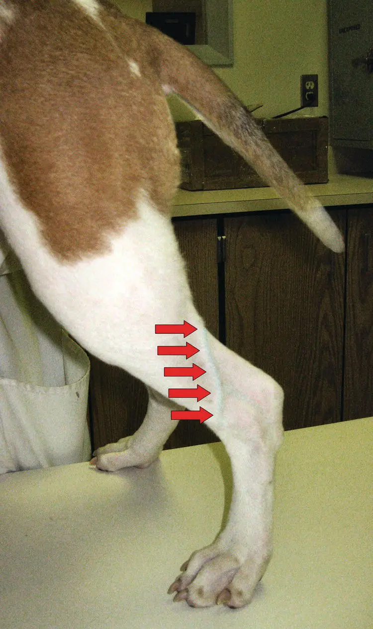
Figure 3.16 Saphenous vein.
Source: Courtesy of Dr Lori Renda‐Francis, LVT.
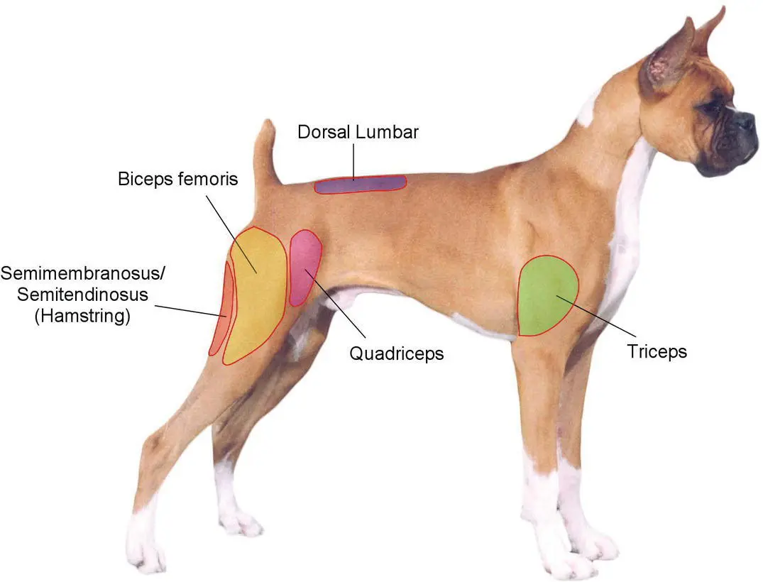
Figure 3.17 Muscles.
Source: Courtesy of Dr Lori Renda‐Francis, LVT.
Internal organs
Digestive system
The most cranial structure is the mouth. The mouth, or oral cavity, is a very important part of the digestive system in dogs. This is where digestion begins. The tongueand front teethhelp a dog pick up pieces of food, and teeth in the back of the mouth grind the food into smaller particles. A dog has a total of 42 teeth, including incisors and canines, located in the front, and the premolars and molars, which are located in the back. As a dog chews, food is broken up into smaller particles for better digestion by enzymes in the stomach and small intestines. It passes through a small tube called the esophagusthat connects the mouth to the stomach via the diaphragm.
The stomachis a sac‐like structure that stores large volumes of food. From the stomach, the food enters the small intestine.There are three parts to the small intestine. The duodenumattaches to the stomach. The middle part is the longest part and is called the jejunum, and the last part is the smallest part and is known as the ileum. It connects to the large intestine. The large intestineconnects the small intestine to the anus. Its primary function is to absorb water from feces as needed in order to keep the animal hydrated. Its other function is to store fecal matter that is awaiting passage from the body. The last structure of the intestine is the rectum, which leads to the most caudal structure – the anus( Figure 3.18).
Читать дальшеИнтервал:
Закладка:
Похожие книги на «Textbook for the Veterinary Assistant»
Представляем Вашему вниманию похожие книги на «Textbook for the Veterinary Assistant» списком для выбора. Мы отобрали схожую по названию и смыслу литературу в надежде предоставить читателям больше вариантов отыскать новые, интересные, ещё непрочитанные произведения.


Обсуждение, отзывы о книге «Textbook for the Veterinary Assistant» и просто собственные мнения читателей. Оставьте ваши комментарии, напишите, что Вы думаете о произведении, его смысле или главных героях. Укажите что конкретно понравилось, а что нет, и почему Вы так считаете.