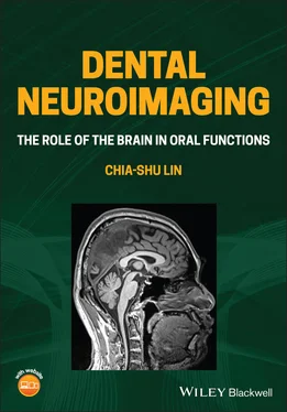
Chia-shu Lin - Dental Neuroimaging
Здесь есть возможность читать онлайн «Chia-shu Lin - Dental Neuroimaging» — ознакомительный отрывок электронной книги совершенно бесплатно, а после прочтения отрывка купить полную версию. В некоторых случаях можно слушать аудио, скачать через торрент в формате fb2 и присутствует краткое содержание. Жанр: unrecognised, на английском языке. Описание произведения, (предисловие) а так же отзывы посетителей доступны на портале библиотеки ЛибКат.
- Название:Dental Neuroimaging
- Автор:
- Жанр:
- Год:неизвестен
- ISBN:нет данных
- Рейтинг книги:4 / 5. Голосов: 1
-
Избранное:Добавить в избранное
- Отзывы:
-
Ваша оценка:
- 80
- 1
- 2
- 3
- 4
- 5
Dental Neuroimaging: краткое содержание, описание и аннотация
Предлагаем к чтению аннотацию, описание, краткое содержание или предисловие (зависит от того, что написал сам автор книги «Dental Neuroimaging»). Если вы не нашли необходимую информацию о книге — напишите в комментариях, мы постараемся отыскать её.
Provides the latest neuroimaging-based evidence on the brain mechanisms of oral functions Dental Neuroimaging: The Role of the Brain in Oral Functions
Dental Neuroimaging: The Role of the Brain in Oral Functions
Dental Neuroimaging — читать онлайн ознакомительный отрывок
Ниже представлен текст книги, разбитый по страницам. Система сохранения места последней прочитанной страницы, позволяет с удобством читать онлайн бесплатно книгу «Dental Neuroimaging», без необходимости каждый раз заново искать на чём Вы остановились. Поставьте закладку, и сможете в любой момент перейти на страницу, на которой закончили чтение.
Интервал:
Закладка:
1.4.5.1 Investigation of the OBB Framework
The OBB framework highlights sensorimotor control of oral functions. The brain mechanisms associated with oral functions can be investigated using a task‐based study, in which brain activities are recorded concurrently when an individual is performing an oral function. The association between sensory or motor processing and oral functions can be modulated by different experimental conditions. For example, when chewing harder food, one should expect stronger sensory feedback from the periodontal tissue than soft food. In this case, changes in brain activation, as shown by functional MRI, would be identified at the somatosensory cortex, and this activity is known to reflect the intensity of sensory inputs (Onozuka et al. 2002; Takahashi et al. 2007). It is noteworthy that the interpretation of an association between task parameters (e.g. the hardness of food) and brain features (e.g. activation of the motor cortex) may be complicated. For example, when chewing a harder piece of food, one may pay more attention to the texture of the food. Therefore, the brain regions with activation may be associated with attention and cognitive control as well as sensory processing (Onozuka et al. 2002, Takahashi et al. 2007).
1.4.5.2 Investigation of the BSA Framework
In contrast to the OBB framework, the BSA framework additionally highlights the importance that the brain will actively adapt to the environment so that even though the functional apparatus is impaired (e.g. losing teeth), individuals can maintain feeding behaviour. A critical step to test this general hypothesis is to focus on the individuals who show structural deficits (e.g. with a higher number of missing teeth) but maintain a good oral function. In this group, the degree of sensorimotor adaptation and learning is assessed using specific tasks (see Chapter 8). The association between task performance and brain activation, particularly in the regions associated with cognitive, affective and motivational processing, is assessed using neuroimaging. Notably, the superiority of neuroimaging is that it can assess brain activation associated with complicated processing of learning and adaptation in human subjects – which may be challenging to perform on animal subjects.
1.4.6 Summary
The OB framework consists of the stomatognathic system as the only functional apparatus for eating and feeding behaviour. According to the OB framework, a sound stomatognathic apparatus means good feeding behaviour.
The OBB framework highlights the role of the exchange of sensorimotor information between the brain and the stomatognathic system in oral functions.
In the BSA framework, the brain is not just a passive translator for the sensorimotor information but also a ‘moderator’ that actively engaged with one's feeding behaviour.
Neuroimaging research on the BSA framework focuses on identifying individual differences in oral functions. The BSA emphasizes that the brain plays a more comprehensive role in sensorimotor and affective–cognitive processing on the stomatognathic functions and feeding behaviour.
Further Readings
1 Please see the Companion Website for Suggested Readings.
References
1 Abrahamsen, R., Dietz, M., Lodahl, S. et al. (2010). Effect of hypnotic pain modulation on brain activity in patients with temporomandibular disorder pain. Pain 151: 825–833.
2 Anderson, T. (1790). Pathological observations on the brain. Lond. Med. J. 11: 182–190.
3 Avivi‐Arber, L., Lee, J., Sood, C. et al. (2015). Long‐term neuroplasticity of the face primary motor cortex and adjacent somatosensory cortex induced by tooth loss can be reversed following dental implant replacement in rats. J. Comp. Neurol. 523: 2372–2389.
4 Avivi‐Arber and Sessle (2018). Jaw sensorimotor control in healthy adults and effects of ageing. J. Oral Rehabil. 45: 50–80.
5 Bandettini, P.A. (2012). Twenty years of functional MRI: the science and the stories. NeuroImage 62: 575–588.
6 Brügger, M., Lutz, K., Brönnimann, B. et al. (2012). Tracing toothache intensity in the brain. J. Dent. Res. 91: 156–160.
7 Carabotti, M., Scirocco, A., Maselli, M.A., and Severi, C. (2015). The gut‐brain axis: interactions between enteric microbiota, central and enteric nervous systems. Ann. Gastroenterol. 28: 203–209.
8 Desouza, D.D., Moayedi, M., Chen, D.Q. et al. (2013). Sensorimotor and pain modulation brain abnormalities in trigeminal neuralgia: a paroxysmal, sensory‐triggered neuropathic pain. PLoS One 8: e66340.
9 Gazzaniga, M.S., Ivry, R.B., and Mangun, G.R. (2019). Cognitive Neuroscience: The Biology of the Mind. W. W. Norton & Company.
10 Gould, D.J., Clarkson, M.J., Hutchins, B., and Lambert, H.W. (2014). How neuroscience is taught to north American dental students: results of the basic science survey series. J. Dent. Educ. 78: 437–444.
11 Gustin, S.M., Peck, C.C., Wilcox, S.L. et al. (2011). Different pain, different brain: thalamic anatomy in neuropathic and non‐neuropathic chronic pain syndromes. J. Neurosci. 31: 5956–5964.
12 Gustin, S.M., Peck, C.C., Cheney, L.B. et al. (2012). Pain and plasticity: is chronic pain always associated with somatosensory cortex activity and reorganization? J. Neurosci. 32: 14874–14884.
13 Habre‐Hallage, P., Dricot, L., Jacobs, R. et al. (2012). Brain plasticity and cortical correlates of osseoperception revealed by punctate mechanical stimulation of osseointegrated oral implants during fMRI. Eur. J. Oral Implantol. 5: 175–190.
14 Hayes, G.B. (1889). Reflex neurosis in relation to dental pathology. Am. J. Dent. Sci. 23: 289–298.
15 Horinuki, E., Shinoda, M., Shimizu, N. et al. (2015). Orthodontic force facilitates cortical responses to periodontal stimulation. J. Dent. Res. 94: 1158–1166.
16 Inamochi, Y., Fueki, K., Usui, N. et al. (2017). Adaptive change in chewing‐related brain activity while wearing a palatal plate: an functional magnetic resonance imaging study. J. Oral Rehabil. 44: 770–778.
17 Iwata, K. and Sessle, B.J. (2019). The evolution of neuroscience as a research field relevant to dentistry. J. Dent. Res. 98: 1407–1417.
18 Jenkinson, M. and Chappell, M. (2018). Introduction to Neuoimaging Analysis. Oxford University Press.Jones, D.K., Knösche, T.R., Turner, R. (2013). White matter integrity, fiber count, and other fallacies: the do’s and don’ts of diffusion MRI. Neuroimage 73: 239–254.
19 Kamer, A.R., Pirraglia, E., Tsui, W. et al. (2015). Periodontal disease associates with higher brain amyloid load in normal elderly. Neurobiol. Aging 36: 627–633.
20 Kamiya, K., Narita, N., and Iwaki, S. (2016). Improved prefrontal activity and chewing performance as function of wearing denture in partially edentulous elderly individuals: functional near‐infrared spectroscopy study. PLoS One 11: e0158070.
21 Kaneko, M., Horinuki, E., Shimizu, N., and Kobayashi, M. (2017). Physiological profiles of cortical responses to mechanical stimulation of the tooth in the rat: an optical imaging study. Neuroscience 358: 170–180.
22 Kimoto, K., Ono, Y., Tachibana, A. et al. (2011). Chewing‐induced regional brain activity in edentulous patients who received mandibular implant‐supported overdentures: a preliminary report. J. Prosthodont. Res. 55: 89–97.
23 Kishimoto, T., Goto, T., and Ichikawa, T. (2019). Prefrontal cortex activity induced by periodontal afferent inputs downregulates occlusal force. Exp. Brain Res. 237: 2767–2774.
24 Lin, C.S. (2018). Revisiting the link between cognitive decline and masticatory dysfunction. BMC Geriatr. 18: 5.
25 Lowell, S.Y., Reynolds, R.C., Chen, G. et al. (2012). Functional connectivity and laterality of the motor and sensory components in the volitional swallowing network. Exp. Brain Res. 219: 85–96.
Читать дальшеИнтервал:
Закладка:
Похожие книги на «Dental Neuroimaging»
Представляем Вашему вниманию похожие книги на «Dental Neuroimaging» списком для выбора. Мы отобрали схожую по названию и смыслу литературу в надежде предоставить читателям больше вариантов отыскать новые, интересные, ещё непрочитанные произведения.
Обсуждение, отзывы о книге «Dental Neuroimaging» и просто собственные мнения читателей. Оставьте ваши комментарии, напишите, что Вы думаете о произведении, его смысле или главных героях. Укажите что конкретно понравилось, а что нет, и почему Вы так считаете.
