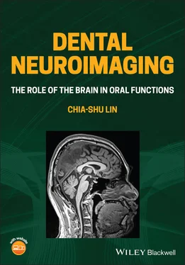
Chia-shu Lin - Dental Neuroimaging
Здесь есть возможность читать онлайн «Chia-shu Lin - Dental Neuroimaging» — ознакомительный отрывок электронной книги совершенно бесплатно, а после прочтения отрывка купить полную версию. В некоторых случаях можно слушать аудио, скачать через торрент в формате fb2 и присутствует краткое содержание. Жанр: unrecognised, на английском языке. Описание произведения, (предисловие) а так же отзывы посетителей доступны на портале библиотеки ЛибКат.
- Название:Dental Neuroimaging
- Автор:
- Жанр:
- Год:неизвестен
- ISBN:нет данных
- Рейтинг книги:4 / 5. Голосов: 1
-
Избранное:Добавить в избранное
- Отзывы:
-
Ваша оценка:
- 80
- 1
- 2
- 3
- 4
- 5
Dental Neuroimaging: краткое содержание, описание и аннотация
Предлагаем к чтению аннотацию, описание, краткое содержание или предисловие (зависит от того, что написал сам автор книги «Dental Neuroimaging»). Если вы не нашли необходимую информацию о книге — напишите в комментариях, мы постараемся отыскать её.
Provides the latest neuroimaging-based evidence on the brain mechanisms of oral functions Dental Neuroimaging: The Role of the Brain in Oral Functions
Dental Neuroimaging: The Role of the Brain in Oral Functions
Dental Neuroimaging — читать онлайн ознакомительный отрывок
Ниже представлен текст книги, разбитый по страницам. Система сохранения места последней прочитанной страницы, позволяет с удобством читать онлайн бесплатно книгу «Dental Neuroimaging», без необходимости каждый раз заново искать на чём Вы остановились. Поставьте закладку, и сможете в любой момент перейти на страницу, на которой закончили чтение.
Интервал:
Закладка:
1.2.3.2 Non‐invasive Methods – Different Focuses of Brain Features
Neuroimaging in the modern days highlights a non‐invasive procedure. For example, no surgical procedure is required for scanning the brain. However, for a non‐surgical approach, subjects may still be exposed to ionizing radiation. The diverse methods can be broadly categorized according to what brain features to be assessed. Computed tomography (CT) and magnetic resonance imaging (MRI) primarily focus on imaging brain structure. As an application of X‐ray imaging, CT may be the tool that dentists are primarily familiar with. It is advantageous in providing a good contrast on the bone tissue which is particularly useful for surgical procedures of dental treatment. In contrast, the MRI assesses the brain based on the water molecules (or strictly speaking, the hydrogen nuclei of the water molecules) in brain tissue. An MRI scanner detects the electromagnetic signals derived from the change of nuclear spinning of hydrogen nuclei. MRI can ‘map’ brain structure because the physical events can be affected by the density of protons (i.e. the hydrogen nuclei) and the relaxation processes associated with the biochemical features of brain tissue (e.g. containing less or more fat). Therefore, different anatomical features (e.g. fat‐containing neural fibres and water‐containing cerebrospinal fluid [CSF]) can be contrasted in MRI images. This advantage enables MRI the primary tool to investigate the morphology, including the size and shape, of the anatomical structure of the brain (Jenkinson and Chappell 2018). In contrast to the structure‐oriented methods, functional approaches focus on detecting the neurophysiological or brain signals associated with mental functions. The approaches include electroencephalogram (EEG), magnetoencephalography (MEG), positron emission tomography (PET), and functional MRI (fMRI), as discussed below.
1.2.3.3 Non‐invasive Methods – Different Sources of Brain Signals
In contrast to structural neuroimaging, functional neuroimaging focuses on the brain signals associated with mental functions. These function‐focusing methods can be categorized into two broad domains. Firstly, EEG and MEG are the methods that directly assess the magneto‐electrical signals from the brain. Both methods rely on the use of an array of strategically deployed sensors on the surface of one's head to collect weak magneto‐electrical signals from the brain. Both methods focus on the magnetic/electrical events of neural activity associated with mental functions. Secondly, PET and fMRI are the methods that assess the metabolic events of the brain, which can be inferred as a surrogated index of neural activity (Gazzaniga et al. 2019). PET assesses the change in metabolic events associated with cerebral blood flow (CBF) by detecting the dynamics of the radioactive‐labelled tracer injected into subjects. fMRI, in contrast, detects the change of the proportion between the oxygenated and deoxygenated haemoglobin as a metabolic index, which is indirectly associated with the change of neural activity (see Chapter 2). Notably, both methods focus on quantifying the relative change of brain signals between different conditions (e.g. when subjects perceive painful vs. non‐painful stimuli). Therefore, the PET and MRI signals do not assess absolute metabolic activity and may not be interpreted as the actual level of neural activity (Gazzaniga et al. 2019).
1.2.4 Structural MRI Methods
Due to the widespread use of MRI in neuroimaging of the human brain, the following section focuses on the application of MRI in the investigation of both structural and functional issues of the brain. The primary goal of structural magnetic resonance imaging (sMRI) methods, in general, is to determine the size and shape of anatomical structures of the brain (Jenkinson and Chappell 2018). Various methods have been developed for investigating grey matter and white matter, two major regions of the CNS.
1.2.4.1 T 1‐Weighted Structural MRI
The most common sMRI data is acquired by T 1‐weighted imaging. Imaging is acquired by weighing on the ‘ T 1’ value, which refers to the time constant for longitudinal relaxation, an index of the rate for protons to return to equilibrium. Critically, this value varies depending on the biochemical components of tissues: the fat‐containing tissue (e.g. neural fibres of white matter) has a shorter T 1 compared to the tissue less rich in fat (e.g. grey matter). In a T 1‐weighted image, the signals collected would preferably show a higher intensity (i.e. brighter) for brain tissue with a higher content of fat and lower intensity for tissue with a lower content of fat. Therefore, the spatial distribution of white matter and grey matter can be differentiated by image intensity. The greatest advantage of T 1‐weighted imaging is that it can be analyzed using different methods to disclose information on brain morphology. For example, tissue‐specific segmentation is a method that separates grey matter, white matter and the space of CSF from the whole‐brain image. Voxel‐based morphometry (VBM) can be used to estimate the amount of grey matter and white matter within each voxel, which can be further used for a group‐based comparison. The method has been widely used for clinical investigation, such as assessing the reduction in hippocampal grey matter between patients with Alzheimer's disease and healthy controls. In addition, the surface‐based analysis is widely used to estimate the thickness of the cortical tissues and the volume of cortical and subcortical regions, which are critical structural features associated with clinical factors (see Section 2.3).
1.2.4.2 Diffusion MRI
While the T 1‐weighted image provides a spatial feature of different brain regions, it provides less information regarding how the brain forms a connectional network. The key to understanding the connection between brain regions is to estimate the orientation of neural fibres. Diffusion magnetic resonance imaging (dMRI) is an MRI method to estimate the distribution of the ‘fibrous’ space in the brain. The method is based on the phenomenon that water molecules spread less freely in the compartment abundant of axons because the freedom to spread is limited by the axons aligned in the same direction. In contrast, the molecules spread more freely in the fluid space, such as the ventricles, where less hindrance exists to restrict the direction of spreading. Diffusion tensor imaging (DTI) is developed to quantify the directionality of diffusion. There are two major applications of dMRI. Firstly, it helps to examine the microstructural properties of the white matter (Jenkinson and Chappell 2018). For example, fractional anisotropy (FA) is a widely used index related to axonal density, the myelination of nerve fibres, and the membrane permeability (Jones et al. 2013). Secondly, dMRI is useful for exploring the structural connectivity of the brain, i.e. how the brain is wired by neural fibres. At present, it is the only tool that can probe the structural connectivity of the human brain in vivo (Jenkinson and Chappell 2018). Tractography has been used to visualize the streamlines that pass between different brain regions. The results provide further information about how brain regions are wired to form a network (see Section 2.3).
1.2.5 Functional MRI Methods
The fMRI methods have two major applications. A task‐based fMRI study investigates the brain signals associated with mental functions. Imaging is conducted when subjects are performing a behavioural task that induces the mental functions of research interest (see Section 2.2). A resting‐state fMRI study investigates the intrinsic activity of the brain, i.e. the spontaneous activity when subjects are not perturbed by external stimuli (see Section 2.4). The fMRI methods can be further categorized according to the nature of the brain signals detected, as discussed below.
Читать дальшеИнтервал:
Закладка:
Похожие книги на «Dental Neuroimaging»
Представляем Вашему вниманию похожие книги на «Dental Neuroimaging» списком для выбора. Мы отобрали схожую по названию и смыслу литературу в надежде предоставить читателям больше вариантов отыскать новые, интересные, ещё непрочитанные произведения.
Обсуждение, отзывы о книге «Dental Neuroimaging» и просто собственные мнения читателей. Оставьте ваши комментарии, напишите, что Вы думаете о произведении, его смысле или главных героях. Укажите что конкретно понравилось, а что нет, и почему Вы так считаете.
