Chia-shu Lin - Dental Neuroimaging
Здесь есть возможность читать онлайн «Chia-shu Lin - Dental Neuroimaging» — ознакомительный отрывок электронной книги совершенно бесплатно, а после прочтения отрывка купить полную версию. В некоторых случаях можно слушать аудио, скачать через торрент в формате fb2 и присутствует краткое содержание. Жанр: unrecognised, на английском языке. Описание произведения, (предисловие) а так же отзывы посетителей доступны на портале библиотеки ЛибКат.
- Название:Dental Neuroimaging
- Автор:
- Жанр:
- Год:неизвестен
- ISBN:нет данных
- Рейтинг книги:4 / 5. Голосов: 1
-
Избранное:Добавить в избранное
- Отзывы:
-
Ваша оценка:
Dental Neuroimaging: краткое содержание, описание и аннотация
Предлагаем к чтению аннотацию, описание, краткое содержание или предисловие (зависит от того, что написал сам автор книги «Dental Neuroimaging»). Если вы не нашли необходимую информацию о книге — напишите в комментариях, мы постараемся отыскать её.
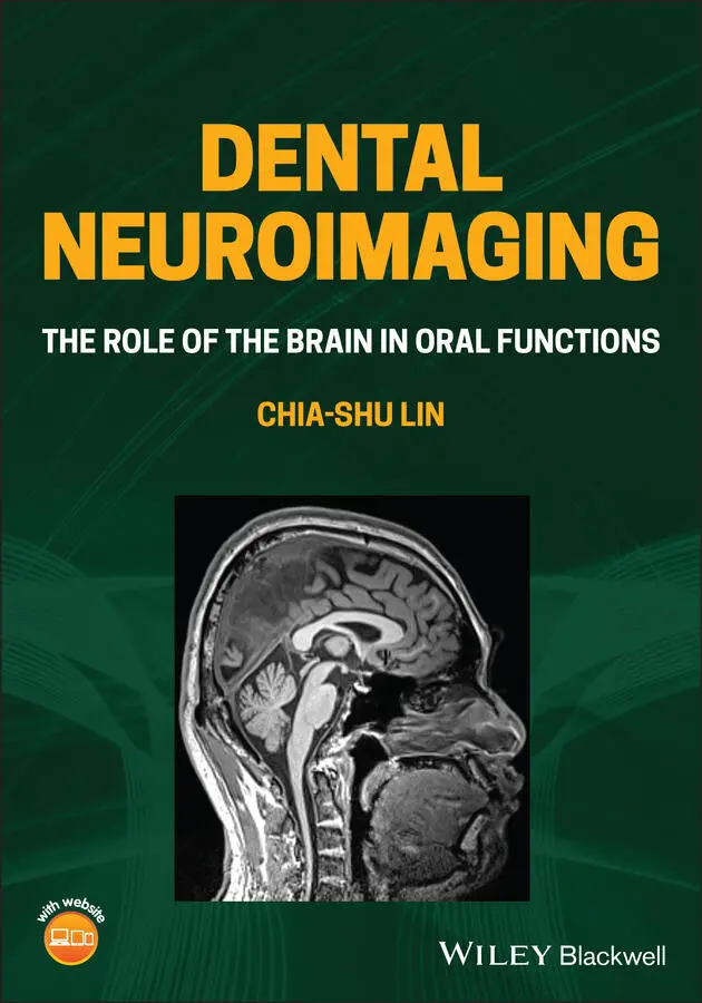
Provides the latest neuroimaging-based evidence on the brain mechanisms of oral functions Dental Neuroimaging: The Role of the Brain in Oral Functions
Dental Neuroimaging: The Role of the Brain in Oral Functions
Dental Neuroimaging — читать онлайн ознакомительный отрывок
Ниже представлен текст книги, разбитый по страницам. Система сохранения места последней прочитанной страницы, позволяет с удобством читать онлайн бесплатно книгу «Dental Neuroimaging», без необходимости каждый раз заново искать на чём Вы остановились. Поставьте закладку, и сможете в любой момент перейти на страницу, на которой закончили чтение.
Интервал:
Закладка:
A second survey was conducted by searching for the past and current research projects funded by the National Institutes of Health (NIH), USA, using the online platform of Research Portfolio Online Reporting Tools (RePORT) report. From 2020 to February 2021, the keywords ‘dental’ and ‘brain’ have led to 106 projects, with 39 projects funded by the National Institute of Dental and Craniofacial Research (NIDCR). This number is almost twice the number of sponsored projects (53) in the whole 1990s when the NIDCR funded 29 projects. The results suggest an increasing trend of cross‐disciplinary research between oral and brain sciences. Critically, not all the projects were granted by the NIDCR, which specializes in orofacial medicine. Several projects were supported by the National Institute of Mental Health and the National Institute on Aging, highlighting the importance of oral issues in cognitive deficits and aging.
1.1.5 Summary
From a functional perspective, the brain, behaviour and oral health are directly linked because the brain plays a crucial role in maintaining oral functions, and the integrity of mental functions is critical to maintaining oral health.
The alliance between the research on dentistry and the brain has a long history. It contributes to tackling unsolved challenges (e.g. orofacial pain) and new challenges (e.g. aging and oral functions).
Topics of neuroscience and the brain are not neglected in dental education. However, many courses are taught by non‐dental school faculties, and the topics were less tailored for dentistry.
Recently, cross‐disciplinary research on oral and brain sciences has quickly emerged in the number of publications and research grants.
1.2 What Is Neuroimaging?
1.2.1 Introduction
In Section 1.1, we have highlighted a significant overlap between dentistry and brain science. Though the two fields are closely linked from a functional perspective, there exists a vast difference between research approaches of the oral cavity and those of the brain. Dentists can visually examine the oral structure, and oral functions can be quantified with a chairside set of assessments. In contrast, brain functions and mental status, sometimes metaphorized as a ‘black box’, can hardly be examined directly at the chairside. Therefore, a pivotal step to facilitate the investigation of the brain is to develop the technology for quantifying brain structure and functions. Neuroimaging, defined as a non‐invasive approach of ‘visualizing the central nervous system, especially the brain, by various imaging modalities’ (MeSH 2012), is such a technological breakthrough that revolutionizes the research approaches of the brain.
A common myth is that neuroimaging or ‘brain scan’ would be a kind of ‘modern magic’. The impression is strengthened by some sci‐fi movies, where ‘peeking into the brain’ is taken as an icon of something futuristic. Contrary to the popular myth, the term ‘neuroimaging’ has been adopted as a common method for regular clinical investigation and research. In the following sections, we outline the primary methods of neuroimaging approaches in brain science, and their roles in brain science and practical implications in dentistry are highlighted.
1.2.2 What Is the Role of Neuroimaging Research in Dentistry
1.2.2.1 Trends of Research Publications in Dental Neuroimaging Research
Table 1.1summarizes the number of dental research publications combined with research on the brain and neuroimaging, according to a PubMed‐based survey. The trend of publication of dental research related to neuroimaging was strikingly similar to that related to the brain. Before 1970, only a few publications were found in these fields. In contrast, after the 1990s, the percentage of these studies showed a pronounced increase. Notably, this trend in publication can be compared with the dental research related to neuroscience in general, as recently reported by Iwata and Sessle (2019). In terms of brain and neuroimaging research, almost half of the dental research on brain and neuroimaging was published last decade (2011–2020) ( Table 1.1). This trend is very different from the general field of neuroscience. It is also noteworthy that more than 90% of the dental research on neuroimaging was published after the mid‐1990s, about the same time when functional magnetic resonance imaging (fMRI) came into practice (Bandettini 2012). The trend reflects that technological innovation of biomedical imaging may facilitate cross‐disciplinary research on dentistry and the brain.
Table 1.1 Trends of the academic publication ain dental research related to brain and neuroimaging.
| Period | Dentistry + Brain | Dentistry + Neuroimaging | ||
|---|---|---|---|---|
| No. of articles | Percentage | No. of articles | Percentage | |
| 2011–2020 | 10004 | 49 | 391 | 65 |
| 2001–2010 | 5119 | 25 | 152 | 25 |
| 1991–2000 | 2773 | 14 | 61 | 10 |
| 1981–1990 | 1719 | 8 | 2 | 0 |
| 1971–1980 | 631 | 3 | 0 | 0 |
| 1961–1970 | 145 | 1 | 0 | 0 |
| 1951–1960 | 53 | 0 | 0 | 0 |
| before 1951 | 11 | 0 | 0 | 0 |
aThe number of articles was surveyed using PubMed with the following combination of keywords: ‘tooth[mesh] OR oral[mesh] OR dental[mesh] OR dentistry[mesh] OR teeth[tiab] OR tooth[tiab] OR oral[tiab] OR dental[tiab] OR dentistry[tiab]’ in conjunction with ‘brain[tiab]’ and ‘neuroimaging[tiab]’ for ‘Dentistry + Brain’ and ‘Dentistry + Neuroimaging’, respectively.
1.2.2.2 The ‘Landmark Discoveries or Concepts’: Past and Future
In their article ‘ The Evolution of Neuroscience as a Research Field Relevant to Dentistry ’ for the Journal of Dental Research (JDR) Centennial Series, Iwata and Sessle enlisted several achievements of orofacial neuroscience in the decades (Iwata and Sessle 2019). Many of these achievements have been made based on clinical, animal and laboratory research. For example, the gate control theory has been widely investigated from the clinical to the molecular levels. Remarkably, animal research has unravelled a complex pattern of bi‐directional projections between the stomatognathic system to the brain ( Figure 1.2). Investigation of the human brain may disclose more insights on this topic. While the ‘gating’ mechanism at the spinal level has been gradually elucidated, how the nociceptive processing is translated to pain, a subjective experience, has remained a challenging issue. As noted in Chapter 6, neuroimaging methods may help extend our current knowledge in pain and its management. Another example is the investigation of neural mechanisms of mastication and swallowing, which significantly impact our understanding of oral physiology and the management of oral dysfunctions. Recent neuroimaging findings, on the one hand, confirm the evidence from animal research (e.g. the role of primary sensorimotor cortices in chewing) ( Table 1.2and Figure 1.2). On the other hand, neuroimaging findings disclose new knowledge about the role of learning and cognitive control in oral motor functions ( Table 1.3). As shown in Table 1.2, many examples reveal how neuroimaging, as an exploratory tool, broadens the frontier of orofacial neuroscience into the uncharted area.
1.2.3 Methods of Neuroimaging
As an approach to visualize the central nervous system (CNS), neuroimaging consists of various methods to image the brain. The methods can be generally categorized by the degree of invasiveness, by the brain features to be quantified (e.g. brain structure or functions), and by the signals to be detected (e.g. neural activity or cerebral flow). A brief introduction of the methods is summarized in the following sections, and more detailed mechanisms are discussed in Chapter 2.
Читать дальшеИнтервал:
Закладка:
Похожие книги на «Dental Neuroimaging»
Представляем Вашему вниманию похожие книги на «Dental Neuroimaging» списком для выбора. Мы отобрали схожую по названию и смыслу литературу в надежде предоставить читателям больше вариантов отыскать новые, интересные, ещё непрочитанные произведения.


Обсуждение, отзывы о книге «Dental Neuroimaging» и просто собственные мнения читателей. Оставьте ваши комментарии, напишите, что Вы думаете о произведении, его смысле или главных героях. Укажите что конкретно понравилось, а что нет, и почему Вы так считаете.