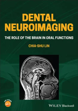
Chia-shu Lin - Dental Neuroimaging
Здесь есть возможность читать онлайн «Chia-shu Lin - Dental Neuroimaging» — ознакомительный отрывок электронной книги совершенно бесплатно, а после прочтения отрывка купить полную версию. В некоторых случаях можно слушать аудио, скачать через торрент в формате fb2 и присутствует краткое содержание. Жанр: unrecognised, на английском языке. Описание произведения, (предисловие) а так же отзывы посетителей доступны на портале библиотеки ЛибКат.
- Название:Dental Neuroimaging
- Автор:
- Жанр:
- Год:неизвестен
- ISBN:нет данных
- Рейтинг книги:4 / 5. Голосов: 1
-
Избранное:Добавить в избранное
- Отзывы:
-
Ваша оценка:
- 80
- 1
- 2
- 3
- 4
- 5
Dental Neuroimaging: краткое содержание, описание и аннотация
Предлагаем к чтению аннотацию, описание, краткое содержание или предисловие (зависит от того, что написал сам автор книги «Dental Neuroimaging»). Если вы не нашли необходимую информацию о книге — напишите в комментариях, мы постараемся отыскать её.
Provides the latest neuroimaging-based evidence on the brain mechanisms of oral functions Dental Neuroimaging: The Role of the Brain in Oral Functions
Dental Neuroimaging: The Role of the Brain in Oral Functions
Dental Neuroimaging — читать онлайн ознакомительный отрывок
Ниже представлен текст книги, разбитый по страницам. Система сохранения места последней прочитанной страницы, позволяет с удобством читать онлайн бесплатно книгу «Dental Neuroimaging», без необходимости каждый раз заново искать на чём Вы остановились. Поставьте закладку, и сможете в любой момент перейти на страницу, на которой закончили чтение.
Интервал:
Закладка:
1.3.3 Links Between Neuroimaging and Clinical Disciplines of Dentistry
Clinically, neuroimaging research may help dental professions to understand the association between mental functions and dental treatment. For example, dentists may need to know the sensory processing of a patient who is chewing with an implant‐supported denture. If a dental implant alters the sensory feedback from occlusion, one should expect to identify changes in cortical activation at the sensory area when subjects are chewing. Neuroimaging would be a suitable tool for the study related to the treatment outcomes of dental practice.
1.3.3.1 Prosthodontics
Restoration of both structural deficits and functional impairment is key to successful prosthodontic treatment. For patients with tooth loss, replacing the missing teeth with a dental prosthesis will restore structural deficits. Moreover, patients should adapt to the prosthesis and improve chewing function. Recent neuroimaging findings have shed light on the mechanisms of adaptation of dental prostheses ( Table 1.3). For example, longitudinal research revealed that adaptation of a new denture was associated with not only the improvement of masticatory performance but also changes in brain activation in the somatosensory cortex (Luraschi et al. 2013). Consistently, tactile stimulation on dental implants was associated with brain activation of the somatosensory areas (Habre‐Hallage et al. 2012). The findings suggest that changes in sensory feedback play a key role in improving oral functions by prostheses. Moreover, in partially edentulous patients, reduced occlusion was associated with reduced activation of the prefrontal cortex, and such reduced activation can be modulated by installing a denture (Kamiya et al. 2016). In patients with maxillary dental implants, tactile stimuli induced brain activation not only in the somatosensory cortex but also in the prefrontal cortex (Habre‐Hallage et al. 2012). Changes in somatosensory and prefrontal activation were also identified in another longitudinal fMRI study that dentated patients were chewing a piece of gum when wearing a plate to cover their palate. The change in brain activation was associated with the recovery of masticatory performance, which was initially impaired (by the palatal plate) and later restored (Inamochi et al. 2017). These novel findings suggest that beyond sensory processing, the attentional and cognitive processing related to wearing a denture, as evidenced by the changes in the prefrontal cortex, may play a vital role in the adaptation of prosthodontic treatment. Thus, the neuroimaging findings provide new clues for prosthodontic treatment by highlighting the patient’s adaptation to dental devices.
1.3.3.2 Periodontics
One of the primary roles of the periodontium is to support sensory feedback via the periodontal ligament. Neuroimaging would help clarify the neural pathway of sensory processing of periodontal stimuli (Kaneko et al. 2017; Kishimoto et al. 2019; Ono et al. 2015). Moreover, neuroimaging may contribute to elucidating the association between periodontal health and other systemic conditions ( Table 1.3). A recently hotly debated issue is the association between dementia and neuroinflammation as well as neurotoxicity, which may relate to periodontal health (Tonsekar et al. 2017). To explore this brain–stomatognathic connection, researchers have directly assessed the association between periodontal health and the Aβ plaques of the brain, a critical feature of Alzheimer's dementia (Kamer et al. 2015). In this study, the pathological feature of Aβ plaque was assessed using PET, and the association between brain pathology and periodontal health (e.g. clinical attachment loss) can be quantified (Kamer et al. 2015). The association between oral health and other pathological brain features, such as lacunar infarction, can also be investigated using neuroimaging methods (Taguchi et al. 2013). Neuroimaging is a valuable tool for investigating the sensory pathway of periodontal inputs and the association between periodontal health and systemic conditions.
1.3.3.3 Orthodontics
Just like prosthodontic treatment, the success of orthodontic treatment is associated with patients' adaptation to the oral appliance. Again, the neuroimaging findings revealed that the use of the oral appliance is associated with an extended area of brain activation, not just confined to the somatosensory cortex (Horinuki et al. 2015; Ozdiler et al. 2019) ( Table 1.3). In rats, experimental tooth movement was associated with changes in brain activity of the secondary somatosensory cortex and the insula (Horinuki et al. 2015). Critically, the animal model revealed that during tooth movement, brain activity change was also associated with inflammation, as identified by the expression of the inflammatory factors and macrophage infiltration in the periodontal tissue (Horinuki et al. 2015). The findings have demonstrated the strength of combined neuroimaging and histological approaches, which help elucidate changes in clinical symptoms and signs related to dental treatment.
1.3.4 Summary
As an in vivo imaging method, neuroimaging has become the crucial method for studying the perceptual and psychosocial aspects of oral functions, which are difficult to approach with animal models.
Recent neuroimaging findings suggest that beyond sensory processing, the attentional and cognitive processing related to wearing a denture may play a vital role in the adaptation of prosthodontic treatment. Neuroimaging research provides new clues for prosthodontic treatment by highlighting the patient’s adaptation to dental devices.
Neuroimaging is a valuable tool for investigating the sensory pathway of periodontal inputs and the association between periodontal health and systemic conditions.
1.4 The Brain–Stomatognathic Axis
1.4.1 Introduction
As addressed in Section 1.1, our understanding of oral functions will not be completed if we overlook brain functions. On the one hand, the CNS plays a crucial role in sensorimotor control of the oral apparatus. On the other hand, all the behaviours related to oral functions, including feeding and oral healthcare, are closely associated with general mental functions, including cognitive and affective processing. Therefore, from the functional perceptive, the brain and the stomatognathic system are both essential for maintaining oral health.
However, the brain–stomatognathic connection stated above is descriptive and does not fully explain the underlying mechanisms. The critical and yet unanswered question is ‘how do the brain and the stomatognathic system work together to maintain our oral health?’. The question is difficult to answer because, before the advent of neuroimaging, researchers have had few tools to directly observe the brain mechanisms associated with oral functions in human subjects. In the following sections, we outline several theoretical frameworks for the brain–stomatognathic connection. The core concepts of the brain–stomatognathic connection are defined. Subsequently, three theoretical frameworks on the brain–stomatognathic connection are discussed. Finally, experimental design to test these theoretical frameworks are discussed.
1.4.2 Core Elements of the Brain–Stomatognathic Connection
Before discussing the brain–stomatognathic connection, we need to define the core elements of the connection. Particularly, we will clarify the functional element, i.e. the brain and the stomatognathic system, and human feeding behaviour.
1.4.2.1 Definition of the Functional Element
Based on the definition from Medical Subject Headings (MeSH) of the National Library of Medicine, USA, the stomatognathic system is defined as ‘the mouth, teeth, jaws, pharynx and related structures as they relate to mastication, deglutition and speech’ (MeSH 1986). By this definition, either intraoral structure (e.g. teeth and the tongue) or extraoral structure (e.g. the masseter) is part of the stomatognathic system since all the structure contributes to maintaining normal oral functions. Notably, from the functional perspective, our brain should also be considered as part of the functional element related to oral functions, even though the brain is not part of the stomatognathic system. Both cortical and subcortical regions are closely associated with oral functions ( Figure 1.2).
Читать дальшеИнтервал:
Закладка:
Похожие книги на «Dental Neuroimaging»
Представляем Вашему вниманию похожие книги на «Dental Neuroimaging» списком для выбора. Мы отобрали схожую по названию и смыслу литературу в надежде предоставить читателям больше вариантов отыскать новые, интересные, ещё непрочитанные произведения.
Обсуждение, отзывы о книге «Dental Neuroimaging» и просто собственные мнения читателей. Оставьте ваши комментарии, напишите, что Вы думаете о произведении, его смысле или главных героях. Укажите что конкретно понравилось, а что нет, и почему Вы так считаете.


