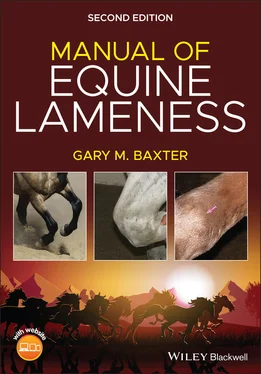
Gary M. Baxter - Manual of Equine Lameness

Здесь есть возможность читать онлайн «Gary M. Baxter - Manual of Equine Lameness» — ознакомительный отрывок электронной книги совершенно бесплатно, а после прочтения отрывка купить полную версию. В некоторых случаях можно слушать аудио, скачать через торрент в формате fb2 и присутствует краткое содержание. Жанр: unrecognised, на английском языке. Описание произведения, (предисловие) а так же отзывы посетителей доступны на портале библиотеки ЛибКат.

Manual of Equine Lameness: краткое содержание, описание и аннотация

Предлагаем к чтению аннотацию, описание, краткое содержание или предисловие (зависит от того, что написал сам автор книги «Manual of Equine Lameness»). Если вы не нашли необходимую информацию о книге — напишите в комментариях, мы постараемся отыскать её.

MANUAL OF EQUINE LAMENESS <p><b>Discover a concise and accessible guide to diagnosing and managing lameness in horses </b> <p>The revised Second Edition of<i> Manual of Equine Lameness </i>offers a concise and accessible manual of lameness diagnosis and treatment in horses. Perfect for use as a quick reference, this book provides straightforward access to the essentials of equine lameness, including the clinical assessment of the horse and commonly performed diagnostic nerve blocks and the most common conditions of the foot, forelimb, and hindlimb that may be contributing to the lameness. Current therapeutic options to treat lameness are also discussed, as well as guidance on how to manage musculoskeletal emergencies. The content has been distilled from the authoritative Seventh Edition of <i>Adams and Stashak’s Lameness in Horses,</i> and this new edition has been re-envisioned to be even quicker and easier to navigate than the previous version. <p>Color photographs and illustrations support the text, which presents lameness information most relevant to equine general practitioners, mixed animal practitioners, and veterinary students. A companion website offers videos that focus on the clinical examination of the horse and select diagnostic blocks and relevant anatomy. Diagnostic and treatment material has been revised from the previous edition to include the most up-to-date information. <p>Readers will find: <ul><li>A thorough introduction to the assessment of the lame horse, including history, visual exam, palpation, subjective and objective assessments of lameness, perineural anesthesia, and intrasynovial anesthesia</li> <li>An exploration of common conditions of the foot, including the navicular region and soft tissue injuries, coffin joint and distal phalanx conditions, and laminitis </li> <li>Discussions of the most common conditions of the forelimb, including the pastern, fetlock, metacarpus/metatarsus, carpus, antebrachium, elbow, and humerus, as well as the shoulder and scapula</li> <li>Discussions of common conditions of the hindlimb and axial skeleton</li> <li>A review of therapeutic options to treat lameness conditions</li> <li>How to manage musculoskeletal emergencies in the horse</li></ul> <p>Ideal for veterinary students, early career equine practitioners, and mixed animal veterinarians, the Second Edition of<i> Manual of Equine Lameness </i>is an indispensable reference for any veterinarian seeking a concise one-stop reference for equine lameness.

Manual of Equine Lameness — читать онлайн ознакомительный отрывок

Ниже представлен текст книги, разбитый по страницам. Система сохранения места последней прочитанной страницы, позволяет с удобством читать онлайн бесплатно книгу «Manual of Equine Lameness», без необходимости каждый раз заново искать на чём Вы остановились. Поставьте закладку, и сможете в любой момент перейти на страницу, на которой закончили чтение.

Тёмная тема
Сбросить

Интервал:

Закладка:

Сделать

3 Chapter 3Figure 3.1 Typical enlargement of the pastern that may be visible in horses ...Figure 3.2 Lateral (a) and dorsopalmar (b) radiographs of the pastern region...Figure 3.3 Lateral (a) and dorsopalmar (b) proton density (pd) MRI images of...Figure 3.4 Lateral (a) and DP radiographs (b) of the pastern following place...Figure 3.5 (a) Lateral radiograph of a horse with a dorsal OC fragment (arro...Figure 3.6 Dorsoplantar radiographs of two different young horses demonstrat...Figure 3.7 Lateral radiographic view (a) and postmortem specimen (b) from a ...Figure 3.8 Visual (a) and radiographic (b) lateral views of a young horse wi...Figure 3.9 Sesamoidean ligaments. Dashed lines indicate positions of the pro...Figure 3.10 Oblique radiograph of the pastern region demonstrating a medial ...Figure 3.11 Lateral radiograph of the pastern demonstrating biaxial eminence...Figure 3.12 Lateral (a), dorsoplantar (b), and two oblique (c and d) radiogr...Figure 3.13 CT images of a comminuted P2 fracture that demonstrate the numer...Figure 3.14 Lateral radiograph one year after the biaxial eminence fracture ...Figure 3.15 Dorsopalmar radiographs of the pastern region revealing short (a...Figure 3.16 Lateral radiograph of the fetlock demonstrating a fracture of th...Figure 3.17 Dorsopalmar (a) and lateral (b) radiographs of a horse with a se...Figure 3.18 Lateral radiographs of a nondisplaced, moderately comminuted P1 ...Figure 3.19 Transverse proton density MRI image showing high signal intensit...Figure 3.20 This horse was 4/5 lame in the left hindlimb and had severe effu...Figure 3.21 Images of the same horse in Figure3.20 .(a) Transverse and (b) ...Figure 3.22 (a) Flexed lateral radiograph of the fetlock demonstrating a sma...Figure 3.23 Lateral (a) and oblique (b) radiographs demonstrating a typical ...Figure 3.24 Oblique radiograph of a Quarter horse mare with a large apical s...Figure 3.25 Flexed lateral (a) and DP (b) radiographs demonstrating a small ...Figure 3.26 (a) DP radiograph of a horse with an abaxial sesamoid fracture (...Figure 3.27 Radiographs of the fetlock in two different horses demonstrating...Figure 3.28 DP radiograph (a) and transverse CT image (b) of two different h...Figure 3.29 DLPMO radiograph of the fetlock (a) of a horse with a large peri...Figure 3.30 Sagittal proton density MR image depicting marked cartilage dama...Figure 3.31 (a) The distal aspect of the third metacarpal condyles demonstra...Figure 3.32 Flexed lateral (a) and dorsopalmar (b) radiographs of two differ...Figure 3.33 (a) DP radiograph of a horse with a subchondral cystic lesion in...Figure 3.34 (a) Lateral radiograph of a horse with disruption of the distal ...Figure 3.35 This horse had swelling of the DFTS and evidence of constriction...Figure 3.36 Tenoscopic view within the DFTS following transection of the ann...Figure 3.37 Bones of the left equine thoracic limb (lateral view).Figure 3.38 A racing Quarter horse with the classical metacarpal profile of ...Figure 3.39 Lateromedial radiograph showing a dorsal cortical fracture of MC...Figure 3.40 Condylar fractures. (a) Incomplete, (b) complete nondisplaced, (...Figure 3.41 Radiographs of a spiraling medial condylar fracture in a racing ...Figure 3.42 This oblique radiograph of the tarsus revealed an incomplete pro...Figure 3.43 (a) Open comminuted mid‐diaphyseal fracture of MTIII in a seven‐...Figure 3.44 Visible enlargement of the medial splint area just distal to the...Figure 3.45 This large exostosis of the medial splint contributed to lamenes...Figure 3.46 Fractures of the distal splint bones such as this rarely heal an...Figure 3.47 Proximal fractures of the fourth metatarsal bone are often commi...Figure 3.48 Chronic middle splint fracture with secondary drainage and perio...Figure 3.49 A dorsomedial‐palmarolateral oblique radiograph of the carpus sh...Figure 3.50 The attachments of the suspensory ligament at the proximal palma...Figure 3.51 (a) Nuclear scintigraphy can identify bone injury at the origin ...Figure 3.52 Transverse proton density image of the proximal metatarsal regio...Figure 3.53 Severe dropping of the fetlock together with a straight hock con...Figure 3.54 Classic appearance of mild (a) and more severe (b) SDF tendiniti...Figure 3.55 A “low bow” of the SDFT, as shown in this image, can be associat...Figure 3.56 Ultrasound longitudinal sections through the midportion of a nor...Figure 3.57 Dorsal view of the left carpus.Figure 3.58 Bilateral symmetrical fluid swellings of the lateral aspects of ...Figure 3.59 An adult horse with synovial effusion of the ECR tendon sheath p...Figure 3.60 Carpal ligaments, medial view (a) and lateral view (b).Figure 3.61 (a) A radiographic image showing a subtle fragment on the distal...Figure 3.62 Radiograph demonstrating osteochondral chip fragments off the pr...Figure 3.63 30° degree flexed dorsoproximal dorsodistal oblique radiograph s...Figure 3.64 Lateromedial (a) and flexed lateromedial (b) radiographs of a fr...Figure 3.65 Aged mare with varus deviation of the left forelimb and swelling...Figure 3.66 Radiographs demonstrating mild signs of osteoarthritis, includin...Figure 3.67 A radiograph demonstrating collapse of the medial aspect of the ...Figure 3.68 Palmaromedial view of carpus with flexor retinaculum cut and ref...Figure 3.69 Carpal canal swelling visible (arrow) and palpable on the latera...Figure 3.70 Lateral radiograph of the carpus demonstrating an osteochondroma...Figure 3.71 Flexed lateral radiograph of the carpus of a horse with a chroni...Figure 3.72 A cranial‐caudal view of a distal diaphyseal comminuted fracture...Figure 3.73 Wound on the medial aspect of the antebrachium from a kick injur...Figure 3.74 A transverse complete midshaft fracture of the radius of a foal....Figure 3.75 A nuclear scintigraphic examination of increased uptake of radio...Figure 3.76 Classification of equine ulnar fractures.Figure 3.77 Lateral view of the left elbow. Dashed lines represent the locat...Figure 3.78 Typical dropped elbow appearance and the inability to extend the...Figure 3.79 (a) Type III ulnar fracture. Courtesy of Lorrie Gaschen. (b) Typ...Figure 3.80 Comminuted ulna fracture in a young foal (a) that healed flowing...Figure 3.81 Subchondral cystic lesion located on the axial aspect of the lat...Figure 3.82 A chronic, infected, acquired capped elbow of the left forelimb....Figure 3.83 (a) Oblique lateral view of a spiral nonarticular fracture of th...Figure 3.84 Nuclear scintigraphic examination of the humerus with increased ...Figure 3.85 Core lesion of the medial branch of the biceps tendon just dista...Figure 3.86 Osseous changes on the tubercles of the cranioproximal humerus (...Figure 3.87 Radiographic manifestations of OCD of the shoulder. (a) The defe...Figure 3.88 Postmortem view of a large OCD lesion of the caudal humeral head...Figure 3.89 Radiographs of a subluxated left shoulder in a miniature horse t...Figure 3.90 The suprascapular nerve courses over the cranial edge of the sca...Figure 3.91 Prominent atrophy of the supraspinatus muscle is evident in this...Figure 3.92 Prominent swelling located over the lateral right shoulder regio...Figure 3.93 Fracture of the supraglenoid tubercle of the scapula with modera...

4 Chapter 4Figure 4.1 Bones of the left pelvic limb, lateral view.Figure 4.2 Dorsal view of the right tarsus.Figure 4.3 Lateromedial radiographic view of the tarsus of a foal showing co...Figure 4.4 (a) Radiographic changes of OA of the distal tarsal joints can va...Figure 4.5 Lateral scintigram (a) and radiograph (b) of the tarsus of a hors...Figure 4.6 Dorsolateral‐plantaromedial radiograph of the tarsus demonstratin...Figure 4.7 Dorsomedial plantarolateral radiographs demonstrating OCD lesions...Figure 4.8 Tarsal radiograph (a; arrow) and CT (b) demonstrating a slab frac...Figure 4.9 A stressed lateromedial radiograph (a) used to help demonstrate o...Figure 4.10 The DP radiographic projection on the left (a) demonstrates disp...Figure 4.11 Oblique radiograph of a horse that had been kicked on the medial...Figure 4.12 This young horse had a history of previous trauma to the tarsus....Figure 4.13 Stay apparatus of the pelvic limb. Please note that the term “fi...Figure 4.14 Well‐circumscribed osteolytic or osseous cyst‐like lesion (arrow...Figure 4.15 Clinical (a) and radiographic (b) appearance of a horse with swe...Figure 4.16 Caudal view of the tarsi in a horse with severe swelling within ...Figure 4.17 Medial view of the left tarsus. The long medial collateral ligam...Figure 4.18 Iodinated contrast material has been injected into the tarsal sh...Figure 4.19 Thoroughpin can involve the lateral (a; arrow) or medial aspect ...Figure 4.20 (a) Oblique radiographs of two different horses demonstrating an...Figure 4.21 Rupture of the peroneus tertius disrupts the reciprocal apparatu...Figure 4.22 Lateral digital extensor myotenectomy illustrating the proximal ...Figure 4.23 Lateral scintigram (a) showing a mild IRU in the distal tibia wi...Figure 4.24 Radiograph demonstrating an incomplete tibial fracture (arrows)....Figure 4.25 Injuries that impart significant energy to the tibia often cause...Figure 4.26 Dorsal view of the right stifle, crus, and tarsus. The long digi...Figure 4.27 Fractures of the tibial tuberosity most often occur in mature sp...Figure 4.28 This small, chronic fracture of the lateral tibial tuberosity wa...Figure 4.29 Line illustration (a) and caudocranial radiograph (b) of the sti...Figure 4.30 Excessive flexion of the hock in a foal with rupture of the left...Figure 4.31 Lateral view of the stifle in a horse with severe femoropatellar...Figure 4.32 Anatomic image of the trochlear ridges of the distal femur. The ...Figure 4.33 Radiographs of three cases of OCD of the lateral trochlear ridge...Figure 4.34 Cranioproximal to craniodistal (skyline) projections showing a t...Figure 4.35 Flexed lateromedial radiograph of the stifle demonstrating fragm...Figure 4.36 Typical stance of a horse with a patella fracture. This horse ha...Figure 4.37 A horse with severe upward fixation of the patella. The limb is ...Figure 4.38 Grades of subchondral cystic lesions (SCLs).Figure 4.39 Deep dissection of the caudal aspect of the right stifle. The jo...Figure 4.40 Caudal‐cranial radiographs of the stifle in a yearling filly (a)...Figure 4.41 Radiograph (a), ultrasound (b), and intraoperative (c) images de...Figure 4.42 A caudal‐cranial radiographic image showing osteophytes on the m...Figure 4.43 (a) Ultrasonographic image of a severe meniscal tear that correl...Figure 4.44 Caudal‐cranial radiographic image of a stifle with a ruptured me...Figure 4.45 (a) Flexed lateromedial radiographic image that demonstrates fra...Figure 4.46 Drawing depicting the muscles in the fibrotic area in the gaskin...Figure 4.47 Images of the adult horse that sustained an acute muscle injury....Figure 4.48 Caudal thigh region where the myotomy procedure is performed to ...Figure 4.49 Caudal view of an adult with severe swelling of the femoral regi...Figure 4.50 (a) Lateromedial oblique view of the stifle of a weanling taken ...Figure 4.51 Lateral radiograph of a weanling with a Type II distal femoral f...Figure 4.52 Recumbent radiographs of the pelvis of two different foals with ...Figure 4.53 Deep dissection of the right hip, ventromedial view.Figure 4.54 Image of the hind end of a pony demonstrating a shorter left hin...Figure 4.55 Ventrodorsal (a) and standing lateral (b) radiographs of the pel...Figure 4.56 Lateral view of an aged pony with the typical toe‐out, hock‐in s...Figure 4.57 An older Quarter horse mare with grade 3/5 hindlimb lameness. Sw...Figure 4.58 Ventrodorsal radiograph of the pelvis of the horse in Figure 4.5...Figure 4.59 Ventrodorsal radiographs of the pelvis of two different three‐mo...

Читать дальше
Тёмная тема
Сбросить

Интервал:

Закладка:

Сделать

Похожие книги на «Manual of Equine Lameness»

Представляем Вашему вниманию похожие книги на «Manual of Equine Lameness» списком для выбора. Мы отобрали схожую по названию и смыслу литературу в надежде предоставить читателям больше вариантов отыскать новые, интересные, ещё непрочитанные произведения.


Stephen Baxter - Project Hades
Stephen Baxter
Gary Braunbeck - Keepers
Gary Braunbeck
Gary Gibson - Nova War
Gary Gibson
Manual of Equine Anesthesia and Analgesia
Неизвестный Автор
Complications in Equine Surgery
Неизвестный Автор
Equine Reproductive Procedures
Неизвестный Автор
Gary Morecambe - Stella
Gary Morecambe
Отзывы о книге «Manual of Equine Lameness»

Обсуждение, отзывы о книге «Manual of Equine Lameness» и просто собственные мнения читателей. Оставьте ваши комментарии, напишите, что Вы думаете о произведении, его смысле или главных героях. Укажите что конкретно понравилось, а что нет, и почему Вы так считаете.

x