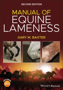
Gary M. Baxter - Manual of Equine Lameness

Здесь есть возможность читать онлайн «Gary M. Baxter - Manual of Equine Lameness» — ознакомительный отрывок электронной книги совершенно бесплатно, а после прочтения отрывка купить полную версию. В некоторых случаях можно слушать аудио, скачать через торрент в формате fb2 и присутствует краткое содержание. Жанр: unrecognised, на английском языке. Описание произведения, (предисловие) а так же отзывы посетителей доступны на портале библиотеки ЛибКат.

Manual of Equine Lameness: краткое содержание, описание и аннотация

Предлагаем к чтению аннотацию, описание, краткое содержание или предисловие (зависит от того, что написал сам автор книги «Manual of Equine Lameness»). Если вы не нашли необходимую информацию о книге — напишите в комментариях, мы постараемся отыскать её.

MANUAL OF EQUINE LAMENESS <p><b>Discover a concise and accessible guide to diagnosing and managing lameness in horses </b> <p>The revised Second Edition of<i> Manual of Equine Lameness </i>offers a concise and accessible manual of lameness diagnosis and treatment in horses. Perfect for use as a quick reference, this book provides straightforward access to the essentials of equine lameness, including the clinical assessment of the horse and commonly performed diagnostic nerve blocks and the most common conditions of the foot, forelimb, and hindlimb that may be contributing to the lameness. Current therapeutic options to treat lameness are also discussed, as well as guidance on how to manage musculoskeletal emergencies. The content has been distilled from the authoritative Seventh Edition of <i>Adams and Stashak’s Lameness in Horses,</i> and this new edition has been re-envisioned to be even quicker and easier to navigate than the previous version. <p>Color photographs and illustrations support the text, which presents lameness information most relevant to equine general practitioners, mixed animal practitioners, and veterinary students. A companion website offers videos that focus on the clinical examination of the horse and select diagnostic blocks and relevant anatomy. Diagnostic and treatment material has been revised from the previous edition to include the most up-to-date information. <p>Readers will find: <ul><li>A thorough introduction to the assessment of the lame horse, including history, visual exam, palpation, subjective and objective assessments of lameness, perineural anesthesia, and intrasynovial anesthesia</li> <li>An exploration of common conditions of the foot, including the navicular region and soft tissue injuries, coffin joint and distal phalanx conditions, and laminitis </li> <li>Discussions of the most common conditions of the forelimb, including the pastern, fetlock, metacarpus/metatarsus, carpus, antebrachium, elbow, and humerus, as well as the shoulder and scapula</li> <li>Discussions of common conditions of the hindlimb and axial skeleton</li> <li>A review of therapeutic options to treat lameness conditions</li> <li>How to manage musculoskeletal emergencies in the horse</li></ul> <p>Ideal for veterinary students, early career equine practitioners, and mixed animal veterinarians, the Second Edition of<i> Manual of Equine Lameness </i>is an indispensable reference for any veterinarian seeking a concise one-stop reference for equine lameness.

Manual of Equine Lameness — читать онлайн ознакомительный отрывок

Ниже представлен текст книги, разбитый по страницам. Система сохранения места последней прочитанной страницы, позволяет с удобством читать онлайн бесплатно книгу «Manual of Equine Lameness», без необходимости каждый раз заново искать на чём Вы остановились. Поставьте закладку, и сможете в любой момент перейти на страницу, на которой закончили чтение.

Тёмная тема
Сбросить

Интервал:

Закладка:

Сделать
.. Figure 1.40 Lateral displacement of the superficial digital flexor tendon fr... Figure 1.41 Horse with fibrotic myopathy of the left hindlimb. There is atro... Figure 1.42 Reciprocal apparatus, lateral view of left hindlimb. Please note... Figure 1.43 Deep dissection of medial aspect of left stifle. The tendon of a... Figure 1.44 Effusion can be seen and palpated within the FP joint cranial to... Figure 1.45 Visible and palpable effusion within the MFT joint is present ju... Figure 1.46 The horse is experiencing upward fixation of the patella. The li... Figure 1.47 Positioning to check for problems with the cranial cruciate liga... Figure 1.48 Test to stress the medial collateral ligaments of the hock and s... Figure 1.49 Typical toe‐out, hock‐in stance that often accompanies problems ... Figure 1.50 Severe atrophy of the left gluteal muscles secondary to a pelvic... Figure 1.51 (a) Palpation of the summits of the dorsal spinous processes to ... Figure 1.52 Firm pressure applied to the back musculature from the withers t... Figure 1.53 Hand and limb positioning to perform distal limb flexion (phalan... Figure 1.54 Upper limb flexion test in which the limb is pulled cranially an... Figure 1.55 Flexion of the upper forelimb can be performed by grasping the a... Figure 1.56 Hock or tarsal flexion (spavin) test. The hindlimb is flexed so ... Figure 1.57 Positioning to perform hock or tarsal flexion of the hindlimb, w... Figure 1.58 Flexion of the stifle is performed by pulling the hindlimb cauda... Figure 1.59 Positioning to perform a full limb flexion of the hindlimb. Figure 1.60 A (10″ × 10″) 15 °wooden wedge block with a nonslip surface... Figure 1.61 Overall vertical head trajectory pattern for horse with impact‐t... Figure 1.62 Two methods of hindlimb lameness detection and evaluation. Verti... Figure 1.63 (A) Fetlock extension and (B) coffin flexion angles during full ... Figure 1.64 Hindlimb protraction is easily seen when viewing from the side o... Figure 1.65 Stationary force plate set for evaluation of lameness in horses.... Figure 1.66 Stationary force plate data (from a single hoof strike) of relev... Figure 1.67 The Q™ (hardware) with Lameness Locator ®(software) on stan... Figure 1.68 Output of Lameness Locator ®indicating right forelimb impac... Figure 1.69 Output of Lameness Locator ®indicating right hindlimb pusho... Figure 1.70 Known compensatory lameness patterns. (a) First part of the law ... Figure 1.71 This image illustrates the positioning to perform a PD nerve blo... Figure 1.72 Injection sites for a PD nerve block. In (A), the needle is inse... Figure 1.73 Injection sites for local anesthesia. A1 and A2. Sites for the p... Figure 1.74 Location of needle insertion to perform a high PD nerve block in... Figure 1.75 Needle location to perform an abaxial sesamoid nerve block in th... Figure 1.76 Low palmar or 4‐point block. a. Site for palmar nerve block; it ... Figure 1.77 Contrast material within the DFTS after a low palmar nerve block... Figure 1.78 Neuroanatomy of the palmar aspect of the carpus illustrating the... Figure 1.79 Needle positioning to perform the lateral approach to block the ... Figure 1.80 The medial approach to desensitize the lateral palmar nerve is l... Figure 1.81 Upper forelimb blocks. A. Site for median nerve block. B. Site f... Figure 1.82 Innervation of the proximal suspensory ligament in the hindlimb ... Figures 1.83 The deep branch of the lateral plantar nerve (DBLPN) can be des... Figure 1.84 Cradling the hindlimb and pulling the flexor tendons medially ca... Figure 1.85 Image illustrating the locations to block the tibial and peronea... Figure 1.86 Dorsolateral approach to the coffin joint. Figure 1.87 The dorsal perpendicular approach to the DIP joint. Figure 1.88 Dorsal parallel approach to the DIP joint. Figure 1.89 Lateral approach to the DIP joint. Figure 1.90 Dorsolateral approach to the PIP joint. Figure 1.91 Palmar/plantar approach to the PIP joint. Figure 1.92 Proximal palmar/plantar approach to the fetlock joint in the sta... Figure 1.93 Injection of the proximal palmar/plantar pouch of the fetlock jo... Figure 1.94 Lateral view of the injection site through the collateral sesamo... Figure 1.95 Distal palmar/plantar approach to the fetlock joint. Figure 1.96 Dorsal injection site for the fetlock joint in the standing hors... Figure 1.97 Dorsal flexed (a) and palmarolateral standing (b) approaches to ... Figure 1.98 Dorsal flexed (a) and palmarolateral standing (b) approaches to ... Figure 1.99 Lateral approaches cranial or caudal to the collateral ligament ... Figure 1.100 Caudolateral approach to the humeral‐ulnar joint. Figure 1.101 The approach to the large caudal outpouching of the elbow joint... Figure 1.102 Craniolateral (left needle) and lateral (right needle) approach... Figure 1.103 Lateral approach to the TMT joint. Figure 1.104 Medial (a) and dorsolateral (b) approaches to the DIT joint. Th... Figure 1.105 Dorsomedial (a), dorsolateral (b), and plantarolateral (b) appr... Figure 1.106 Medial approaches (a and b) to the MFT joint. Figure 1.107 Cranial (a) and lateral (b) approaches to the femoropatellar jo... Figure 1.108 Injection sites for the lateral femorotibial joint just proxima... Figure 1.109 Lateral view of the injection site for the coxofemoral joint. T... Figure 1.110 The proximal approach to the DFTS can be performed with the lim... Figure 1.111 Distal approaches to the DFTS. Figure 1.112 Injection site for the DFTS on the axial surface of the proxima... Figure 1.113 Injection site for the tarsal sheath. Figure 1.114 Lateral view of the foot demonstrating the correct angulation o... Figure 1.115 Lateral (abaxial) approach to the navicular bursa that avoids p... Figure 1.116 Injection sites for the calcaneal bursa are located either abov... Figure 1.117 Distal (a) and proximal (b) approaches to the bicipital bursa f...

2 Chapter 2Figure 2.1 Sagittal section of equine fetlock and digit.Figure 2.2 Attachments of deep digital flexor tendon and collateral sesamoid...Figure 2.3 Front foot (a) and lateral radiograph (b) of a horse with a rever...Figure 2.4 Cross sections of the navicular bone at necropsy demonstrating cy...Figure 2.5 Single, large cystic lesion (a; arrow) and multiple cystic lesion...Figure 2.6 Skyline radiographs of the same horse as in Figure 2.9 (a) demons...Figure 2.7 Multiple abnormalities within the navicular bone as seen on an ob...Figure 2.8 Fracture fragment from the distal border of the navicular bone (a...Figure 2.9 (a) Lateral STIR MR image demonstrating abnormal signal within th...Figure 2.10 Heel elevation may be helpful in treating select horses with nav...Figure 2.11 Distal sesamoid (navicular) bone. (a) Proximal view. (b) Distal ...Figure 2.12 Bipartite navicular bone as seen on a skyline radiograph in a yo...Figure 2.13 A wing fracture of the navicular bone (arrows) as seen on the ob...Figure 2.14 This oblique radiograph of the navicular bone was taken 23 month...Figure 2.15 MRI images demonstrating a single, large DDFT lesion (a; arrow) ...Figure 2.16 Transverse T1‐weighted fast low angle shot (FLASH) image with fa...Figure 2.17 Transverse fast low angle shot (FLASH) image with fat saturation...Figure 2.18 Sagittal proton density image of the central part of the foot of...Figure 2.19 The medial and lateral collateral ligaments of the DIP joint ari...Figure 2.20 Transverse proton density image with fat saturation oriented par...Figure 2.21 Bony proliferation on the dorsomedial aspect of P2 on this obliq...Figure 2.22 Effusion within the DIP joint can be seen and palpated as swelli...Figure 2.23 The calcification of the extensor tendon seen on this lateral ra...Figure 2.24 Classification of P3 fractures in horses.Figure 2.25 Two variable sized Type II “wing” fractures of the P3. These are...Figure 2.26 Small (a) and large (b) Type IV P3 fractures involving the exten...Figure 2.27 CT image of a Type V, comminuted fracture of P3. Fracture lines ...Figure 2.28 Lateral radiograph of the same horse in Figure 2.26b following a...Figure 2.29 Standing dorsopalmar radiograph demonstrating a large uniaxial s...Figure 2.30 Type II articular fracture that was associated with a large side...Figure 2.31 Normal solar surface of the forefoot showing anatomic structures...Figure 2.32 This horse was non‐weight‐bearing lame with no evidence of a hoo...Figure 2.33 This horse had what was thought to be a routine abscess at the t...Figure 2.34 Type VI fractures are also referred to as solar margin fractures...Figure 2.35 Dorsopalmar radiographs of the distal phalanx in two adult horse...Figure 2.36 This horse had a draining tract at the dorsal coronet, but no ab...Figure 2.37 This horse presented for an acute onset hindlimb lameness. A nai...Figure 2.38 Taking radiographs of the foot prior to removing the foreign bod...Figure 2.39 This horse had a history of stepping on a farriery nail with its...Figure 2.40 Lateromedial (LM) projection of the distal limb after injection ...Figure 2.41 Three‐dimensional dissection of the coronary region of the hoof ...Figure 2.42 Dorsopalmar radiograph of P3 demonstrating a smooth margined lyt...Figure 2.43 Single CT image of a foot (a) demonstrating a defect within the ...Figure 2.44 Hoof wall removal directly over the keratoma that was located wi...Figure 2.45 Extensive separation under lateral hoof wall causing a shift of ...Figure 2.46 Dorsal hoof wall resection to treat a horse with WLD. Note the s...Figure 2.47 A is thrush. Note the deterioration of the frog that is recessed...Figure 2.48 Topography of the solar surface of the hoof. The right half has ...Figure 2.49 Small, pale, demarcated growth along the caudal aspect of the fr...Figure 2.50 In symmetrical distal displacement, the distal phalanx descends ...Figure 2.51 In medial or lateral asymmetrical displacement (a), one side of ...Figure 2.52 (a and b) In horses with early chronic laminitis, the surface of...Figure 2.53 Photomicrograph of laminae of the equine hoof. In the top image,...Figure 2.54 Several measurements obtained from lateral radiographs of the di...Figure 2.55 For assessment of rotation of the distal phalanx, the clinician ...Figure 2.56 (a) At rest, the foot is stable with respect to the ground. The ...Figure 2.57 This horse has medial asymmetrical distal displacement, evidence...Figure 2.58 Tenotomy of the DDFT is most commonly performed while standing a...Figure 2.59 Schematic drawings showing the ideal hoof‐pastern axis (a), exte...Figure 2.60 (a) Lateral radiograph demonstrating the center of rotation that...Figure 2.61 Heels of the hoof capsule trimmed to the base of the frog. Note ...Figure 2.62 (a) Lateral radiograph that illustrates hoof‐pastern axis (yello...Figure 2.63 Long‐toe low heel foot conformation. Note the broken back hoof‐p...Figure 2.64 Example of a moderate club foot. Note the disparity of the growt...Figure 2.65 (a) Dorsopalmar view of a foot with sheared heels. Note the disp...Figure 2.66 Weanling quarter horses with typical club foot appearances. The ...Figure 2.67 Corrective trimming and shoeing of a horse affected with sheared...Figure 2.68 A partial‐thickness toe crack that developed in a horse with a c...Figure 2.69 A full‐thickness quarter crack just after debridement down to th...Figure 2.70 (a) Acute quarter crack with hemorrhage. Note the tightly packed...Figure 2.71 Straight‐bar shoe (a) with the medial heel unloaded (b). Note th...Figure 2.72 Placement of wires for a quarter crack repair .

Читать дальше
Тёмная тема
Сбросить

Интервал:

Закладка:

Сделать

Похожие книги на «Manual of Equine Lameness»

Представляем Вашему вниманию похожие книги на «Manual of Equine Lameness» списком для выбора. Мы отобрали схожую по названию и смыслу литературу в надежде предоставить читателям больше вариантов отыскать новые, интересные, ещё непрочитанные произведения.


Stephen Baxter - Project Hades
Stephen Baxter
Gary Braunbeck - Keepers
Gary Braunbeck
Gary Gibson - Nova War
Gary Gibson
Manual of Equine Anesthesia and Analgesia
Неизвестный Автор
Complications in Equine Surgery
Неизвестный Автор
Equine Reproductive Procedures
Неизвестный Автор
Gary Morecambe - Stella
Gary Morecambe
Отзывы о книге «Manual of Equine Lameness»

Обсуждение, отзывы о книге «Manual of Equine Lameness» и просто собственные мнения читателей. Оставьте ваши комментарии, напишите, что Вы думаете о произведении, его смысле или главных героях. Укажите что конкретно понравилось, а что нет, и почему Вы так считаете.

x