Adrian Becker - Orthodontic Treatment of Impacted Teeth
Здесь есть возможность читать онлайн «Adrian Becker - Orthodontic Treatment of Impacted Teeth» — ознакомительный отрывок электронной книги совершенно бесплатно, а после прочтения отрывка купить полную версию. В некоторых случаях можно слушать аудио, скачать через торрент в формате fb2 и присутствует краткое содержание. Жанр: unrecognised, на английском языке. Описание произведения, (предисловие) а так же отзывы посетителей доступны на портале библиотеки ЛибКат.
- Название:Orthodontic Treatment of Impacted Teeth
- Автор:
- Жанр:
- Год:неизвестен
- ISBN:нет данных
- Рейтинг книги:4 / 5. Голосов: 1
-
Избранное:Добавить в избранное
- Отзывы:
-
Ваша оценка:
Orthodontic Treatment of Impacted Teeth: краткое содержание, описание и аннотация
Предлагаем к чтению аннотацию, описание, краткое содержание или предисловие (зависит от того, что написал сам автор книги «Orthodontic Treatment of Impacted Teeth»). Если вы не нашли необходимую информацию о книге — напишите в комментариях, мы постараемся отыскать её.
Provides protocols for common cases as well as complex and rare presentations Contains individual chapters on the specific aspects of the diagnosis and treatment of impaction in each of the different types of teeth Covers prevalence, etiology, diagnosis, attitudes to treatment, treatment timing, treatment methods, and prognosis Features more than 1,000 high-quality color images and illustrations
remains essential reading for all specialist orthodontists, academic researchers and instructors, oral and maxillofacial surgeons, and advanced students in orthodontics.
Orthodontic Treatment of Impacted Teeth — читать онлайн ознакомительный отрывок
Ниже представлен текст книги, разбитый по страницам. Система сохранения места последней прочитанной страницы, позволяет с удобством читать онлайн бесплатно книгу «Orthodontic Treatment of Impacted Teeth», без необходимости каждый раз заново искать на чём Вы остановились. Поставьте закладку, и сможете в любой момент перейти на страницу, на которой закончили чтение.
Интервал:
Закладка:
What is recommended is a simple, logical but systematic approach that can be employed chairside to reach a similar conclusion in just a few minutes, even at the initial orthodontic consultation visit. This method will rely on the same criteria of establishing the development of crown and root, but must do so in a step‐by‐step manner, starting from a different point of departure. The ‘starting point’ of this systematic approach has, for reasons that will be explained in the following paragraphs, been set at the cut‐off dental age of 9 years.
Assessing dental age in the clinical setting – the Jerusalem method
There are several criteria that are appropriate to the appraisal of tooth development when using full‐mouth periapical radiographs or a panoramic film. The information that is available regarding the ages at which the various stages of dental development occur is based on the classic random studies that have been carried out over many decades of the local populations of the researchers involved. The figures for the mean ages at which these stages occur, in the hypothetical child, are as follows:
1 The first signs of the presence of a tooth are discernible radiographically with the initiation of calcification of incisal edges and cusp tips. Thereafter, one may observe the formation of the completed crown as well as progressive degrees of root formation (usually expressed in fractions), and thence the fully closed root apex. Since orthodontic treatment is largely performed on a relatively older section of the child population, the stages of actual formation of the root become the only relevant factors.
2 The accuracy with which one may assess fractions of an incomplete, immeasurable and merely ‘expected’ final root length is not reliable and is very much a matter of individual observer variation.
3 The stage of tooth development that is easiest to define with confidence and with accuracy is that which relates to the closure of the root apex. So long as the dental papilla at the root end remains discernible, the apex is open and Hertwig’s root‐forming, epithelial sheath is in an active stage of increasing root length. However, once fully closed, the papilla disappears and a continuous lamina dura will be seen on a periapical radiograph, closely following the root outline. These are the specific diagnostic signs of that landmark event. Apexification is therefore the most important single factor upon which a system of assessment may be faithfully and easily made of the dental age of a given patient in the clinical environment.
4 From population studies, we learn that the first permanent tooth to erupt in the mouth is the mandibular central incisor, closely followed by the first permanent molars, and this occurs at the age of 6 years.
5 Root development of the permanent teeth is completed approximately 2.5–3 years after their normal eruption [4]. This allows us to conclude that, at the age of 8.5–9 years, the child’s mandibular incisors will be the first teeth to exhibit closed apices and will usually be closely followed by the four first permanent molars. This being the case, it is clear that the age of 9 years must be the basic starting point from which to commence the evaluation of the child’s dental age. If mandibular incisors or molars demonstrate root closure, then the tentative diagnosis would be that the patient has a dental age of at least 9 years. If the apices are still open, then the conclusion would be that the child has a lower dental age.
It should be emphasized, however, that the exercise is aimed at ranking a specific child’s dental development vis‐à‐vis the above hypothetical mean. Whether or not the evaluated tooth has actually erupted is entirely irrelevant to this equation.
Let us examine the progressive diagnostic path in its correct order (see also Table 1.1):
1 If the mandibular central incisor roots are complete, we may presume that the patient is at least 9 years old (dental age), i.e. this figure is derived from 6 years (the normal age of eruption as determined by two‐thirds root length development), with the addition of 2.5–3 years to apexification.
2 We may then proceed and check for closed apices of the first molars (9–9.5 years).
3 At 9.5 years, the mandibular lateral incisors roots will have completely developed.
4 The next teeth in the expected eruption series are the maxillary central incisors, whose closed apices would indicate a dental age of 10 years.
5 Because their rate of development is variable, it would be wise to bypass the assessment of the maxillary lateral incisors at this point in the diagnostic process and move on to examine the later teeth.
6 Apexification of the mandibular canines and first premolars (12–13 years).
7 Thereafter, the maxillary first premolars (13–14 years).
8 In common with the maxillary lateral incisors, the mandibular second premolars are also developmentally variable teeth and their assessment should also be bypassed for the present calculation.
9 Next there are the maxillary canines (14–15 years).
10 The final stage of development relates to the four second molars (15 years).
Table 1.1 Apexification age of individual tooth types.
| 9 years | Mandibular central incisors |
| 9–9.5 years | First molars and mandibular lateral incisors |
| 10 years | Maxillary central incisors |
| 11 years | Maxillary lateral incisors |
| 12–13 years | Mandibular canines |
| 13–14 years | Maxillary first premolars |
| 14–15 years | Second premolars and maxillary canines |
| 15 years | Second molars |
This stage‐by‐stage apexification determination will lead us to the last tooth in this sequence with a closed apex ( Figure 1.4), which indicates the dental age of the patient. Once the determination is completed, it is valuable to return to the maxillary lateral incisor and the mandibular second premolar. If these are developing normally, then their age of eruption would be 8 years and 11 years, respectively, with an apexification date of 11 and 14 years, respectively. Retarded development of these individual teeth may be age assessed according to the above criteria for calcification. An illustration of this situation would be where the overall dental age assessment is diagnosed as 12 years, yet the right maxillary lateral incisor might match a 9‐year‐old child and the left mandibular second premolar might even be characteristic of someone 8 years of age.
In contrast to the above process of examination and assessment and in the case of a dental age less than 9 years, none of the permanent teeth will have completed their root development. Here clinicians will have no choice but to rely on their own estimation of the degree of root development, of the degree of crown completion and, in the very young, of the stage of initiation of crown calcification ( Figure 1.5). This is most conveniently carried out by working backwards from the expected development at age 9 years and, with this as a base, comparing the dental development status of the patient, beginning with the mandibular central incisors and the first permanent molars.
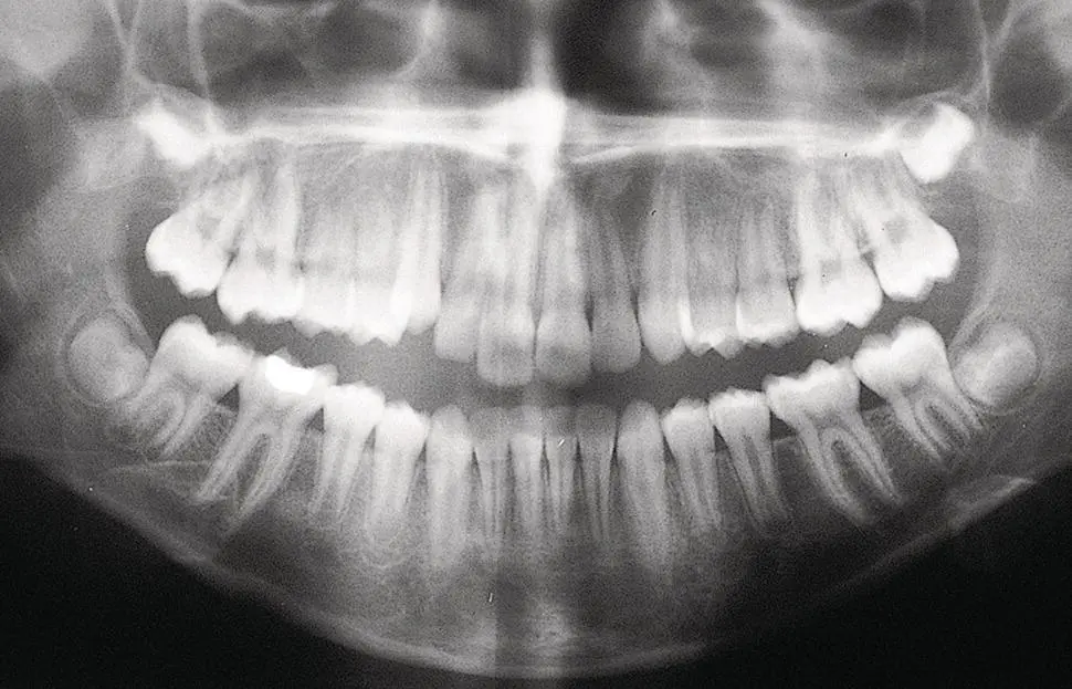
Fig. 1.4 Root apices are closed in all first molars, all mandibular and three of the maxillary incisors, excluding the left lateral incisor. Canine and premolar apices are open.
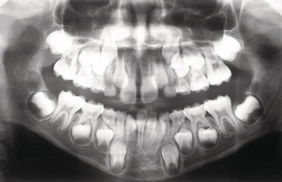
Fig. 1.5 No closed apices. Dental age assessment 7–7.5 years.
Читать дальшеИнтервал:
Закладка:
Похожие книги на «Orthodontic Treatment of Impacted Teeth»
Представляем Вашему вниманию похожие книги на «Orthodontic Treatment of Impacted Teeth» списком для выбора. Мы отобрали схожую по названию и смыслу литературу в надежде предоставить читателям больше вариантов отыскать новые, интересные, ещё непрочитанные произведения.


Обсуждение, отзывы о книге «Orthodontic Treatment of Impacted Teeth» и просто собственные мнения читателей. Оставьте ваши комментарии, напишите, что Вы думаете о произведении, его смысле или главных героях. Укажите что конкретно понравилось, а что нет, и почему Вы так считаете.