Essential Cases in Head and Neck Oncology
Здесь есть возможность читать онлайн «Essential Cases in Head and Neck Oncology» — ознакомительный отрывок электронной книги совершенно бесплатно, а после прочтения отрывка купить полную версию. В некоторых случаях можно слушать аудио, скачать через торрент в формате fb2 и присутствует краткое содержание. Жанр: unrecognised, на английском языке. Описание произведения, (предисловие) а так же отзывы посетителей доступны на портале библиотеки ЛибКат.
- Название:Essential Cases in Head and Neck Oncology
- Автор:
- Жанр:
- Год:неизвестен
- ISBN:нет данных
- Рейтинг книги:5 / 5. Голосов: 1
-
Избранное:Добавить в избранное
- Отзывы:
-
Ваша оценка:
Essential Cases in Head and Neck Oncology: краткое содержание, описание и аннотация
Предлагаем к чтению аннотацию, описание, краткое содержание или предисловие (зависит от того, что написал сам автор книги «Essential Cases in Head and Neck Oncology»). Если вы не нашли необходимую информацию о книге — напишите в комментариях, мы постараемся отыскать её.
Essential Cases in Head and Neck Oncology This textbook also includes:
Covers the full spectrum of head and neck surgeries, including reconstructive procedures Discusses ethics related to cancer treatments, medical research, and other care issues Promotes multidisciplinary critical thinking, clinical problem-solving, communication, and collaboration Helps medical students and trainees evaluate their learning and contextualize their knowledge Features high-quality images and succinct explanatory text throughout
is an indispensable study aid for trainee clinicians, residents, and fellows studying for board certification and other exams, and an excellent reference guide for oncologists, otolaryngologists, surgeons, and other practitioners working in medical oncology, radiation oncology, and oromaxillofacial surgery.
Essential Cases in Head and Neck Oncology — читать онлайн ознакомительный отрывок
Ниже представлен текст книги, разбитый по страницам. Система сохранения места последней прочитанной страницы, позволяет с удобством читать онлайн бесплатно книгу «Essential Cases in Head and Neck Oncology», без необходимости каждый раз заново искать на чём Вы остановились. Поставьте закладку, и сможете в любой момент перейти на страницу, на которой закончили чтение.
Интервал:
Закладка:
Question: What course of treatment would you recommend?
Answer:Treatment of advanced oral cavity SCC requires extirpation of the primary tumor with negative margins and addressing the lymphatics of the neck. Segmental mandibulectomy is required to obtain negative primary site surgical margins. The mandibular defect will require free tissue transfer reconstruction. The ipsilateral involved neck will require a neck dissection, level I–IV (skip metastases to level V in oral cavity carcinoma are uncommon). As the lesion crosses midline anteriorly, it is appropriate to consider or perform a contralateral selective (level I–III) neck dissection.
Question: What are the potential functional deficits after surgical treatment?
Answer:While rare, injuries to the spinal accessory nerve (CN XI) resulting in shoulder weakness, and marginal mandibular branch of the facial nerve (CN VII) resulting in lower lip depressor weakness, can occur but are not an expected outcome in experienced settings. Left chin numbness is to be expected after a segmental mandibulectomy as the inferior alveolar nerve is transected during the extirpation. Injury to the hypoglossal nerve (CN XII) is also rare and would be considered a complication. Treatment of alveolar cancer is not expected to result in significant dysphagia or aspiration postoperatively. However, when resection extends across the midline anteriorly, resulting in detachment of the genial muscular attachments, some loss of function can be observed.
Final pathology shows a 4.2 cm tumor centered in the left retromolar trigone with mandibular invasion, extensive perineural invasion, and a deep positive margin. In the right neck dissection specimen, 0/8 lymph nodes are involved with the tumor. In the left neck dissection specimen, 6/32 lymph nodes are involved with the tumor. There is a 3.1 cm level 1b lymph node that shows extranodal extension into the submandibular gland.
Question: Based on the final pathology, what is the pathologic stage and your recommended adjuvant treatment regimen?
Answer:pT4aN3bM0, stage IVb. Mandibular involvement results in pT4a staging. Any node with extranodal extension (ENE) results in an upstaging to N3b. There are many indications for radiation, including positive margins, perineural invasion (PNI), and multiple nodes involved. There are two indications for chemotherapy: positive surgical margins at the primary site and extranodal extension. In this case, considering that a large resection was performed and the defect is reconstructed with fibula flap, the expectation of re‐resection is not realistic. Therefore, continuing with concurrent adjuvant chemoradiation with platinum‐based agents is appropriate.
Key Points
Evaluation of oral cavity cancer starts with a biopsy and typically a CT scan of the face and neck with IV contrast to assess the extent of the primary lesion and to evaluate for regional lymphadenopathy. An MRI may be indicated if there is concern for significant perineural invasion, deep tongue invasion, or extension near the orbit, skull base, or parapharyngeal space. The use of PET/CT should be in patients with stage III or IV disease.
Management of tumors of the oral cavity typically involves upfront surgery with removal of the primary tumor with clear surgical margins. A neck dissection should be performed for pathologic lymphadenopathy. Contralateral selective neck dissections should be performed if the tumor crosses the midline.
Adjuvant radiation therapy should be considered for advanced primary tumors, the presence of lymph node metastases, perineural invasion, lymphovascular invasion, or close surgical margins.
Adjuvant chemoradiation therapy should be recommended in instances of positive surgical margins and/or the presence of extracapsular spread in cervical lymph nodes.
CASE 5
Arnaud Bewley
History of Present Illness
A 62‐year‐old white female is seen in the office with a 3‐month history of a gradually enlarging lower lip mass. She has not had any previous biopsies, imaging, or treatment.
Her past medical history includes hypercholesterolemia, hypertension, appendectomy, and tonsillectomy. She takes Lisinopril and atorvastatin. She has a history of 30 pack‐year smoking but quit 10 years ago. She drinks a glass of wine with dinner every night.
Question: What are the other important points in history?
Answer:
Pain and tenderness. This is an important question, as pain that is out of proportion with exam can be suggestive of perineural invasion.
The presence of neck masses is an important finding, as advanced lower lip cancers have a high propensity for spread to the regional lymphatics.
Voice change. It is important to screen for other head and neck cancer primaries in patients who are high risk.
Weight loss, which is often associated with the duration and severity of symptoms.
The lesion is exquisitely painful, particularly along the right lower lip. She has not experienced any voice changes or weight loss and has not noticed any neck masses.
Physical Examination
An image of the patient’s lower lip lesion is shown in Figure 5.1. The tumor is very tender to palpation and extends from the right commissure to about 3 mm from the left commissure. Intraorally, it is free from the fixed gingiva by about 1 cm. There is subcutaneous extension palpable at the right lateral aspect of the tumor. There is no palpable lymphadenopathy. The remainder of her head and neck examination is unremarkable.
Management
Question: What would you recommend next?
Answer:Punch biopsy of the lower lip lesion. Pathologic diagnosis is imperative and is the most appropriate next step in management. This would most easily be obtained with a punch biopsy at the time of her initial visit.
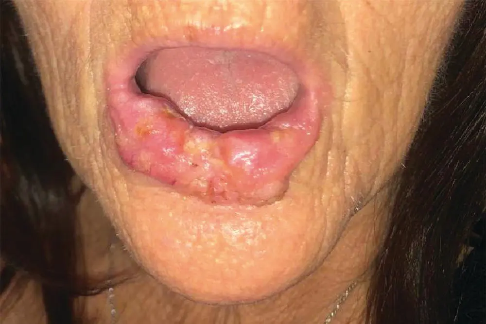
FIGURE 5.1 The patient’s lower lip mass. The tumor extends from the right Commissure to about 3mm from the left Commissure.
Question: What other tests or studies would you consider, if any?
Answer:A CT of the neck with IV contrast is an important next step in evaluating regional adenopathy. MRI of the neck could be considered, given the acute tenderness noted along the right lower lip. An MRI would be superior to a CT in evaluating for enhancement of the right mental nerve. An MRI would also allow for evaluation of the regional nodes, though less cost‐effective than a CT scan. PET/CT scan could be considered. Also, a potential method of evaluating the regional nodal basin while also ruling out distant metastatic disease. For early stage disease (T1/T2) without clinical or radiographic evidence of regional disease, a PET/CT is usually unnecessary. If a PET/CT is considered, it is recommended that CT is performed with contrast and with adequate detail to delineate the neck anatomy.
A punch biopsy is performed and demonstrates SCC.
A contrast‐enhanced CT scan is obtained with representative images shown below. No abnormal lymph nodes are reported. The tumor is measured around 5 cm on the CT scan (see Figure 5.2).
Question: Based on the patient's examination and radiographic findings, how would you stage this disease?
Answer:T3N0M0, stage III. Cancers of the lip mucosa continue to be staged as cancers of the oral cavity, while cancers of the external vermillion lip are now staged as cutaneous carcinomas, per AJCC 8th Edition. However, in advanced tumors, this can be a difficult distinction to make when tumors involve both the mucosal and external vermillion surface. In these cases, staging should be based on the tumor's historical origin when this can be deduced. In this case, the patient reports the tumor originated on the inner aspect of the lip, and this is corroborated by the greater degree of extension noted on the mucosal surface. The tumor is greater than 4 cm in diameter, therefore, it meets the criteria for a T3 primary. It would also likely meet the 1 cm depth of invasion criteria for T3 tumor. There is no clinical or radiographic evidence of regional metastatic spread; therefore, the patient should be staged as a T3N0M0, stage III.
Читать дальшеИнтервал:
Закладка:
Похожие книги на «Essential Cases in Head and Neck Oncology»
Представляем Вашему вниманию похожие книги на «Essential Cases in Head and Neck Oncology» списком для выбора. Мы отобрали схожую по названию и смыслу литературу в надежде предоставить читателям больше вариантов отыскать новые, интересные, ещё непрочитанные произведения.


Обсуждение, отзывы о книге «Essential Cases in Head and Neck Oncology» и просто собственные мнения читателей. Оставьте ваши комментарии, напишите, что Вы думаете о произведении, его смысле или главных героях. Укажите что конкретно понравилось, а что нет, и почему Вы так считаете.