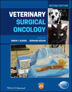
Veterinary Surgical Oncology
Здесь есть возможность читать онлайн «Veterinary Surgical Oncology» — ознакомительный отрывок электронной книги совершенно бесплатно, а после прочтения отрывка купить полную версию. В некоторых случаях можно слушать аудио, скачать через торрент в формате fb2 и присутствует краткое содержание. Жанр: unrecognised, на английском языке. Описание произведения, (предисловие) а так же отзывы посетителей доступны на портале библиотеки ЛибКат.
- Название:Veterinary Surgical Oncology
- Автор:
- Жанр:
- Год:неизвестен
- ISBN:нет данных
- Рейтинг книги:5 / 5. Голосов: 1
-
Избранное:Добавить в избранное
- Отзывы:
-
Ваша оценка:
- 100
- 1
- 2
- 3
- 4
- 5
Veterinary Surgical Oncology: краткое содержание, описание и аннотация
Предлагаем к чтению аннотацию, описание, краткое содержание или предисловие (зависит от того, что написал сам автор книги «Veterinary Surgical Oncology»). Если вы не нашли необходимую информацию о книге — напишите в комментариях, мы постараемся отыскать её.
The new edition of the most comprehensive resource on surgical oncology, covering both basic and advanced surgical oncology procedures in small animals Veterinary Surgical Oncology
Veterinary Surgical Oncology
Veterinary Surgical Oncology, Second Edition
Veterinary Surgical Oncology — читать онлайн ознакомительный отрывок
Ниже представлен текст книги, разбитый по страницам. Система сохранения места последней прочитанной страницы, позволяет с удобством читать онлайн бесплатно книгу «Veterinary Surgical Oncology», без необходимости каждый раз заново искать на чём Вы остановились. Поставьте закладку, и сможете в любой момент перейти на страницу, на которой закончили чтение.
Интервал:
Закладка:
Early tissue biopsies for histology and immunochemistry are recommended for progressive edematous lesions of unknown origin as cytology consisted of mild inflammation in dogs diagnosed with lymphangiosarcoma (Curran et al. 2016).
The cell‐specific immunohistochemical lymphatic vessel endothelial receptor‐1 (LYVE‐1) has been successfully used as a marker to diagnose lymphangiosarcoma in cats (Galeotti et al. 2004). LYVE‐1 in conjunction with Prospero‐related homeobox gene‐1 (PROX‐1) is recommended by Halsey et al. (2016) to differentiate between lymphangiosarcoma and HSA in dogs.
The prognosis is poor, with reported survival times being only a few months (Lenard et al. 2007; Williams 2005) up to over one year. In case series of 12 dogs, survival ranged from 60 to 876 days for 3 dogs with palliation; 90 days with prednisone in 1; 182 days with chemotherapy in 1; 240–941 days for 5 dogs receiving surgery; and 574 days for 1 receiving surgery, radiation, and chemotherapy. One dog is alive with recurrence at 243 days following surgery and carboplatin chemotherapy (Curran et al. 2016).
A five‐year‐old Giant schnauzer, suffering from chylothorax and a lymphangiosarcoma involving the whole left sublumbar area was treated surgically by mass resection, pleural omentalization, and pericardiectomy followed by mitoxantrone administration. The dog was still alive 10 months after surgery (Sicotte et al. 2012).
Doxorubicin treatment resulted in a six‐month recurrence‐free interval in a five‐year‐old female Boxer after incomplete local resection in the area of the caudal mammary gland. At relapse Toceranib resulted in almost complete regression of a lymphangiosarcoma, leaving just a skin plaque. Metronomic chemotherapy using chlorambucil and meloxicam had failed to adequately control the disease in this dog (Marcinowska et al. 2013).
Leiomyoma and Leiomyosarcoma
Cutaneous smooth muscle is present in three separate locations: arrector pili muscles, blood vessel walls, and genital/areolar skin. Benign or malignant smooth muscle neoplasms may arise from each of these locations. In dogs, leiomyomas of arrector pili muscles have been described (Cooper and Valentine 2002; Goldschmidt and Shofer 1998).
Leiomyosarcoma are malignant tumors of smooth muscle cells. They mainly are associated with the digestive tract, but they can occur in any part of the body as firm lobulated masses (Cohen et al. 2003; Kapatkin et al. 1992; Pierini et al. 2017). Leiomyosarcomas of the skin do not tend to metastasize, in contrast to hepatic leiomyosarcomas (100%). The prognosis in dogs with leiomyosarcoma of the spleen, stomach, small intestine, and especially the cecum is good to excellent if surgery is performed. In dogs with leiomyosarcoma of the liver, the prognosis is poor (Kapatkin et al. 1992).
Surgical removal of the lesions is the treatment of choice (Liu and Mikaelian 2003).
Doxorubicin has been used as adjunctive therapy after surgical removal of a primary leiomyosarcoma involving the jugular vein in a dog. The dog received five doses of intravenous doxorubicin, and there was no recurrence of the tumor 30 months post treatment (Pierini et al. 2017).
Melanoma
Benign melanocytic tumors comprise 3–4% of all skin tumors in dogs and 0.6–1.3% in cats. Malignant melanocytic tumors generally termed melanoma or malignant melanoma (MM), account for 0.8–2% of all cutaneous tumors in dogs and 0.4–2.8% in cats (Gross et al. 2005; Vail and Withrow 2007). MMs are more common in dogs with heavily pigmented skin, whereas cats with black or gray hair coats may be predisposed (Gross et al. 2005). Cutaneous and ocular melanomas in dogs usually behave in a benign manner, whereas amelanotic melanoma arising from mucocutaneous junctions, oral cavity, digits, and the nail bed tends to be malignant, with the tendency to rapidly invade surrounding tissue and to metastasize. Benign melanomas are usually small, pigmented, firm, dome‐shaped tumors of the skin that are freely moveable over deeper tissue structures. MMs tend to grow more rapidly with an invasive growth pattern; more commonly ulcerate, and appear brown, gray, or black, depending on the amount of melanin production (Goldschmidt and Hendrick 2002; Gross et al. 2005). Non‐pigmented or amelanotic melanomas can occur in the skin, but they are more commonly seen in the oral cavity (Vail and Withrow 2007).
In a study of 87 cases, canine cutaneous MM, postsurgery median progression‐free survival time, and median overall survival time have been reported to be 1282 days and 1363 days, respectively. With a post‐surgery metastatic rate of 21.8% and a local recurrence rate of 8%. Increasing mitotic index was predictive of a significantly decreased survival time on multivariable analysis (Laver et al. 2018).
In cats, 42–68% of cutaneous melanocytic tumors show malignant behavior (Gross et al. 2005). They more often involve the head (pinna, nose, and eyelids) or the digits and the eye.
Melanomas can be diagnosed easily by FNA cytology, but histologic evaluation is important to evaluate the malignancy and prognosis. Transformation from benign to malignant melanoma often occurs in humans, and the influence of ultraviolet radiation in the pathogenesis is well documented (Leiter and Garbe 2008). Such transformation has sporadically been reported in dogs. The treatment of choice for local cutaneous melanoma in both the cat and dog is surgical excision.
In cats, prognosis after surgical resection of non‐ocular cutaneous MMs is reasonable, with reported metastatic rates of 5–25% (Luna et al. 2000).
The prognosis for malignant‐behaving melanomas, especially located on the lip or oral cavity, is guarded to poor. In a study of Bostock (1979) 38 of 42 (90%) dogs with a histologically malignant melanoma of the lip or oral cavity died because of the tumor, compared to only 15 of 33 (45%) of malignant skin melanomas. A total of 6 of 59 (10%) dogs with a tumor of mitotic index of 2 or less died from tumor‐associated disease 2 years after surgery compared to 19 of 26 (73%) of dogs having a tumor with a mitotic index of 3 or more (Bostock 1979). WHO’s staging scheme for dogs with oral melanoma is based on diameter size: less than 2 cm tumor (stage I), 2 cm to less than 4 cm tumor (stage II), 4 cm or larger tumor and/or lymph node metastasis (stage III), and distant metastasis (stage IV).
Indoleamine 2,3‐dioxygenase (IDO) per high‐power field was identified as an independent marker for increased risk of death in dogs with primary melanocytic tumors (Porcellato et al. 2019).
As with human MM, response rates to chemotherapy, including mitoxantrone, intravenous melphalan, cisplatin, doxorubicin, carboplatin, and dacarbazine, are low and short‐lived in dogs (Bergman 2007; Vail and Withrow 2007). Carboplatin is reported to have activity against MM according to Rassnick et al. (2001). Smaller dogs were more likely to develop gastrointestinal toxicosis after treatment, and prophylactic gastrointestinal protectants should be considered when small dogs are treated (Rassnick et al. 2001).
There is no evidence that immunotherapy with liposome‐encapsulated muramyl tripeptide phosphatidylethanolamine (L‐MTP‐PE) administered after surgery alone or combined with recombinant canine granulocyte macrophage colony‐stimulating factor (rcGM‐CSF) has antitumor activity in treating advanced stage canine (oral) melanoma, but L‐MTP‐PE administration in early stage (I) may prolong survival time (MacEwen et al. 1999).
Bergman et al. (2003, 2006; Bergman 2007) reported the promising results of a canine melanoma vaccine. Thirty‐three stage II to III dogs with surgical locoregionally controlled melanoma had a median survival time of 569 days. This vaccine evoked a specific anti‐tyrosinase humoral immune response (Bergman et al. 2006). Since then several studies have been conducted on this vaccine, retrospectively, but have not delivered an unequivocal outcome of its effectiveness. Ottnod et al. (2013) reported the vaccine did not achieve a greater progression‐free survival, disease‐free interval, or median survival time for vaccinated compared to non‐vaccinated dogs. Verganti et al. (2017) reported that the vaccine could be considered as palliative treatment in dogs with stage IV disease.
Читать дальшеИнтервал:
Закладка:
Похожие книги на «Veterinary Surgical Oncology»
Представляем Вашему вниманию похожие книги на «Veterinary Surgical Oncology» списком для выбора. Мы отобрали схожую по названию и смыслу литературу в надежде предоставить читателям больше вариантов отыскать новые, интересные, ещё непрочитанные произведения.
Обсуждение, отзывы о книге «Veterinary Surgical Oncology» и просто собственные мнения читателей. Оставьте ваши комментарии, напишите, что Вы думаете о произведении, его смысле или главных героях. Укажите что конкретно понравилось, а что нет, и почему Вы так считаете.
