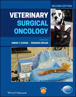
Veterinary Surgical Oncology
Здесь есть возможность читать онлайн «Veterinary Surgical Oncology» — ознакомительный отрывок электронной книги совершенно бесплатно, а после прочтения отрывка купить полную версию. В некоторых случаях можно слушать аудио, скачать через торрент в формате fb2 и присутствует краткое содержание. Жанр: unrecognised, на английском языке. Описание произведения, (предисловие) а так же отзывы посетителей доступны на портале библиотеки ЛибКат.
- Название:Veterinary Surgical Oncology
- Автор:
- Жанр:
- Год:неизвестен
- ISBN:нет данных
- Рейтинг книги:5 / 5. Голосов: 1
-
Избранное:Добавить в избранное
- Отзывы:
-
Ваша оценка:
- 100
- 1
- 2
- 3
- 4
- 5
Veterinary Surgical Oncology: краткое содержание, описание и аннотация
Предлагаем к чтению аннотацию, описание, краткое содержание или предисловие (зависит от того, что написал сам автор книги «Veterinary Surgical Oncology»). Если вы не нашли необходимую информацию о книге — напишите в комментариях, мы постараемся отыскать её.
The new edition of the most comprehensive resource on surgical oncology, covering both basic and advanced surgical oncology procedures in small animals Veterinary Surgical Oncology
Veterinary Surgical Oncology
Veterinary Surgical Oncology, Second Edition
Veterinary Surgical Oncology — читать онлайн ознакомительный отрывок
Ниже представлен текст книги, разбитый по страницам. Система сохранения места последней прочитанной страницы, позволяет с удобством читать онлайн бесплатно книгу «Veterinary Surgical Oncology», без необходимости каждый раз заново искать на чём Вы остановились. Поставьте закладку, и сможете в любой момент перейти на страницу, на которой закончили чтение.
Интервал:
Закладка:
PWTs commonly arise in soft tissue. Their histopathological features are quite similar to those of canine malignant PNSTs, making their differential diagnosis challenging. Suzuki et al. suggest that NGFR and Olig2 are useful to distinguish between PWTs and malignant PNSTs as PWT cells displayed significantly weaker immunoreactivity than MPNSTs to these markers (Suzuki et al. 2014).
In cases of undifferentiated canine PWTs further evaluation has been reported by transmission electron microscopy. By electron microscopy, ultrastructural findings support a perivascular wall origin for all cases with 4 categories of differentiation: myopericytic, myofibroblastic, fibroblastic, and still undifferentiated (Palmieri et al. 2013).
Proteins of the VEGF‐, PDGFB‐, and bFGF‐mediated pathways have been reported to be highly expressed in 47.5–76.5 %, and in 10–35% of TGFβ1‐ and COX2‐mediated pathways of PWTs (Avallone et al. 2015). No association with the Ki67 labeling index was found in this study. Blockade of tyrosine kinase receptors after surgery could represent a promising therapy with the aim to reduce the PWT relapse rate and prolong the time to relapse (Avallone et al. 2015).
PWT can locally recur after a long time after surgery. Recurrence was reported in 2, 8, 20, and 24% at six months, one, two, and three years, respectively. Size of the tumor was a significant prognostic factor. An early diagnosis of PWT associated with small tumor size (<5 cm) and clean surgical margins ensures a good prognosis independently of histological grade (Stefanello et al. 2011). Major prognostic factors for perivascular wall tumors reported by Avallone et al. (2014) are tumor size, depth of growth, and pathological profiles. Smoothelin, heavy caldesmon, desmin, myosin, calponin, and CMG‐3G5 were the most valuable markers to differentially diagnose canine PWT (Avallone 2007).
Hemangiopericytomas (HEPs) are derived from pericytes, spindle cells surrounding blood vessels. Distinguishing these pericytes from endothelial cells, fibroblasts, and other spindle cells surrounding blood vessels is difficult based on morphologic and immunophenotypic assessment. Canine HEP has been proposed to be a diagnosis of immunohistochemical exclusion (Schulman et al. 2009; Mazzei et al. 2002; Williamson and Middleton 1998). The biological behavior of HEPs resembles that of PNSTs, and they stain similarly positive for the histochemical marker s100 (Chijiwa et al. 2004). Reported prevalence in dogs varies from 2 to 7% (Goldschmidt and Shofer 1992; Kaldrymidou et al. 2002; Mukaratirwa et al. 2005; Pakhrin et al. 2007), although they may have been formerly over diagnosed. The tumors are predominantly located on the extremities; however, other regions including the perineal area have been described. Only one case of HEP is reported in a cat (Baldi and Spugnini 2006). HEPs grow slowly and are locally aggressive because of infiltration into the surrounding tissues. Dogs with HEP showed higher serum VEGF levels compared to the patients with malignant PNSTs (De Quieroz et al. 2013). In a study by Stefanello et al. (2008), 35 dogs with marginal excision of low‐grade spindle cell sarcoma incurred a local recurrence rate of 10.8% and a metastatic rate of 0% (Avallone et al. 2007; Bostock and Dye 1980; Chijiwa et al. 2004; Fossum et al. 1988; Graves et al. 1988; Mayr et al. 1990; Stefanello et al. 2008).
Myxosarcoma
Myxosarcoma are tumors of fibroblast origin that characteristically produce myxoid matrix, resembling mucus, containing mucopolysaccharides. Myxosarcoma develops commonly as cyst‐like masses filled with mucoid fluid. They are rare and mainly occur on the chest or limbs. Myxosarcomas have also been reported in the heart, eye, spinal canal, temporomandibular joint, brain, and bone. Biologically, they behave like any STS, with aggressive first surgery after diagnosis being the treatment of choice (Dennis 2008; Foale et al. 2003; Kirby et al. 2014; Levy et al. 1997; Manfredi et al. 2015; Parslow 2016). Pulmonary metastasis of a myxosarcoma in a dog has been reported (Headley et al. 2011).
Cutaneous Hemangiosarcoma
Hemangiosarcoma (HSA) is a malignant neoplasm arising from vascular endothelial cells (Liptak 2007). HSAs may be associated with ultraviolet exposure (Hargis et al. 1992). Prevalence appears to be higher in tropical and subtropical areas (Mukaratirwa et al. 2005). Skin HSAs account for less than 1% of all (sub)cutaneous tumors in dogs and 2–3% in cats (Goldschmidt and Shofer 1992; Miller et al. 1991; Schultheiss 2004). In dogs, 17–35% of all HSAs (including both visceral and nonvisceral) are cutaneous (Schultheiss 2004; Brown et al. 1985), and 50–77% of all HSAs in cats are (sub)cutaneous (Johannes et al. 2007; Scavelli et al. 1985). Dermal HSA is commonly observed in shorthaired dogs on the poorly pigmented skin of the ventral abdomen, medial thigh, inguinal region, and scrotum (Trappler et al. 2014). This is typically seen in “sun bathers” dogs that lie on their back in the sun. Cutaneous HSA has been reported in a Golden retriever appearing from the left elbow to the digits (Tsuji et al. 2013).
Subcutaneous HSAs are more commonly observed in dogs with variable hair coat and pigmentation. The most common primary sites for cutaneous HSAs in the cat are the pinna, lateral face, and the inguinal or abdominal subcutis.
Skin HSAs vary in size, are poorly circumscribed, dark and soft, and may resemble bruises or ecchymosis (Gross et al. 2005). Alopecia, ulceration, and hemorrhage are common, and animals may present with a hematoma.
Elevated serum big ET‐1 can be used as a diagnostic marker for canine HSAs. Serum big ET‐1 levels in dogs with HSA have been found to be significantly (P < 0.01) higher than in other dogs. High sensitivity (100%) and specificity (95%), for HSAs diagnosis were obtained using a cut‐off of 17 pg/mL (Fukumoto et al. 2015).
Canine cutaneous HSAs are staged according to tumor depth and invasion: superficial tumors, confined to the dermis (stage I), tumors extending into subcutaneous tissues (stage II), and tumors invading muscle and fascia (stage III) (Ward et al. 1994). Tumor‐free surgical margins are more likely to be achieved in cutaneous than subcutaneous lesions and are therefore associated with longer survival times. Completeness of excision was the most important prognostic factor in a large retrospective study, with disease‐free intervals (DFIs) of at least 1 year for completely excised skin HSAs. Similarly, dermal location was associated with more complete resections compared to the subcutaneous location of HSAs (Schultheiss 2004). Subcutaneous HSA (stage II) are more biologically aggressive than the cutaneous (stage I) form and are more likely to recur locally and result in euthanasia or death.
After attempted wide surgical excision of solitary HSAs in 10 cats, a favorable long‐term prognosis was achieved. In this study, complete or incomplete surgical excision of feline cutaneous HSA resulted in long survival times (mean survival time 622 days, range 90–1460 days) regardless of age, tumor size, or location. If the tumor was in the subcutaneous regions of the trunk or proximal pelvic limb, the mass was excised with a surface margin extending 1–3 cm beyond the tumor and a deep margin of at least one fascial plane. All the cats that had complete surgical excision were alive with no evidence of disease at their last evaluation. These five cats had a mean disease‐free interval of 479 days (median 420 days; range 120–1186 days). None of the cats with clean surgical margins received chemotherapy postoperatively. Cats that had surgery had a significantly longer median survival time (>1460 days) than cats that did not undergo surgery (60 days). In a study of 53 cats, subcutaneous tumors were associated with longer survival than visceral tumors, and cutaneous tumors were associated with longer survival than subcutaneous tumors. Completely excised tumors were associated with longer survival than incompletely excised tumors, and cats with incompletely excised tumors had longer survival times than those for which surgical resection was not attempted (Johannes et al. 2007; McAbee et al. 2005).
Читать дальшеИнтервал:
Закладка:
Похожие книги на «Veterinary Surgical Oncology»
Представляем Вашему вниманию похожие книги на «Veterinary Surgical Oncology» списком для выбора. Мы отобрали схожую по названию и смыслу литературу в надежде предоставить читателям больше вариантов отыскать новые, интересные, ещё непрочитанные произведения.
Обсуждение, отзывы о книге «Veterinary Surgical Oncology» и просто собственные мнения читателей. Оставьте ваши комментарии, напишите, что Вы думаете о произведении, его смысле или главных героях. Укажите что конкретно понравилось, а что нет, и почему Вы так считаете.
