Joe Mayhew - Large Animal Neurology
Здесь есть возможность читать онлайн «Joe Mayhew - Large Animal Neurology» — ознакомительный отрывок электронной книги совершенно бесплатно, а после прочтения отрывка купить полную версию. В некоторых случаях можно слушать аудио, скачать через торрент в формате fb2 и присутствует краткое содержание. Жанр: unrecognised, на английском языке. Описание произведения, (предисловие) а так же отзывы посетителей доступны на портале библиотеки ЛибКат.
- Название:Large Animal Neurology
- Автор:
- Жанр:
- Год:неизвестен
- ISBN:нет данных
- Рейтинг книги:5 / 5. Голосов: 1
-
Избранное:Добавить в избранное
- Отзывы:
-
Ваша оценка:
Large Animal Neurology: краткое содержание, описание и аннотация
Предлагаем к чтению аннотацию, описание, краткое содержание или предисловие (зависит от того, что написал сам автор книги «Large Animal Neurology»). Если вы не нашли необходимую информацию о книге — напишите в комментариях, мы постараемся отыскать её.
delivers a practical and complete reference for veterinarians, veterinary trainees and scientists dealing with large animal neurology. The book is vividly illustrated in full colour and contains many clinical photographs and detailed line drawings to highlight the concepts discussed within.
Organised into three parts,
offers practitioners and students straightforward guides on how to perform neurologic examinations for domestic large animal species, including neonates. It also discusses the presenting clinical syndromes caused by common nervous system diseases, as well as giving details of the specific neurologic diseases of large domestic animals.
The book includes:
A thorough introduction to the evaluation of large animal neurologic patients, including discussions of neuroanatomy, neurologic evaluation, ancillary diagnostic aids, and the important pathologic responses of the nervous system Comprehensive exploration of 26 presenting clinical problems, including behaviour disorders, seizures, epilepsy, sleep disorders, blindness, strabismus, monoplegia, wobblers, tetraplegia, pruritus and cauda equina syndrome Detailed coverage of the specific diseases, including those of genetic, infectious, nutritional, toxic and metabolic cause, and the many diseases with multifactorial and with unknown cause Perfect for all equine and farm animal veterinarians, veterinary neurologists, as well as trainees in the field, is also an ideal resource for undergraduate veterinary students, animal pathologists, and neuroscience researchers.
Large Animal Neurology — читать онлайн ознакомительный отрывок
Ниже представлен текст книги, разбитый по страницам. Система сохранения места последней прочитанной страницы, позволяет с удобством читать онлайн бесплатно книгу «Large Animal Neurology», без необходимости каждый раз заново искать на чём Вы остановились. Поставьте закладку, и сможете в любой момент перейти на страницу, на которой закончили чтение.
Интервал:
Закладка:
Animals with various diffuse cerebellar diseases have been observed to have bilaterally deficient menace responses. These animals are not blind, do not have facial paralysis that would explain the menace deficit, and they pull their head away from the menacing gesture, thus demonstrating degrees of amblyopia. It has been assumed that in large animals the pathway for the menace response passes from the occipital cortex contralateral to the visual threat, back across the midline and through the ipsilateral cerebellum, before targeting the facial nucleus ipsilateral to the visual stimulus. It is perhaps as likely that the cerebellum exerts both stimulatory and inhibitory influences on many cerebral functions, including visual responses. 17Cerebellar disease may well thus interfere with such modulating influences, thereby effecting an altered, suppressed, menace response. Foals and calves appear to be able to see by a few hours of age but do not blink in response to a menacing gesture until several days to 2 weeks of age. 18They do blink in response to a dazzling bright light and pull their heads away from a strong menacing gesture, often in a jerky manner. These normal findings in the neonatal period are reminiscent of those found with cerebellar disease in more mature animals.
Oculomotor nerve—CN III (parasympathetic)
The diameter of the pupillary aperture is controlled by the constrictor muscles of the pupil, which are innervated by the parasympathetic fibers in the oculomotor nerve ( Figure 2.7), and by the dilator muscles of the pupil, which are innervated by the sympathetic fibers from the cranial cervical ganglion ( Figure 2.8). These autonomic innervations originate from centers in the brainstem, and change pupil diameter in response to light via the oculomotor nerve, and from fear and excitement via the sympathetic innervation ( Figure 2.9).
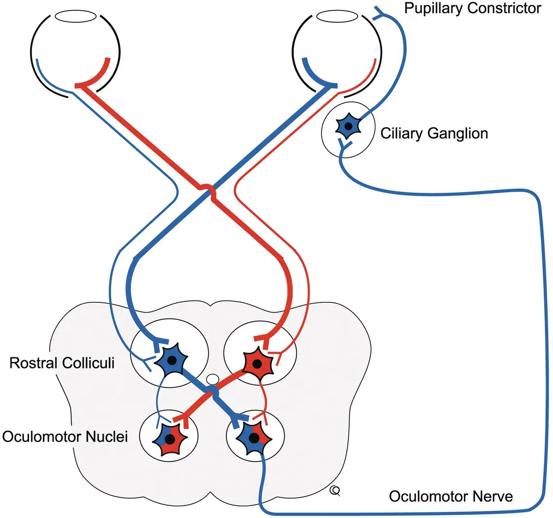
Figure 2.7 Pupillary light pathway.
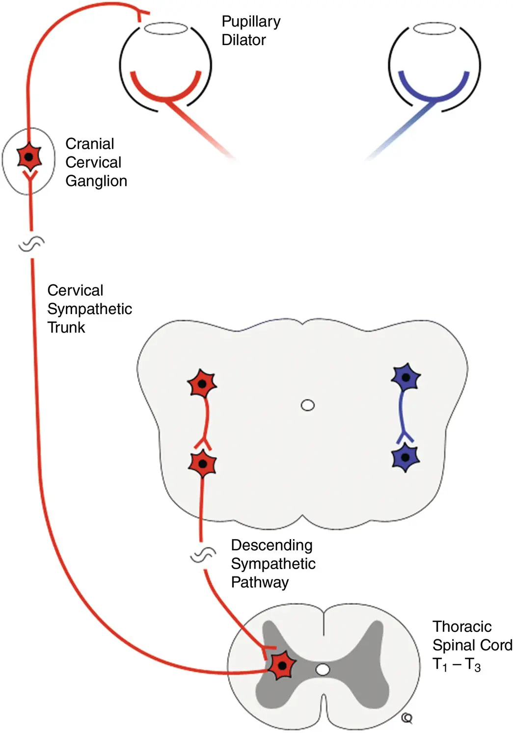
Figure 2.8 Ocular sympathetic pathway.
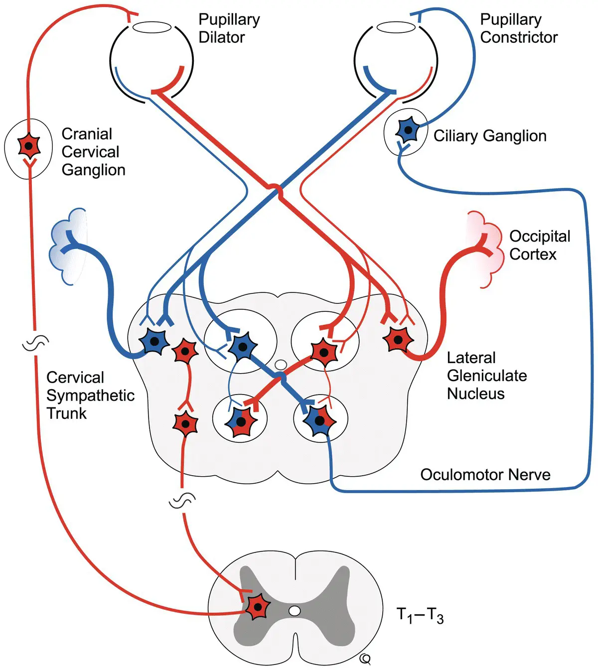
Figure 2.9 Visual and light pathways.
The first observation of CN III function to be made is the size and symmetry of the pupillary apertures, considering the amount of ambient light and the emotional status of the patient. The response of the pupils to light directed into each eye, the pupillary light responses, can be noted after observing the eyeballs for any lesions that may interfere with the response of the pupil such as iritis, iris atrophy, or adhesions. The normal response to light directed into one eye is constriction of both pupils. This is referred to as a direct response in the same or ipsilateral eye in which the light is shone and a slightly less pronounced consensual (indirect) response in the other, contralateral eye. The incoming or afferent pathway for this reflex is similar to that for the menace response to the level of the brainstem. The pathway goes via the optic nerve with most axons crossing within the optic chiasm, then through the optic tracts lateral and dorsal to the thalamus, and finally passing ventrally into the midbrain. Interconnections between left and right sides occur via the caudal commissure so that impulses arrive at each parasympathetic oculomotor nucleus in the midbrain on both sides. The motor, efferent pathway is from these nuclei, via the oculomotor nerves to the ciliary ganglia behind the eyeball and to the constrictor muscles of the pupils.
Because the observation of pupillary light responses in both eyes simultaneously is awkward to perform alone in large animals, the swinging light test is a useful alternative. The use of this test essentially removes the need to do consensual pupillary light reflex testing. The swinging light test is performed as follows. A bright focused source of light is shone into one eye, and after pupillary constriction is completed, the light is quickly transferred to the opposite eye in which further pupillary constriction is expected to occur if the eye is normal. This test is then performed in reverse and repeated with swinging movements of the light source, starting at say 30 cm from the eyes and moving step‐wise closer to say 5 cm from the eyeball. It is best to perform this test in dim light, and in performing the test in this manner the patient becomes accustomed to the swinging bright light so that it does not blink with a dazzle response when the light reaches the retina, which often obscures the initial fast pupillary response. If the pupil dilates when the light reaches the second eye in this test, there must be an afferent optic lesion involving that second eye.
The reason for further pupillary constriction occurring when the light reaches the second, normal eye is that the direct pupillary light reflex is stronger in its effect than the indirect or consensual pupillary light reflex. This fact should be recalled when interpreting some complex results of testing vision and pupillary responses, and two other issues are also worthy of iteration. First, the mydriatic effect of atropine can last for days if not weeks, producing confusion in evaluation and interpretation of pupillary responses to light. Second, it appears to take a more complete optic nerve or chiasmal lesion to cause observable mydriasis even with poor vision and a poor or even absent menace response, most particularly with bilateral lesions. Consider for example a yearling colt with obvious bilateral blindness when in its environment, that followed head trauma occurring after rearing and falling over backward onto the poll and suffering from bilateral, focal, traumatic optic neuropathy. The colt may still have essentially appropriate, mid‐sized pupils that even respond somewhat to a bright light. Evaluation of partial, especially asymmetric lesions such as this can be problematic unless this factor is appreciated.
These pupillary light reflexes are within the brainstem and thus are not affected by lesions involving the cerebral visual cortex ( Figure 2.9). A widely dilated, mydriatic pupil in an eye that has normal vision suggests an oculomotor nerve lesion; the pupil being unresponsive to light directed into either eye. The pupil in the contralateral eye, with normal oculomotor function, responds to light directed into both the ipsilateral eye (direct response) and the contralateral eye (consensual response). The oculomotor nerves are subject to damage following diffuse brain swelling and from space‐occupying lesions in the forebrain. Both processes can cause pressure to be exerted ventrally onto the brainstem and these nerves can be damaged as they lay between the midbrain and the basilar bones. With asymmetric swelling of cerebral tissue, greater pressure may be applied to one oculomotor nerve resulting in unequal pupils or anisocoria, which is usually evident as ipsilateral pupillary dilation. A severe brainstem contusion can produce various pupillary abnormalities in association with coma or semicoma, and these can change rapidly in the first few hours following injury. Progressive, bilateral, pupillary dilation following cranial injury warrants a grave prognosis.
When reporting results of pupillary function and light responses, it removes considerable confusion to describe what happens to each pupil when light is shone in each eye in turn and not to use the terms direct and consensual or indirect responses. Thus, with a left‐sided retrobulbar abscess involving left CN III parasympathetic fibers or ciliary ganglion or nerve, as well as the left optic nerve, the unambiguous description would be as follows. The patient is blind in the left eye only and has no menace response in the left eye with normal facial nerve function and normal palpebral reflexes. The left pupil is widely dilated, and light directed into the left eye causes no response in either pupil. Light directed into the right eye causes the right pupil to constrict but the left pupil remains dilated.
Читать дальшеИнтервал:
Закладка:
Похожие книги на «Large Animal Neurology»
Представляем Вашему вниманию похожие книги на «Large Animal Neurology» списком для выбора. Мы отобрали схожую по названию и смыслу литературу в надежде предоставить читателям больше вариантов отыскать новые, интересные, ещё непрочитанные произведения.


Обсуждение, отзывы о книге «Large Animal Neurology» и просто собственные мнения читателей. Оставьте ваши комментарии, напишите, что Вы думаете о произведении, его смысле или главных героях. Укажите что конкретно понравилось, а что нет, и почему Вы так считаете.