Alan Gunn - Parasitology
Здесь есть возможность читать онлайн «Alan Gunn - Parasitology» — ознакомительный отрывок электронной книги совершенно бесплатно, а после прочтения отрывка купить полную версию. В некоторых случаях можно слушать аудио, скачать через торрент в формате fb2 и присутствует краткое содержание. Жанр: unrecognised, на английском языке. Описание произведения, (предисловие) а так же отзывы посетителей доступны на портале библиотеки ЛибКат.
- Название:Parasitology
- Автор:
- Жанр:
- Год:неизвестен
- ISBN:нет данных
- Рейтинг книги:3 / 5. Голосов: 1
-
Избранное:Добавить в избранное
- Отзывы:
-
Ваша оценка:
Parasitology: краткое содержание, описание и аннотация
Предлагаем к чтению аннотацию, описание, краткое содержание или предисловие (зависит от того, что написал сам автор книги «Parasitology»). Если вы не нашли необходимую информацию о книге — напишите в комментариях, мы постараемся отыскать её.
Highly detailed textbook on parasites and parasite relationships Parasitology: An Integrated Approach
Parasitology: An Integrated Approach, 2nd edition
Parasitology — читать онлайн ознакомительный отрывок
Ниже представлен текст книги, разбитый по страницам. Система сохранения места последней прочитанной страницы, позволяет с удобством читать онлайн бесплатно книгу «Parasitology», без необходимости каждый раз заново искать на чём Вы остановились. Поставьте закладку, и сможете в любой момент перейти на страницу, на которой закончили чтение.
Интервал:
Закладка:
4.2 Phylum Kinetoplastida
The Kinetoplastida is a large diverse group of protozoa that includes both plant and animal parasites ( Table 4.1). Some authors consider the Kinetoplastida to be a phylum, while others refer to it as a class or an order. They are commonly known as the trypanosomes from the genus Trypanosoma that includes the causative agents of Human African Trypanosomiasis (HAT) and several other parasites of medical and veterinary importance. The genus name Trypanosoma derives from the Greek words trypano ( τρύπανο ) = an auger [a device for boring holes in wood] and soma ( σώμα ) = body that refer to their corkscrew‐like locomotion. Magez and Radwanska (2014) provide a comprehensive review of all aspects of trypanosome biology and their transmission.
Table 4.1 Examples of kinetoplastid parasites of medical, veterinary, and agricultural importance and the diseases they cause.
| Genus | Example | Host | Vector/transmission | Disease |
|---|---|---|---|---|
| Leishmania | Leishmania donovani | Humans, dogs, rats | Phlebotomus sandflies | Kala‐azar (visceral leishmaniasis) |
| Leishmania major | Humans, monkeys, dogs, rodents | Phlebotomus sandflies | Cutaneous leishmaniasis | |
| Leishmania tropica | Humans, monkeys, dogs, rodents | Phlebotomus sandflies | Cutaneous leishmaniasis | |
| Leishmania braziliensis | Humans, sloths, monkeys, opossums, and many others | Lutzomyia sandflies | Cutaneous/mucocutaneous leishmaniasis | |
| Trypanosoma | Trypanosoma brucei gambesiense | Humans | Tsetse flies (Glossina spp.) | African trypanosomiasis (sleeping sickness) |
| Trypanosoma congolense | Cattle | Tsetse flies (Glossina spp.) | African trypanosomiasis (nagana) | |
| Trypanosoma equiperdum | Horses | venereal | Dourine | |
| Trypanosoma cruzi | Humans, dogs, cats, rats, and many others | Triatomid bugs | Chagas disease | |
| Phytomonas | Phytomonas staheli | Coconut palms | Lincus lobuliger (Pentatomid bug) | Hartroot |
The Kinetoplastida are characterised by their possession of a flagellum and a unique intracellular structure called a kinetoplast ( Figure 4.1). The kinetoplast is a disk of interlocking DNA circles (kDNA) located within a large mitochondrion. The structure of kinetoplast DNA is unlike that found in any other organism and its complex replication involves special proteins (da Silva et al. 2017). It may therefore be possible to design drugs to interfere with the replication of kinetoplast DNA. The position of the mitochondrion is such that the kinetoplast is just underneath the kinetosome that is itself situated underneath the base of the flagellum. The kinetosome (sometimes called the basal body), is a structure found in many organisms and is homologous with the centriole; it is involved in the formation of the flagellum. The Kinetoplastida always have a flagellum that may be long and free, incorporated into the cell surface to form an undulating membrane, or small and enclosed within a pocket. The inner core of the flagellum is the axoneme. Alongside this, and connecting to it, is the paraxial rod that consists of a lattice‐like crystalline array of structural proteins. In the promastigote, epimastigote, and trypomastigote stages, the flagellum emerges at the anterior end of the cell and therefore acts as a propeller that pulls the cell along rather than pushing it from behind ( Figure 4.2).
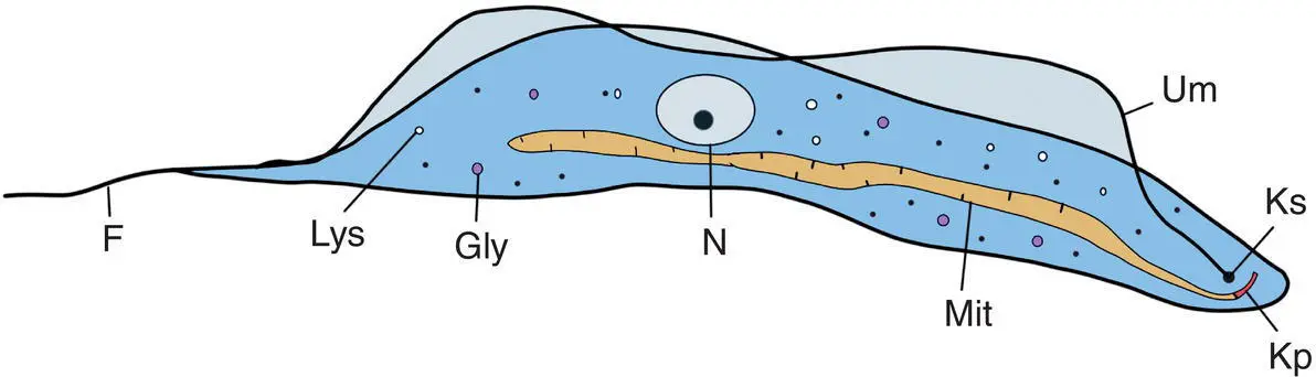
Figure 4.1 Diagram of a typical trypanosome. Kp: kinetoplast; Ks: kinetosome; F: flagellum; Mit: mitochondrion; Gly: glycosome; N: nucleus; Lys: lysosome; Um: undulating membrane.
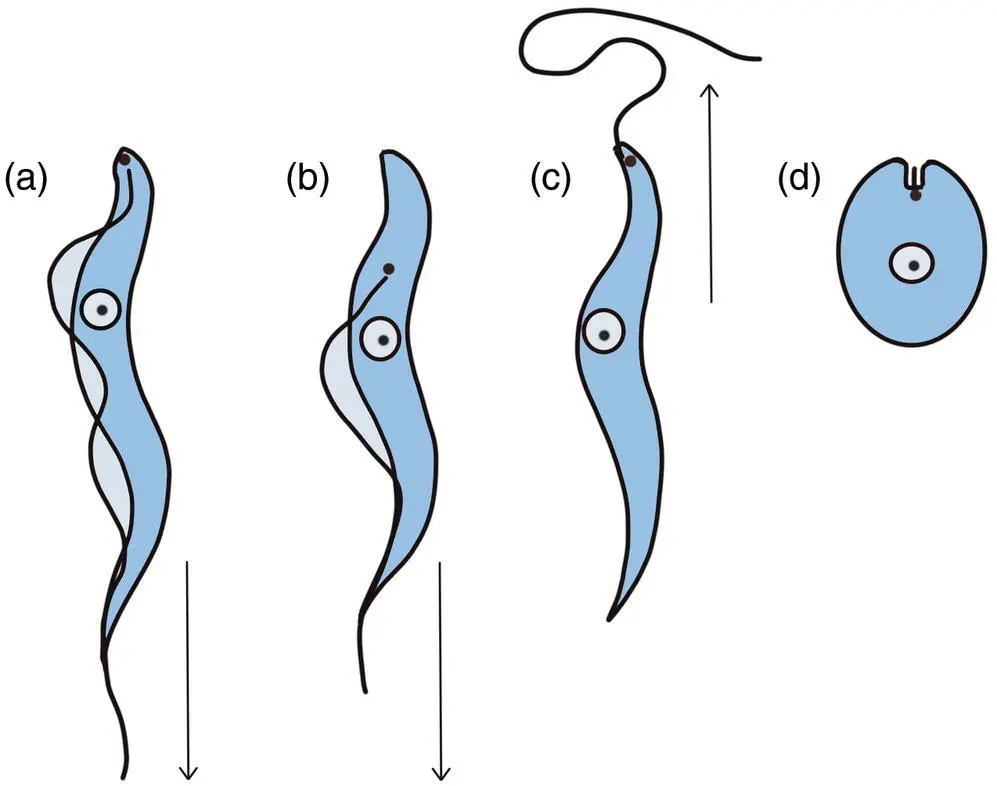
Figure 4.2 Morphological forms of trypanosomes. (a) Trypomastigote. (b) Epimastigote. (c) Promastigote. (d) Amastigote. The flagellum pulls the cell forwards rather than pushes it from behind. Arrows: direction of movement.
The Kinetoplastida have a unique organelle called the glycosome. This may be related to the peroxisomes (which they do not have) found in other organisms. The glycosomes are about 0.25 μm in diameter and contain glycolytic enzymes that are normally present in the cytoplasm of other organisms. The bloodstream forms of trypanosomes are extremely metabolically active and soon die if they run out of glucose to metabolise. The glycosomes are the site of glucose metabolism, and therefore, like the kinetoplast DNA, they are a potential target for antiparasitic drug design.
Many species within the Kinetoplastida are only parasitic in insects. For example, members of the genus Leptomonas live in the gut of various insects including blood‐feeding reduviid bugs (Kaufer et al. 2017). Because they have only a single host, these species are referred to as monoxenous although there are rare case reports of human infections in patients who are HIV+ve. There are also accounts of Leptomonas co‐infections with visceral leishmaniasis and post kala‐azar dermal leishmaniasis caused by Leishmania donovani (Thakur et al. 2020).
Many of the Kinetoplastida alternate between an invertebrate host such as a blood‐feeding insect or leech, and a vertebrate host with development occurring in both. Parasites that have more than one type of host are called heteroxenous. Heteroxenous Kinetoplastida species often express two or more morphological forms with one form present in the invertebrate and the other in the vertebrate ( Table 4.2). Some members of the Kinetoplastida exhibit sexual reproduction, or something similar (Berry et al. 2019; Gibson and Peacock 2019), but it is uncertain whether it is a widespread phenomenon in the group.
4.2.1 Genus Leishmania
Members of the genus Leishmania exhibit two distinct morphologies: the amastigote form that occurs in the vertebrate host, and the promastigote form that occurs in the invertebrate vector. The vertebrate hosts are mostly mammals, whilst the invertebrates are various species of sandflies. Those species parasitic in reptiles belong to the subgenus Sauroleishmania and do not cause zoonotic infections. Perhaps counter‐intuitively, molecular evidence indicates that the Leishmania evolved in the Neotropical regions during the Mesozoic era as parasites of mammals and those species parasitizing reptiles, the Sauroleishmania , subsequently evolved from them (Noyes et al. 2000).
Table 4.2 Morphological forms of Kinetoplastida parasitic in humans and domestic animals.
| Morphological form | Description | Example |
|---|---|---|
| Amastigote | Kinetoplast and kinetosome above the nucleus, flagellum short and confined in pocket. Cell shape globular | Leishmania donovani inside vertebrate macrophage Trypanosoma cruzi in human spleen, liver, muscle, and other cell types |
| Promastigote | Kinetoplast and kinetosome at anterior end of cell, flagellum free, and long. Cell shape elongate | Leishmania donovani in sandfly gut |
| Epimastigote | Kinetoplast and kinetosome close and anterior to the nucleus. There is a short undulating membrane before the flagellum emerges at the anterior of the cell. Cell shape elongate | Trypanosoma cruzi in triatomid gut |
| Trypomastigote | Kinetoplast and kinetosome at posterior end of cell. Flagellum forms an undulating membrane that runs the length of the cell and may continue free when it reaches the anterior end. Cell shape elongate | Trypansoma cruzi in human bloodstream |
Trypanosomes that are parasitic insects exhibit other morphological forms, such as choanomastigote, opisthomastigote, and paramastigote.
Читать дальшеИнтервал:
Закладка:
Похожие книги на «Parasitology»
Представляем Вашему вниманию похожие книги на «Parasitology» списком для выбора. Мы отобрали схожую по названию и смыслу литературу в надежде предоставить читателям больше вариантов отыскать новые, интересные, ещё непрочитанные произведения.


Обсуждение, отзывы о книге «Parasitology» и просто собственные мнения читателей. Оставьте ваши комментарии, напишите, что Вы думаете о произведении, его смысле или главных героях. Укажите что конкретно понравилось, а что нет, и почему Вы так считаете.