Gerald E. McDonnell - Antisepsis, Disinfection, and Sterilization
Здесь есть возможность читать онлайн «Gerald E. McDonnell - Antisepsis, Disinfection, and Sterilization» — ознакомительный отрывок электронной книги совершенно бесплатно, а после прочтения отрывка купить полную версию. В некоторых случаях можно слушать аудио, скачать через торрент в формате fb2 и присутствует краткое содержание. Жанр: unrecognised, на английском языке. Описание произведения, (предисловие) а так же отзывы посетителей доступны на портале библиотеки ЛибКат.
- Название:Antisepsis, Disinfection, and Sterilization
- Автор:
- Жанр:
- Год:неизвестен
- ISBN:нет данных
- Рейтинг книги:4 / 5. Голосов: 1
-
Избранное:Добавить в избранное
- Отзывы:
-
Ваша оценка:
Antisepsis, Disinfection, and Sterilization: краткое содержание, описание и аннотация
Предлагаем к чтению аннотацию, описание, краткое содержание или предисловие (зависит от того, что написал сам автор книги «Antisepsis, Disinfection, and Sterilization»). Если вы не нашли необходимую информацию о книге — напишите в комментариях, мы постараемся отыскать её.
is well suited as a textbook and is outstanding as a reference book for facilities managers and application engineers in manufacturing plants, hospitals, and food production facilities. It is also essential for public health officials, healthcare professionals, and infection control practitioners.
Antisepsis, Disinfection, and Sterilization — читать онлайн ознакомительный отрывок
Ниже представлен текст книги, разбитый по страницам. Система сохранения места последней прочитанной страницы, позволяет с удобством читать онлайн бесплатно книгу «Antisepsis, Disinfection, and Sterilization», без необходимости каждый раз заново искать на чём Вы остановились. Поставьте закладку, и сможете в любой момент перейти на страницу, на которой закончили чтение.
Интервал:
Закладка:
Figure 1.6also shows that other structures can be present on the surfaces of bacteria. Of particular interest in the consideration of biocidal processes are external barriers that can protect the cell from its environment. Many bacteria produce an external layer of highmolecular-weight polysaccharides, as well as associated lipids and proteins, which is referred to as a glycocalyx. This can be a simple, loosely associated slime layer or a more rigid, thicker, and firmly attached capsule structure. Capsules can range in structure and size, typically from one-half to five times the cell diameter in thickness. Glycocalyx production plays an important role in the development of bacterial biofilms, which are a further intrinsic resistance mechanism (see section 8.3.8). Glycocalyx structures are found in both gram-positive and gram-negative bacteria. Examples are Streptococcus mutans (in dental plaque), Streptococcus pneumoniae (in nasopharyngeal colonization), and E. coli (enteropathogenic strains that attach to epithelial cells in the intestine). In addition to direct cell protection, they can also play roles in pathogenesis, in bacterial attachment to surfaces, and in preventing drying of the cell. Other bacteria (including archaea) produce an S-layer, similar to polysaccharide capsules, which is composed of protein and glycoproteins to form an external crystalline structure; an example is the external surface of Bacillus anthracis , which produces an S-layer consisting of two protein types that is itself covered by a unique protein (poly-D-glutamic acid) capsule layer.
TABLE 1.10 Examples of gram-negative bacteria
| General type | Key characteristics | Example(s) | Significance |
| Spirochetes | Thin; helical or spiral shaped | Borrelia | Cause what are often described as tick-borne diseases in animals, humans, and birds (e.g., B. burgdorferi , implicated in Lyme disease) |
| Treponema | Cause human and animal diseases; T. pallidum causes syphilis, a persistent sexually transmitted disease | ||
| Helical, vibroid | Usually mobile; vibroid shaped | Campylobacter Helicobacter | C. jejuni causes gastroenteritis H. pylori causes peptic ulcers due to gastritis |
| Aerobic or microaerophilic rods and cocci | Diverse group of rods or cocci that use oxygen for growth | Acetobacter Bordetella Legionella | Cause food spoilage B. pertussis causes whooping cough, a respiratory disease Associated with water or moist environments; L. pneumophila causes a form of pneumonia known as Legionnaires’ disease |
| Neisseria | Most strains are nonpathogenic and found on mucous membranes. N. gonorrhoeae causes the sexually transmitted disease gonorrhoea, and N. meningitidis can cause meningitis in young adults. | ||
| Pseudomonas, Burkholderia | Common environmental contaminants in water and soil; some strains are plant pathogens. P. aeruginosa and B. cepacia are frequently implicated in hospital-acquired infections, usually associated with proliferation in moist environments and water lines. Pseudomonads can cause biofilm fouling in industrial water lines. | ||
| Facultative anaerobes | Rod-shaped; can grow in the presence or absence of oxygen | Enterobacteriaceae | |
| Erwinia | Plant saprophytes and pathogens | ||
| Escherichia | E. coli is the most prevalent microorganism in the lower intestinal tract and a common cause of intestinal and urinary tract infections. It is also widely used as a cloning host in molecular biology. | ||
| Salmonella | Leading cause of gastroenteritis, mostly food or water borne; examples are S. enterica serovarTyphi (causing typhoid fever) and S enterica serovarTyphimurium (causing gastroenteritis and enteric fever) | ||
| Yersinia | Zoonotic infections; Y. pestis causes plague | ||
| Vibrionaceae | |||
| Vibrio | Gastrointestinal diseases, including cholera ( V. cholerae ) and food poisoning ( V. parahaemolyticus ) | ||
| Pasteurellaceae | |||
| Haemophilus | Commonly found in the upper respiratory tracts of humans and some animals; H. infiuenzae is a leading cause of meningitis in children | ||
| Pasteurella | Can cause septicemia in animals and humans | ||
| Other gram-negative bacteria | Various shapes and growth requirements | Bacteroides | Anaerobic rods; commonly found in the intestine and as opportunistic pathogens in wounds |
| Veillonella | Anaerobic cocci; human and animal parasites | ||
| Rickettsia, Chlamydia, Chlamydophila | Obligate intracellular pathogens | ||
| Cyanobacteria | Free-living in water; photosynthetic; can be unicellular or filamentous | ||
| Myxobacteria | Waterborne bacteria that are motile by a gliding mechanism | ||
| Leptothrix | Sheathed, filamentous bacteria associated with polluted water |
Bacteria can also have a variety of other proteinaceous cell surface appendages, including pili, fimbriae, and flagella ( Fig. 1.6). For example, flagellar filaments are composed primarily of flagellin protein subunits and have other proteins that interact with the cell membrane and/or cell wall structure. Flagella are specifically involved in bacterial motility. Fimbriae and pili play important roles in surface, including cell surface, interactions.
A further cell wall structure that deserves separate consideration is the mycobacterial cell wall ( Fig. 1.7). Mycobacteria are aerobic, slow-growing, rod-shaped bacteria (for example, Mycobacterium tuberculosis [ Fig. 1.9]), which typically stain gram positive and can be further differentiated by acid-fast stain (staining with fuchsin, which resists acid and alcohol decolorization) due to their unique hydrophobic cell wall structure.
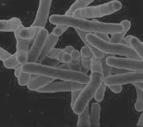
FIGURE 1.9 Cells of Mycobacterium tuberculosis . Courtesy of Clifton Barry, NIAID.
This mycobacterial cell wall structure presents a strong permeability barrier and is responsible for the higher level of resistance to antibiotics and biocides in comparison to other bacteria. The cell membrane is similar to that described in other bacteria, which can be linked to the cell wall by glycolipids. The cell wall has a three-layer structure, consisting of a peptidoglycan layer external to the cell membrane, which is covalently linked to a specific polysaccharide (known as arabinogalactan), and finally an external mycolic acid layer. The peptidoglycan is similar to that in other bacteria, but N -acetylmuramic acid is replaced with N -glycoylmuramic acid and cross-linked by three- and four-amino-acid peptides. Arabinogalactan is a polysaccharide of arabinose and galactose. The mycolic acids are attached to arabinose residues of the arabinogalactan and are some of the longest fatty acids known in nature. In mycobacteria, they typically range in carbon length from C 60to C 90and can make up >50% of the cell weight. In addition, the mycobacterial cell wall can contain a variety of proteins (including enzymes), short-chain fatty acids, waxes, and LPSs. Examples are the LPS lipoarabinomannan, which plays a role in host interactions during M. tuberculosis infections, and porin proteins, with a function in molecule transport similar to that seen in the outer membranes of gram-negative bacteria. In some disease-causing mycobacteria, these may also form an external capsule containing enzymes and adherence factors that play roles in mycobacterial pathogenesis. Similar basic cell wall structures have been identified in other bacteria, including actinomycetes ( Nocardia ) and gram-positive rods ( Corynebacterium ), with notably shorter-chained mycolic acids of C 46to C 60and C 22to C 32, respectively, and in some cases ( Amycolatopsis ) no mycolic acids. Examples of bacteria with mycobacterium-like cell wall structures are given in Table 1.11.
Читать дальшеИнтервал:
Закладка:
Похожие книги на «Antisepsis, Disinfection, and Sterilization»
Представляем Вашему вниманию похожие книги на «Antisepsis, Disinfection, and Sterilization» списком для выбора. Мы отобрали схожую по названию и смыслу литературу в надежде предоставить читателям больше вариантов отыскать новые, интересные, ещё непрочитанные произведения.


Обсуждение, отзывы о книге «Antisepsis, Disinfection, and Sterilization» и просто собственные мнения читателей. Оставьте ваши комментарии, напишите, что Вы думаете о произведении, его смысле или главных героях. Укажите что конкретно понравилось, а что нет, и почему Вы так считаете.